Field Guide to Venomous and Medically Important Invertebrates ...
Field Guide to Venomous and Medically Important Invertebrates ...
Field Guide to Venomous and Medically Important Invertebrates ...
You also want an ePaper? Increase the reach of your titles
YUMPU automatically turns print PDFs into web optimized ePapers that Google loves.
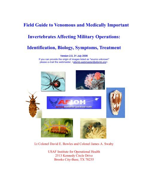
<strong>Field</strong> <strong>Guide</strong> <strong>to</strong> <strong>Venomous</strong> <strong>and</strong> <strong>Medically</strong> <strong>Important</strong><br />
<strong>Invertebrates</strong> Affecting Military Operations:<br />
Identification, Biology, Symp<strong>to</strong>ms, Treatment<br />
Version 2.0, 31 July 2006<br />
If you can provide the origin of images listed as "source unknown"<br />
please e-mail the webmaster, .<br />
Lt Colonel David E. Bowles <strong>and</strong> Colonel James A. Swaby<br />
USAF Institute for Operational Health<br />
2513 Kennedy Circle Drive<br />
Brooks City-Base, TX 78235
Preface<br />
This guide is intended <strong>to</strong> be a primary <strong>and</strong> expeditious information source for aiding deployed<br />
military personnel in the initial steps of pest identification related <strong>to</strong> surveillance <strong>and</strong> public<br />
health matters associated with invertebrates of medical importance. It is not intended <strong>to</strong> be a<br />
definitive or exhaustive treatise on the subject material. Furthermore, the contents of this manual<br />
represent only a compilation of the available literature, <strong>and</strong> it is not intended in any fashion <strong>to</strong><br />
represent an original research paper. This document is intended <strong>to</strong> be a starting point for<br />
obtaining information <strong>and</strong> not an end point. Readers seeking additional information on a<br />
particular <strong>to</strong>pic addressed herein should refer <strong>to</strong> the referenced material or other sources of<br />
information as appropriate.<br />
Treatment guidelines, where presented, are based on current available information at the time<br />
this document was written. Practitioners have the sole responsibility <strong>to</strong> ensure the correct<br />
dosages of medicines are provided, <strong>and</strong> they also should ensure correct treatment regimes by<br />
consulting the most recent appropriate information sources.<br />
Use of trade or br<strong>and</strong> names in this publication is for the sole purpose of illustration <strong>and</strong> does not<br />
imply endorsement by the United States Air Force.<br />
THIS FIELD GUIDE IS INTENDED FOR USE BY MILITARY AND CIVILIAN<br />
EMPLOYEES OF THE UNITED STATES DEPARTMENT OF DEFENSE ONLY AND<br />
IS NOT AUTHORIZED FOR PUBLIC DISSEMINATION OR SALE IN ANY FORM<br />
Contents
Introduction<br />
Types of negative interactions with arthropods<br />
Envenomation<br />
Myiasis<br />
Bites <strong>and</strong> Piercing<br />
Urtication<br />
Allergic reactions<br />
Delusory Parasi<strong>to</strong>sis <strong>and</strong> En<strong>to</strong>mophobia<br />
Dangerous <strong>Invertebrates</strong> of Military Importance<br />
Arachnids<br />
Spiders<br />
Banana spiders<br />
Black widow spiders <strong>and</strong> their relatives<br />
Brown recluse spiders<br />
Funnel web spiders<br />
Hobo spiders<br />
Megalopmorph spiders<br />
Six-eyed s<strong>and</strong>/crab spiders<br />
W<strong>and</strong>ering spiders<br />
White-tailed spiders<br />
Yellow sac spiders<br />
Other potentially dangerous spiders<br />
Tarantulas<br />
Scorpions<br />
Dangerous scorpions<br />
Effects of scorpion venom<br />
Treatment of envenomation by scorpions<br />
Mites<br />
Chiggers or harvest mites<br />
Scabies mites<br />
Other medically important mites that bite people<br />
Dust mites<br />
Flour <strong>and</strong> grain mites<br />
Ticks<br />
Camel Spiders<br />
Insects<br />
Collembola<br />
Pubic louse<br />
Human lice<br />
Head louse<br />
Body louse<br />
Cockroaches<br />
True bugs<br />
Assassin <strong>and</strong> kissing bugs<br />
Bed bugs
Other biting Hemiptera<br />
Ants, wasps, <strong>and</strong> bees<br />
Honey bees<br />
Bumble <strong>and</strong> carpenter bees<br />
Other bees<br />
Wasps <strong>and</strong> hornets<br />
Velvet ants<br />
Other wasps<br />
Ants<br />
Bulldog <strong>and</strong> jumper ants<br />
Bullet ants<br />
Fire ants<br />
Harvester ants<br />
Other ants<br />
Lepidoptera (moths)<br />
Beetles<br />
Blister beetles<br />
Rover beetles<br />
Dermestids<br />
Flies<br />
Flies that cause myiasis<br />
Human bot fly<br />
Tumbu <strong>and</strong> Lund’s fly<br />
Congo floor maggot<br />
Sheep bot fly<br />
Sheep maggot<br />
Old World screw worm<br />
New World screw worm<br />
Wohlfahrtia magnifica<br />
Other flies that cause myiasis<br />
Treatment of myiasis<br />
Biting flies<br />
Mosqui<strong>to</strong>es<br />
Biting midges<br />
S<strong>and</strong> flies<br />
Black flies<br />
Tsetse flies<br />
Stable <strong>and</strong> dog flies<br />
Horse <strong>and</strong> deer flies<br />
Filth flies<br />
Fleas<br />
Human flea<br />
Oriental rat flea<br />
Northern rat flea<br />
Western hen flea<br />
Tunga flea or chigoe
Other Arthropods<br />
Centipedes<br />
Millipedes<br />
Sticktight flea<br />
Rabbit flea<br />
Squirrel flea<br />
Rodent flea<br />
Ground squirrel flea<br />
―S<strong>and</strong> <strong>and</strong> beach fleas‖<br />
Other <strong>Invertebrates</strong><br />
Porifera<br />
Coelenterates<br />
Jellyfish<br />
Moon jellyfish<br />
Sea Nettles<br />
Sea Wasp or box jellyfish<br />
Small box jellyfish<br />
False box jellyfish<br />
Pink jellyfish<br />
Thimble jellyfish<br />
Lion’s mane jellyfish<br />
Portuguese Man O’ War<br />
Sea anemones<br />
Sea Ferns<br />
Corals<br />
Bryozoans<br />
Echinoderms<br />
Sea stars <strong>and</strong> brittle stars<br />
Crown of thorns starfish<br />
Mosaic sea star<br />
Chain-link brittle star<br />
Sea urchins<br />
Sea cucumbers<br />
Annelid worms<br />
Leeches<br />
Polychetes or bristleworms<br />
Mollusks<br />
Squids, oc<strong>to</strong>puses <strong>and</strong> cuttlefishes<br />
Cone shells<br />
Pteropods<br />
Nudibranchs or sea slugs<br />
Snails as hosts of schis<strong>to</strong>somiasis<br />
Swimmer’s itch
SECTIONS<br />
Introduction<br />
The purpose of this guide is <strong>to</strong> present the reader with a basic yet sound underst<strong>and</strong>ing of the<br />
dangerous types of invertebrates that may be encountered during military operations worldwide.<br />
Brief descriptions of the physical <strong>and</strong> behavioral characteristics of these animals are presented.<br />
This field guide only considers those animals which pose a threat from direct contact <strong>and</strong> does<br />
not address <strong>to</strong>xic responses from food or contact allergies, or consumption of certain poisonous<br />
animals. In addition, a few invertebrates are included here not because they present a danger <strong>to</strong><br />
people, but because they are often incorrectly presumed <strong>to</strong> be dangerous.<br />
A considerable amount of literature was reviewed, digested <strong>and</strong> consolidated <strong>to</strong> create this field<br />
guide. In an effort <strong>to</strong> make a concise guide with streamlined text <strong>to</strong> facilitate ease of reading<br />
under the operational conditions it was intended <strong>to</strong> serve, individual information sources have<br />
not been cited in the text. To the maximum extent possible the information extracted from these<br />
references has been paraphrased. Some suggested references for obtaining additional<br />
information on these animals are included at the end of this document.<br />
An attempt was made <strong>to</strong> use common language in this document <strong>to</strong> describe symp<strong>to</strong>ms<br />
associated with arthropod attacks. However, for the sake of brevity in some situations, or where<br />
specific terms were essential <strong>to</strong> avoid confusion, we used medical <strong>and</strong> technical terminology.<br />
Those terms are defined in Appendix 1.
Threats from invertebrates encompass two broad categories: point source threats <strong>and</strong><br />
psychological threats. Point source threats are those that can cause physical injury or death in a<br />
brief period of time. The sting of a wasp, <strong>and</strong> transmission of deadly disease agents are two<br />
examples of point source threats. Psychological threats, by comparison, are those that do not kill<br />
or directly threaten health, but rather present unpleasant situations for people <strong>to</strong> the extent that<br />
routine functioning is impaired. Both point source <strong>and</strong> psychological threats have the real<br />
potential <strong>to</strong> disrupt or even halt military operations, <strong>and</strong> they present serious concerns that<br />
comm<strong>and</strong>ers <strong>and</strong> the military medical community must address during both peacetime <strong>and</strong><br />
contingencies.<br />
There are several types of potential negative interactions associated with invertebrates including<br />
physical pain, disease, envenomation, myiasis, allergic reactions, psychological disorders, <strong>and</strong><br />
death.<br />
Physical pain- Bites, piercings, <strong>and</strong> stings caused by a wide variety of invertebrates can produce<br />
varying amounts of suffering among victims. Symp<strong>to</strong>ms can range from mild annoyance <strong>to</strong><br />
incapacitation. Although such physical trauma generally is not lethal, it may render a victim<br />
incapable of normal activity, <strong>and</strong> it can result in psychological disturbance among certain<br />
individuals.<br />
Disease- Transmission of arthropod- or vec<strong>to</strong>rborne disease agents represents the most<br />
substantial <strong>and</strong> continuous non-combat threat <strong>to</strong> military members during deployments. The<br />
World Health Organization has estimated there are 10 million cases annually of vec<strong>to</strong>rborne<br />
diseases worldwide with many being fatal. His<strong>to</strong>rically, vec<strong>to</strong>rborne diseases have produced far
more morbidity <strong>and</strong> mortality (greater than 60%) among U.S. military forces during modern<br />
wars than battle injury <strong>and</strong> non-battle injury combined. In addition, vec<strong>to</strong>rborne disease<br />
epidemics can serve as a severe detriment <strong>to</strong> force morale.<br />
Envenomation, the injection of venom in<strong>to</strong> the body through either bites <strong>and</strong>/or stings, is perhaps<br />
the most rapid <strong>and</strong> immediate deleterious response invertebrates can inflict on humans. The<br />
response of such envenomations can range from mild irritation <strong>and</strong> limited necrosis of tissue <strong>to</strong><br />
systemic failure <strong>and</strong> death. The venoms producing these conditions are broadly grouped as<br />
either neuro<strong>to</strong>xic or necrotic. Neuro<strong>to</strong>xic venoms are those that negatively affect the nervous<br />
system while necrotic venoms are those that destroy blood <strong>and</strong> tissue. Occasionally, the venom<br />
of some invertebrates contains both neuo<strong>to</strong>xic <strong>and</strong> necrotic properties. In addition <strong>to</strong> injecting<br />
venom, some caterpillars <strong>and</strong> beetles produce <strong>to</strong>xins that cause dermatitis when contacted.<br />
Myiasis is the invasion of the tissue of man or animals with the larvae (maggots) of certain flies<br />
(Diptera) that consume flesh or body fluids for sustenance. Such invasions may be benign or<br />
even asymp<strong>to</strong>matic, or they may result in more destructive disturbances. Myiasis is often<br />
described in terms associated with the site of entry. Types of myiasis recognized in humans<br />
include urogenital, gastrointestinal, ocular, auricular, <strong>and</strong> cutaneous which is the traumatic<br />
invasion of tissue <strong>and</strong> the most significant form of myiasis. Most instances of myiasis are<br />
accidental or opportunistic (facultative) rather than obligate. Although flesh-eating dipteran<br />
larvae can be successfully used <strong>to</strong> debride necrotic tissue from wounds under controlled medical<br />
conditions, myiasis under operational conditions potentially can damage healthy tissue <strong>and</strong><br />
produce severe psychological distress in victims. When wounds are involved, the term
―traumatic‖ may be applied, <strong>and</strong> when the lesion is boil like, it is referred <strong>to</strong> as furuncular<br />
myiasis. When larvae burrow in the skin in such a way that the progress may be followed as the<br />
larva advances, the term ―creeping myiasis‖ (creeping eruption) is applied. Myiasis has<br />
tremendous potential for psychological disturbance among afflicted military personnel.<br />
Urtication is a physiological response <strong>to</strong> contact with <strong>to</strong>xins of certain invertebrate body parts,<br />
such as the setae of certain moth larvae, <strong>and</strong> nema<strong>to</strong>cysts (stinging cells) of jellyfish <strong>and</strong> corals.<br />
Urtication can cause a painful burning <strong>and</strong> itchy skin eruption, or hives, at the point of contact.<br />
Although rarely fatal, urtication can be debilitating <strong>and</strong> may result in systemic shock in some<br />
individuals.<br />
Allergic Reactions occur primarily through contact with venom, saliva, or certain body parts of<br />
invertebrates such as setae. Reactions can be either localized (wheals, swelling) or systemic<br />
(anaphylactic shock), <strong>and</strong> the range of severity, including death, is broad.<br />
Delusory Parasi<strong>to</strong>sis <strong>and</strong> En<strong>to</strong>mophobia are psychological disorders stemming from contact with<br />
insects <strong>and</strong> their relatives. Psychological threats posed by invertebrates often are cumulative in<br />
their effect. In other words, the more experience an individual has the greater the negative<br />
impact on health <strong>and</strong> welfare. The importance of such cumulative encounters is a function of the<br />
number <strong>and</strong> diversity of pests in an area, the quality of living conditions, ability <strong>to</strong> escape the<br />
pests, fatigue, <strong>and</strong> stress. Under certain conditions, such as an extended deployment, nuisance<br />
pests can become a more substantial threat <strong>to</strong> mission success than disease, especially when pest<br />
densities are high <strong>and</strong> disease incidence is low. Delusory parasi<strong>to</strong>sis often is an intensely
emotional psychological disorder characterized by the unfounded belief that parasites, usually<br />
insects, are living on or in the body. This condition, although very rare, can become sufficiently<br />
severe in some individuals <strong>to</strong> be incapacitating, <strong>and</strong> these individuals often require professional<br />
mental health care. En<strong>to</strong>mophobia, by comparison, is simply an irrational fear of insects <strong>and</strong><br />
their relatives, or the damage or diseases they are capable of inflicting. For example, some<br />
individuals may develop an irrational fear of bees after being stung. The primary difference<br />
between en<strong>to</strong>mophobia <strong>and</strong> delusory parasi<strong>to</strong>sis is that the former occurs only in the presence of<br />
certain insects while the latter encompasses a near constant state of agitation <strong>and</strong> distress.<br />
Pesticide Use for Controlling Invertebrate Vec<strong>to</strong>rs <strong>and</strong> Pests<br />
Pesticides for controlling vec<strong>to</strong>rs <strong>and</strong> pests should be applied only by qualified pest management<br />
personnel. Department of Defense guidance on pesticide selection <strong>and</strong> integrated pest<br />
management can be found in the Contingency Pest Management <strong>Guide</strong> (Technical <strong>Guide</strong> No. 24,<br />
Armed Forces Pest Management Board), <strong>and</strong> <strong>Guide</strong> <strong>to</strong> Operational Surveillance of <strong>Medically</strong><br />
<strong>Important</strong> Vec<strong>to</strong>rs <strong>and</strong> Pests (―Operational En<strong>to</strong>mology‖) available from the USAF School of<br />
Aerospace Medicine. Additional information <strong>and</strong> instruction on pest management issues of<br />
military importance can be found on the website of the Armed Forces Pest Management Board<br />
(www.afpmb.org).<br />
Personal Protective Measures Against Arthropods<br />
Guidance on personal protection from arthropods <strong>and</strong> other invertebrates can be found in<br />
Personal Protective Measures Against Insects <strong>and</strong> Other Arthropods of Military Significance<br />
(Technical <strong>Guide</strong> No. 36, Armed Forces Pest Management Board), <strong>and</strong> <strong>Guide</strong> <strong>to</strong> Operational
Surveillance of <strong>Medically</strong> <strong>Important</strong> Vec<strong>to</strong>rs <strong>and</strong> Pests (―Operational En<strong>to</strong>mology‖). Additional<br />
information <strong>and</strong> instruction on personal protection from arthropods can be found on the website<br />
of the Armed Forces Pest Management Board (www.afpmb.org).<br />
Dangerous <strong>Invertebrates</strong> of Military Importance<br />
Appendix 1 contains a list of the dangerous invertebrates of military importance including their<br />
geographic distributions.<br />
Arachnids<br />
(Spiders, Scorpions, Ticks, Mites, Camel Spiders)<br />
Envenomation by arachnids causes significant medical illness worldwide. Among the most<br />
important groups of spiders are the widow spiders (Latrodectus spp.), recluse spiders (Loxosceles<br />
spp.), the Australian funnel web spiders (Atrax <strong>and</strong> Hadronyche spp.) <strong>and</strong> the w<strong>and</strong>ering or<br />
banana spiders (Phoneutria spp.) of Brazil. Scorpions are widely distributed worldwide, but<br />
only a few species primarily distributed in Africa, the Middle East, <strong>and</strong> Latin America can inflict<br />
fatal stings. However, scorpion stings represent the most important source of arachnid<br />
envenomation in many of these areas occasionally causing morbidity among adults <strong>and</strong> death<br />
among children. Ticks <strong>and</strong> mites are no<strong>to</strong>rious vec<strong>to</strong>rs of serious human disease <strong>and</strong> irritation.<br />
Finally, some arachnids such as camel spiders may be harmless if left alone, but their appearance<br />
can cause psychological distress among people.<br />
Spiders
The vast majority of the approximately 25,000 species of spiders known worldwide are<br />
completely harmless <strong>to</strong> people. However, a few species are capable of causing substantial pain,<br />
suffering, <strong>and</strong> even death <strong>to</strong> their victims. Even the potentially dangerous species are shy <strong>and</strong><br />
secretive, <strong>and</strong> contact with them is normally accidental. Because of the difficulty in accurately<br />
identifying spiders, all types should be avoided.<br />
Banana spiders<br />
Some banana spiders (Phoneutria fera, Phoneutria ochracea, Phoneurtria spp.) distributed in<br />
South America are aggressive <strong>and</strong> have been implicated in human envenomations leading <strong>to</strong><br />
death. These spiders are also commonly referred <strong>to</strong> as w<strong>and</strong>ering spiders in South America, but<br />
they are not related <strong>to</strong> the w<strong>and</strong>ering spiders of Africa. Banana spiders do not spin a web. These<br />
spiders bite hundreds of South Americans yearly <strong>and</strong> most often during the winter months. The<br />
bites are painful, <strong>and</strong> after a few hours, the pain becomes deeply seated <strong>and</strong> generalized, <strong>and</strong> the<br />
area around the bite becomes swollen. The venom is a potent neuro<strong>to</strong>xin that affects both the<br />
central <strong>and</strong> peripheral nervous system. Envenomation may involve a variety of symp<strong>to</strong>ms<br />
including altered pulse rates, irregular heartbeat, temporary blindness, sweating, fever, <strong>and</strong><br />
increased gl<strong>and</strong>ular functions, especially the kidneys. Roughly 24 hours following the bite, the<br />
victim may suffer from general muscle pain <strong>and</strong> prostration. Fatalities are not common <strong>and</strong><br />
children under 6 years of age are the most vulnerable. There is no antivenom available, <strong>and</strong><br />
treatment may include use of analgesics <strong>and</strong> antihistamines although they are not always<br />
effective.<br />
Figure 1. Banana spider (Phoneutria fera), South America. Pho<strong>to</strong>: Danne Rydgren.<br />
Black widows spiders <strong>and</strong> their relatives
Black widow spiders <strong>and</strong> their relatives (Latrodectus spp.) are among the most dangerous spiders<br />
<strong>to</strong> humans. Although timid <strong>and</strong> reclusive in behavior, they can inflict painful <strong>and</strong> potentially<br />
deadly bites when provoked or accidentally contacted. These spiders often are shiny black in<br />
appearance <strong>and</strong> approximately one inch or less in body length. Most widow spiders have the<br />
ventral (bot<strong>to</strong>m) side of the abdomen is variously marked with red spots or other shapes, <strong>and</strong><br />
some species may also have similar markings on the dorsum (<strong>to</strong>p side). The red hourglass<br />
marking of the southern black widow, Latrodectus mactans, in North America, <strong>and</strong> the red<br />
dorsal spot of the redback, Latrodectus hasselti, in the Austro-Asian region are perhaps the most<br />
well known of such markings among these spiders. Approximatley 40 species of ―black<br />
widows‖ occur worldwide. <strong>Medically</strong> important species occur in the Middle East, Europe,<br />
Madagascar, Africa, Asia, Australia, <strong>and</strong> throughout the Western Hemisphere. Geographically<br />
unique common names applied <strong>to</strong> the black widows include shoe-but<strong>to</strong>n spider (South Africa),<br />
katipo (New Zeal<strong>and</strong>), redback (Australia), <strong>and</strong> malmignatte <strong>and</strong> karakurt (Europe). Other<br />
species of Latrodectus of concern that are not black in color include the brown widow (tropical<br />
areas worldwide; common in the southern United States), red widow (central <strong>and</strong> southern<br />
Florida, Africa), <strong>and</strong> northern widow (northern Florida <strong>to</strong> Canada).<br />
Figure 2. Southern black widow (Latrodectus mactans), North America. Pho<strong>to</strong>: Texas Parks &<br />
Wildlife Department.<br />
Figure 3. Web of southern black widow (Latrodectus mactans) showing irregular pattern of silk<br />
threads. Pho<strong>to</strong>: David Bowles & Mark Pomerinke.<br />
Figure 4. Red back (Latrodectus hassleti), Australia. Pho<strong>to</strong>: source unknown.<br />
Figure 5. Brazilian black widow (Latrodectus curacaviensis), South America. Pho<strong>to</strong>: source<br />
unknown.
Figure 6. African black widow (Latrodectus indistinctus), South Africa. Pho<strong>to</strong>: source<br />
unknown.<br />
Figure 7. Red widow, (Latrodectus bishopi). Pho<strong>to</strong>: source unknown.<br />
Figure 8. Brown widow (Latrodectus geometricus). Pho<strong>to</strong>: Invasive Species Council.<br />
Figure 9. Brown widow (Latrodectus geometricus). Pho<strong>to</strong>: F. J. Santana.<br />
Figure 10. Brown widow (Latrodectus geometricus) showing red hourglass on ventral side of<br />
abdomen. Pho<strong>to</strong>: F. J. Santana.<br />
Although bites from black widows are relatively rare, <strong>and</strong> the <strong>to</strong>xicity of their neuro<strong>to</strong>xic venom<br />
varies widely, envenomation by these spiders can be dangerous. Widow spider bites can cause a<br />
clinical condition referred <strong>to</strong> as latrodectism. The most significant feature of latrodectism is<br />
severe <strong>and</strong> persistent pain, although some bites may cause only minor effects. Although the bite<br />
itself is often painless initially, significant systemic symp<strong>to</strong>ms may ensue in a matter of minutes,<br />
beginning with severe localized pain of increasing intensity that may become generalized.<br />
Symp<strong>to</strong>ms include severe muscle pain, rigid ―boardlike‖ abdominal cramping, tightness through<br />
the chest, difficulty breathing, <strong>and</strong> nausea. The derma<strong>to</strong>logic responses of Latrodectus bites may<br />
be mild, <strong>and</strong> include localized redness of the skin, sweating, <strong>and</strong> erection or bristling of hair at<br />
the wound site within the first half hour. The nodes draining the wound site may become<br />
palpable <strong>and</strong> painful. In addition, cyanosis may develop around the bite site <strong>and</strong> there may be<br />
various derma<strong>to</strong>logical eruptions such as hives or itchy wheals. Although death is rare, mortality<br />
can be 4-5% without treatment. Bite victims usually require medical treatment, including<br />
antivenom for non-sensitive individuals, <strong>and</strong> hospitalization. Black widow bites have been<br />
occasionally misdiagnosed as ruptured ulcers, acute appendicitis, kidney problems, or food<br />
poisoning.
There are several commercially available widow spider antivenoms. These antivenoms include<br />
those for the black widows (L. mactans, L. indistinctus) of North America (Merck), the red-back<br />
spider (L. hasselti) in Australia, <strong>and</strong> brown widow (L. geometricus) spiders of South Africa, the<br />
Argentinian L. mactans, <strong>and</strong> the Mexican widow spider. European widow spider (L.<br />
tredecimguttatus) antivenom is no longer produced. Although these antivenoms produce few<br />
allergic responses, <strong>and</strong> they have been shown effective under labora<strong>to</strong>ry conditions in addition <strong>to</strong><br />
having cross-reactivity between many species, they are seldom used. Treatments for<br />
envenomation by black widows may include use of antivenom for high-risk patients, but muscle<br />
relaxants such as calcium gluconate, magnesium sulfate, <strong>and</strong> diazepam are more commonly used<br />
treatments. His<strong>to</strong>rically, an effective treatment included use of muscle relaxants <strong>and</strong> an<br />
intravenous solution of 10% calcium gluconate. Recommendations for pain control include<br />
intravenous morphine sulfate for severe cases <strong>and</strong> aspirin <strong>and</strong> acetaminophen for milder<br />
envenomations.<br />
Brown recluse spiders<br />
Brown recluse spiders (Loxosceles spp.), also known as fiddlebacks, are widely distributed<br />
throughout the world with over 100 described species in the genus. Envenomation by some of<br />
these species has well documented dangerous effects on people. Not all of the known species<br />
have been shown <strong>to</strong> be dangerous, but it is possible that many pose a potential health threat <strong>to</strong><br />
people. Antivenoms are available for Loxosceles spp., but there is little evidence <strong>to</strong> support their<br />
effectiveness, particularly against local effects. The known <strong>and</strong> potentially dangerous species of<br />
Loxosceles are shown in Appendix 1.
Figure 11. Brown recluse (Loxosceles reclusa), North America. Pho<strong>to</strong>: David Bowles & Mark<br />
Pomerinke.<br />
Figure 12. Loxosceles valida, South Africa. Pho<strong>to</strong>: Museums of Cape Town.<br />
Figure 13. Fiddle-shaped marking on the cephalothorax of a brown recluse. Pho<strong>to</strong>: Dr. Harold<br />
J. Harlan.<br />
The fiddle-shaped mark on the cepahalothorax, long legs <strong>and</strong> sleek, brown <strong>to</strong> gray coloration are<br />
characteristic of all members of this genus. However, the fiddle-shaped mark is not well defined<br />
in some species. Loxosceles reclusa, a North American species, is perhaps the most recognized<br />
species in this group. The Chilean recluse, Loxosceles laeta, <strong>and</strong> the Mediterranean recluse,<br />
Loxosceles rufescens have been introduced <strong>to</strong> the United States, Canada, <strong>and</strong> likely other areas of<br />
the world as well. Although some of these introduced populations are thought <strong>to</strong> have been<br />
exterminated, others almost certainly have become naturalized <strong>and</strong> continue <strong>to</strong> exist. Generally,<br />
these spiders are shy <strong>and</strong> reclusive by nature, but they will bite when harassed or accidentally<br />
contacted, <strong>and</strong> multiple bites are not uncommon.<br />
Considerable myth <strong>and</strong> misinformation surround the bites of brown recluse spiders. Many<br />
―bites‖ by these spiders reported by physicians are misdiagnosed, <strong>and</strong> the ―bite‖ is often due <strong>to</strong><br />
other fac<strong>to</strong>rs. In reality, brown recluse bites are rare relative <strong>to</strong> their population densities in<br />
structures shared with people. Most bites by brown recluses are asymp<strong>to</strong>matic or self-resolving.<br />
Although the bite of a brown recluse produces little pain initially, the potent necrotic venom is<br />
capable of rapidly destroying living human tissue around the bite site. This phenomenon is<br />
known as necrotising arachnidism syndrome, but it occurs in only about 10% of victims.
Approximately 12-24 hours following envenomation, the area around the bite site becomes<br />
painful <strong>and</strong> swollen, the skin becomes reddened or mottled purplish-red, there may be some<br />
areas of hardened tissue, <strong>and</strong> a blister or pimple may form. In certain cases, there is further<br />
progression of the venom’s action <strong>to</strong> include, a light-colored halo around the bite site with the<br />
central area appearing gray-blue in color, <strong>and</strong> the surrounding area become reddened. When<br />
these later symp<strong>to</strong>ms appear, the patient will often develop deep tissue necrosis, <strong>and</strong> a typical<br />
lesion is about the size of a dime or smaller, raised around the edges <strong>and</strong> sunken in the center.<br />
While some bite victims do not show any reaction the range of responses in those that do varies<br />
from a small pimple-like lesion, <strong>to</strong> severe, full tissue necrosis <strong>and</strong> formation of ulcers that may<br />
take months <strong>to</strong> heal or require surgical intervention. Rarely, additional systemic reactions may<br />
occur including disseminated intravascular coagulation, passing blood in the urine, acute kidney<br />
failure, convulsions, coma, <strong>and</strong> rarely death.<br />
Figure 14. Mild necrosis from a probable brown recluse bite in a deployed soldier. Pho<strong>to</strong>:<br />
James A. Swaby.<br />
Figure 15. First day response in a patient bitten by a brown recluse. Pho<strong>to</strong>: source unknown.<br />
Figure 16. Late stage necrosis in the same patient 10 days following a brown recluse bite.<br />
Pho<strong>to</strong>: source unknown.<br />
Treatment for brown recluse bites can include, <strong>to</strong> the extent possible, immediate immobilization<br />
<strong>and</strong> elevation of the affected area <strong>and</strong> use of cold compresses. Under direct care of a physician,<br />
analgesics can be used for pain relief, <strong>and</strong> the victim should have a tetanus booster when<br />
necessary. Corticosteroids can be injected in<strong>to</strong> the bite wound <strong>to</strong> reduce pain <strong>and</strong> inflammation.<br />
Also, 100 mg of Dapsone given daily can limit cutaneous necrosis. Systemic antibiotics may be
used <strong>to</strong> treat secondary infections. In extreme cases of necrosis, surgical excision of the wound<br />
<strong>and</strong> skin grafts may be necessary, but only after the necrosis has completely s<strong>to</strong>pped.<br />
Funnel web spiders<br />
Roughly 35 species are known among the genera Atrax <strong>and</strong> Hadronyche, the Australian funnel<br />
web spiders. Funnel web spiders are of moderate size with some species approaching 1.5 inches<br />
(40 mm) in length. Several of the funnel web spiders are known <strong>to</strong> envenomate humans with<br />
serious consequences, <strong>and</strong> bites of some species can be fatal. The genus Atrax is distributed in<br />
eastern <strong>and</strong> southern Australia <strong>and</strong> New Zeal<strong>and</strong>, while Hadronyche is generally distributed<br />
throughout Australia <strong>and</strong> Tasmania. The distribution of Atrax robutsus (Sydney funnel web<br />
spider) is restricted <strong>to</strong> an area in a radius of approximately 100 miles (160 km) around Sidney,<br />
Australia. Atrax robustus <strong>and</strong> Atrax formidabilis arguably are the most venomous <strong>and</strong> dangerous<br />
spiders in the world. Several species of funnel web spiders have very serious bites, but there<br />
have been no reported fatalities. Less than a couple of dozen human deaths from funnel web<br />
spider envenomations have been recorded since the 1920’s.<br />
Figure 17. Funnel web spider (Atrax robustus) Australia. Pho<strong>to</strong>: Danne Rydgren.<br />
Figure 18. Funnel web spider in a defensive posture with fangs exposed. Pho<strong>to</strong>: Marc Birat.<br />
Unlike black widows where the bites of males are inconsequential, the neuro<strong>to</strong>xic venom of male<br />
funnel web spiders is much more virulent than that of the female even though the female secretes<br />
larger quantities of venom. Females normally have a limited terri<strong>to</strong>ry while the males w<strong>and</strong>er<br />
about seasonally searching from females. Thus, w<strong>and</strong>ering males are responsible for the<br />
majority of bites attributed <strong>to</strong> these spiders. One unusual aspect of funnel web venom is that it<br />
appears <strong>to</strong> be <strong>to</strong>xic only <strong>to</strong> primates (monkeys <strong>and</strong> humans) which lack a naturally occurring<br />
inhibi<strong>to</strong>r. When attacking, funnel web spiders grip the victim <strong>and</strong> bite repeatedly.
Reactions <strong>to</strong> envenomation by funnel web spiders includes skeletal muscle spasms <strong>and</strong><br />
twitching, weakness, excessive salivation <strong>and</strong> sweating, bristling of hairs, rapid heartbeat, high<br />
blood pressure, irregular heartbeat, abdominal pain, nausea, vomiting, pulmonary edema, leaking<br />
of capillaries, kidney failure, unconsciousness, shock, <strong>and</strong> death. Once injected, venom can<br />
reach the circula<strong>to</strong>ry system in as little as 2 minutes, <strong>and</strong> death can result in as little as 15<br />
minutes, but fatalities can occur up <strong>to</strong> 3 days following the bite. Antivenom for the various<br />
dangerous funnel web spiders is available in Australia.<br />
Hobo Spiders<br />
The hobo spider (Tegenaria agrestis) was introduced <strong>to</strong> the United States from Europe <strong>and</strong> has<br />
been implicated in human envenomations in the Pacific Northwest, Alaska, <strong>and</strong> Idaho, <strong>and</strong><br />
additionally throughout their natural distribution. Symp<strong>to</strong>ms include a slight prickling sensation<br />
following the bite <strong>and</strong> a small insensitive hard area that appears within 30 minutes surrounded by<br />
an exp<strong>and</strong>ing reddened area of up <strong>to</strong> 6 inches in diameter. This area will become blistered<br />
between 15 <strong>and</strong> 35 hours after the bite, <strong>and</strong>, about a day later, the blisters break <strong>and</strong> ooze.<br />
Necrosis sometimes continues even after the wound starts <strong>to</strong> scab over. The necrotic lesion can<br />
vary from 1/2 <strong>to</strong> 1 inch (12-25 mm) or more in diameter <strong>and</strong> may take several months <strong>to</strong> heal.<br />
Painful headaches also have been associated with hobo spider envenomation. An effective<br />
antivenom is available for treating bites of this species, but it is not given <strong>to</strong> all patients.<br />
Figure 19. Hobo spider (Tegenaria agrestis). Pho<strong>to</strong>: Maxence Salomon.
Megalomorph spiders<br />
Harpactirella lightfooti of South Africa is a large spider that is seldom encountered by most<br />
people, but the severity of their bites makes them of medical interest. One of the most<br />
distinguishing features of these spiders is that their spinnerets are very long <strong>and</strong> protrude well<br />
beyond the posterior margin of the body. The body length may exceed 30 mm. <strong>and</strong> the body <strong>and</strong><br />
legs are thickly covered with brownish-gray hair-like setae. The exact distribution of H.<br />
lightfooti in Africa is not well established, but it appears <strong>to</strong> be restricted <strong>to</strong> the Southwestem<br />
Cape region. They reside in silk-lined tunnels beneath rocks <strong>and</strong> logs.<br />
Figure 20. Megalomorph spider (Harpactirella lightfooti), South Africa. Pho<strong>to</strong>: Ansie<br />
Dippenaar-Schoeman.<br />
The severity of envenomation by H. lightfooti is still matter of speculation. However, bites of<br />
megalomorph spiders have been reported <strong>to</strong> produce symp<strong>to</strong>ms that include a burning pain<br />
experienced at the bite site. After a latent period of 2 hours, patients may vomit continuously<br />
<strong>and</strong> show marked signs of shock, collapse, <strong>and</strong> be unable walk. No discoloration or swelling is<br />
usually visible at the site of the bite. Latrodectus antivenom has shown some promise for<br />
treating bites of this species in mice.<br />
Six-eyed s<strong>and</strong>/crab spiders<br />
Members of the genus Sicarius are medium-sized spiders with a body length up <strong>to</strong> 0.6 inches (15<br />
mm), <strong>and</strong> the width across the legs is about 2 inches (50 mm). Most species are reddish-brown<br />
<strong>to</strong> yellow in color without any distinct patterns. They often camouflage themselves with s<strong>and</strong><br />
particles wedged between body hairs in order <strong>to</strong> blend in<strong>to</strong> the background of their specific<br />
habitat. These spiders are shy <strong>and</strong> secretive, but they will bite when accidentally contacted.
Figure 21. Six-eyed s<strong>and</strong> spider (Siciarius sp.), South Africa. Pho<strong>to</strong>: Genevieve.<br />
Figure 22. Six-eyed s<strong>and</strong> spider (Siciarius sp.), South Africa. Pho<strong>to</strong>: Museums of Cape Town.<br />
There are 22 known species in the genus Sicarius which are broadly distributed in Zimbabwe,<br />
South Africa, Central <strong>and</strong> South America, <strong>and</strong> the Galapagos Isl<strong>and</strong>s. They are arguably the<br />
most venomous group of spiders in southern Africa. Six-eyed s<strong>and</strong> spiders have a virulent<br />
cy<strong>to</strong><strong>to</strong>xic poison capable of destroying tissue around the site of the bite <strong>and</strong> throughout the body,<br />
causing massive internal bleeding. Tissue damage from a bite can be extensive <strong>and</strong> severe, but<br />
bites <strong>to</strong> humans are not well documented. However, under experimental conditions, rabbits<br />
envenomated with Sicarius venom died within 4-6 hours <strong>and</strong> au<strong>to</strong>psies revealed extensive<br />
damage <strong>to</strong> subdermal tissue <strong>and</strong> skeletal muscle. Also, there was swelling of the liver <strong>and</strong><br />
damage <strong>to</strong> heart <strong>and</strong> kidney tissues as well as blocked arteries in the lungs. The severity of the<br />
damage depended on the amount of venom delivered by the spider, the health of the patient, or if<br />
the patient has allergies, the age of the patient <strong>and</strong> the site of the bite. Small children <strong>and</strong> the<br />
elderly appear <strong>to</strong> be the most adversely affected. Some patients display symp<strong>to</strong>ms of stress. No<br />
antivenom is available.<br />
W<strong>and</strong>ering spiders<br />
The various common names applied <strong>to</strong> these South African spiders include w<strong>and</strong>ering<br />
spider, lizard-eating spider <strong>and</strong> dwaalspinaekop. The most common species implicated in human<br />
bites is Palystes natalius of South Africa. This is the largest spider in the region <strong>and</strong> females<br />
reach up <strong>to</strong> 1.6 inches (40 mm) in length with the male being only slightly smaller than the<br />
female. They are the only spiders which might be confused with the baboon spiders (Family<br />
Theraphosidae), but they can be distinguished from baboon spiders in having the eyes arranged
in two sets of four rather than clustered in single, small clump. Other distinguishing<br />
characteristics of P. natalius include having a brownish-gray colored body while the ventral<br />
surfaces of the legs are bright yellow with transverse black b<strong>and</strong>s, <strong>and</strong> a reddish oral region.<br />
These free-living spiders are often found running on the walls of houses. Palystes natalius is<br />
medically important because its venom causes convulsions <strong>and</strong> death in guinea pigs under<br />
experimental conditions. However, some researchers have argued that the guinea pigs died from<br />
shock from being pierced by the spider’s large chelicerae, <strong>and</strong> not from the venom. In one<br />
human case, the bite from this species produced a burning pain at the site of the bite<br />
accompanied by slight swelling, which persisted for few days.<br />
Figure 23. W<strong>and</strong>ering spider (Palystes natalius), South Africa. Pho<strong>to</strong>: Museums of Cape<br />
Town.<br />
White-tailed spiders<br />
The bite of white-tailed spider (Lampona cylindrata, Lampona murina) of Australia reportedly<br />
can cause a burning pain followed by swelling <strong>and</strong> itching. Whether or not there may be<br />
formation of necrotic lesions similar <strong>to</strong> those of the brown recluse is an area of active scientific<br />
debate, however. Although necrosis has been recorded for white-tailed spider bites, some<br />
suspect the necrosis actually stems from contamination of the bite wound with bacteria<br />
(Mycobacterium ulcerans) carried on the fangs of the spider. However, such necrosis is rarely<br />
reported. White-tailed spiders can grow <strong>to</strong> 0.8 inch (20 mm) in length, <strong>and</strong> they typically inhabit<br />
cool, outdoor locations such as under bark, rocks <strong>and</strong> leaf litter, <strong>and</strong> in houses. They are widely<br />
distributed in Australia <strong>and</strong> Tasmania.<br />
Figure 24. White tail spider (Lampona cylindrata), Australia. Pho<strong>to</strong>: source unknown.
Yellow Sac spiders<br />
More than 200 species of yellow sac spiders in the genus Cheiracanthium are distributed<br />
worldwide. Some of the known dangerous species of Cheiracanthium are shown in Appendix 1.<br />
These spiders are relatively small (0.4 inch or 10 mm, body length), <strong>and</strong> yellowish in color. Sac<br />
spiders construct sack-like, silken tubes in foliage or under bark or s<strong>to</strong>nes in which they hide.<br />
Although fairly reclusive in nature, sac spiders will occasionally enter houses <strong>and</strong> other<br />
structures. Yellow sac spiders are aggressive <strong>and</strong> will bite defensively. The clinical significance<br />
of these spiders is not well known, but they have been shown capable of causing a painful bite<br />
with associated necrosis <strong>and</strong> occasionally systemic effects. However, several species of<br />
Cheiracanthum have been implicated in human envenomations, <strong>and</strong> they reportedly are<br />
responsible for upwards of 90% of all dangerous spider bites in South Africa.<br />
The number of species of yellow sac spiders which can inflict dangerous bites is not known, but<br />
because some species are considered dangerous, all sac spiders should be considered a potential<br />
threat. Many reported ―brown recluse‖ bites outside the known range of Loxosceles reclusa in<br />
the United States may be due <strong>to</strong> envenomation by yellow sac spiders or perhaps other spiders. In<br />
the United States C. inclusum is native while C. mildei is introduced. Cheiracanthium mildei<br />
was first identified as a cause of necrotic arachnidism in 1970, when it was linked with skin<br />
lesions in the Bos<strong>to</strong>n, Massachusetts area where it is the most common spider found in houses.<br />
This species also is common in houses in New York City, <strong>and</strong> may well be the cause of "brown<br />
recluse‖ bites rumors mistakenly reported from that area. In the late 1970's <strong>and</strong> early 1980's C.<br />
mildei produced a significant number of bites in the Provo, Utah area. Similarly, C. inclusum is<br />
reportedly responsible for bites in Georgia <strong>and</strong> southwestern Canada. Bites by C. inclusum are
probably far more common <strong>and</strong> widespread than reported, <strong>and</strong> it is likely that more reports will<br />
surface as yellow sac spiders become better known as clinically significant species.<br />
Figure 25. Yellow sac spider (Cheiracanthium sp.). Pho<strong>to</strong>: Darwin Vest.<br />
Figure 26. Hobo spider (Cheiracanthium mildei). Pho<strong>to</strong>: Jeff Barnes.<br />
Figure 27. Hobo spider (Cheiracanthium mildei) showing the eyes. Pho<strong>to</strong>: Peter DeVries.<br />
The bites of yellow sac spiders are not life threatening, but they can result in substantial necrosis<br />
due <strong>to</strong> their cy<strong>to</strong><strong>to</strong>xic venom. Bites are generally characterized as producing instant, intense<br />
stinging pain, similar <strong>to</strong> that of the sting of a wasp or hornet. Following the initial sting there<br />
may be localized redness, swelling <strong>and</strong> itching; <strong>and</strong> eventual formation of a necrotic lesion.<br />
Healing of the necrotic lesions typically is complete within eight weeks. Systemic effects are<br />
usually not severe, but may include chills, fever, headache, dizziness, nausea, loss of appetite,<br />
<strong>and</strong> sometimes shock. Treatment of the local lesion should follow the same pro<strong>to</strong>cols as outlined<br />
for the hobo <strong>and</strong> brown recluse spiders. Corticosteroid therapy may be beneficial when systemic<br />
effects are present.<br />
Other potentially dangerous spiders<br />
Other spiders that have been implicated in arachnidism include Argiope spp. (garden spiders), of<br />
which representatives can be found worldwide, <strong>and</strong> Phidippus spp. (jumping spiders) found<br />
primarily in the Western Hemisphere. However, the responses <strong>to</strong> the necrotic envenomations<br />
from these species are generally mild, although victims may exhibit localized distress. Similarly,<br />
some species of Lycosa (wolf spiders) distributed in the Western Hemisphere have cyanotic<br />
venom that may produce localized necrosis. Most envenomations by wolf spiders involve<br />
intense pain <strong>and</strong> reddening at the bite site, with variable amounts of swelling. In some instances<br />
there is bleeding at the puncture sites because of the powerful jaws of these spiders. The bites of
one South American species, Lycosa rap<strong>to</strong>ria, have been shown <strong>to</strong> produce necrotic lesions, <strong>and</strong><br />
victims may experience swollen lymph vessels around the bite area with eventual eschar<br />
formation <strong>and</strong> sloughing of the wound.<br />
Figure 28. Garden spider (Argiope sp.), North America. Pho<strong>to</strong>: Jeff Barnes.<br />
Figure 29. Jumping spider (Phidippus sp.), Thail<strong>and</strong>. Pho<strong>to</strong>: John Moore.<br />
Figure 30. Wolf spider (Lycosa avida). Source uknown.<br />
Other spiders from different regions of the world may also produce mild arachnidism, but such<br />
cases are seldom reported <strong>and</strong> are not generally considered medically significant. Treatment for<br />
these milder cases of necrosis should include immobilization <strong>and</strong> elevation of the bitten area,<br />
cold compresses, analgesics, tetanus boosters, <strong>and</strong> systemic antibiotics for secondary<br />
infections.<br />
Tarantulas<br />
Though widely feared, tarantulas (Family Theraphosidae) are not particularly dangerous <strong>to</strong><br />
people. Bites from their long, needle-like fangs, can be quite painful, <strong>and</strong> the setae shed from<br />
their bodies can be a painful urticarial irritant when introduced in<strong>to</strong> the eyes or mucous<br />
membranes. However, their venom produces a reaction comparable in physical character <strong>to</strong> that<br />
of bees <strong>and</strong> wasps. Localized reactions for a tarantula bite can be treated with <strong>to</strong>pical<br />
corticosteroids, systemic antihistamines, <strong>and</strong> cold compresses.<br />
Figure 31. Tarantula. Pho<strong>to</strong>: David Bowles & Mark Pomerinke.<br />
Scorpions
Scorpions (Order Scorpiones) are a largely nocturnal, secretive group of animals widely<br />
distributed in tropical, subtropical, <strong>and</strong> desert habitats worldwide generally located south of 45 o N<br />
latitude. Although all scorpions are venomous, only a few of the over 1000 known species are<br />
dangerous <strong>to</strong> humans. The stings of most species are similar <strong>to</strong> that of a bee or wasp. Most<br />
scorpions are not aggressive <strong>and</strong> stinging incidences usually occur only accidentally. However,<br />
scorpions stings remain a serious public health menace in many areas of the world. For example,<br />
approximately 200,000 people are stung by scorpions yearly in Mexico with 700-800 deaths. In<br />
Tunisia, data collected from 1986 <strong>to</strong> 1992 showed 30,000-45,000 cases per year of people stung<br />
by scorpions, <strong>and</strong> the number of deaths varied from 35 <strong>to</strong> 105 per year, largely among children.<br />
Some medically important scorpions <strong>and</strong> their geographic distributions are shown in Appendix<br />
1.<br />
Figure 32. Stinger of Parabuthtus granulatus. Pho<strong>to</strong>: Museums of Cape Town.<br />
Dangerous Scorpions<br />
Most potentially lethal scorpions belong <strong>to</strong> the Family Buthidae which primarily is distributed in<br />
Africa <strong>and</strong> Southeast Asia. However, all scorpion stings, regardless of geographic location<br />
should be treated as potentially dangerous unless the scorpion can be positively identified. For<br />
example, several species of Centruroides distributed from Mexico southward in the Americas<br />
have stings with serious medical consequences but other species in this genus only produce<br />
painful encounters. Among the most dangerous scorpions in the world are Centroides sufussus<br />
in Mexico, Tityus serrulatus in Brazil, <strong>and</strong> the infamous yellow scorpion of the Middle East,<br />
Leiurus quinquestriatus. Of the 86 species of scorpions known from India, only two species
Mesobuthus tamulus, the common red scorpion, <strong>and</strong> Palamneits swammerdami, are potentially<br />
lethal. Indeed, the common red scorpion has killed many people with a his<strong>to</strong>ric mortality rate<br />
around 30%. In the western Cape of Africa, Parabuthus granulatus is the most important<br />
venomous species while Androc<strong>to</strong>nus australis <strong>and</strong> Buthus occitanus in northern Africa are<br />
regularly implicated in stinging humans with serious consequences. Opis<strong>to</strong>phthalmus<br />
glabrifrons, Family Scorpionidae, is widespread in southern Africa, <strong>and</strong> is able <strong>to</strong> produce a<br />
variety of dangerous systemic symp<strong>to</strong>ms, but no deaths have yet been attributed <strong>to</strong> this species.<br />
Androc<strong>to</strong>nus crassicauda <strong>and</strong> Buthus occitanus generally are considered <strong>to</strong> be the two most<br />
dangerous scorpions in Jordan. Similarly, A. crassicauda is the second most frequent source of<br />
scorpion sting in southwest Iran where it is considered <strong>to</strong> be a significant social hazard. This<br />
species is responsible for many deaths annually, mostly among children. Of 2,534 patients in<br />
one study in southwest Iran, three scorpion species accounted for nearly all of the stings, i.e.,<br />
Androc<strong>to</strong>nus crassicauda (41%) <strong>and</strong> Mesobuthus eupeus (45%) (Family Buthidae), <strong>and</strong><br />
Hemiscorpion lepturus (13%) (Family Scorpionidae). In the United States, the only scorpion<br />
capable of inflicting a fatal sting is Centruroides exilicauda (=C. sculpturatus, C. gertschi)<br />
which is distributed in Arizona, California, Utah, <strong>and</strong> western Mexico. However, no deaths in<br />
the United States have been attributed <strong>to</strong> this species since 1968.<br />
Figure 33. Androc<strong>to</strong>nus crassicauda, Middle East. Pho<strong>to</strong>: Al Sirhan.<br />
Figure 34. Buthus occitanus, Middle East. Pho<strong>to</strong>: Danne Rydgren.<br />
Figure 35. Centruroides exilcauda, North America. Pho<strong>to</strong>: Kelly Swift.<br />
Figure 36. Hotten<strong>to</strong>tta jayakari, Middle East. Pho<strong>to</strong>: Eric Ythier.<br />
Figure 37. Yellow scorpion, (Leiurus quinquestriatus), Middle East. Pho<strong>to</strong>: W. Wüster.<br />
Figure 38. Mesobuthus eupeus, Middle East. Pho<strong>to</strong>: G. Witt.
Figure 39. Red scorpion (Mesobuthus tamulus), India. Pho<strong>to</strong>: Eric Ythier.<br />
Figure 40. Thick-clawed scorpion (Opisophthalmus glabifrons), South Africa. Pho<strong>to</strong>: R. David<br />
Gabon.<br />
Figure 41. Parabuthus transvaalicus, South Africa. Pho<strong>to</strong>: Kelly Swift.<br />
Figure 42. Tityus stigmurus. Pho<strong>to</strong>: W. Wüster.<br />
Most dangerously venomous scorpions have long <strong>and</strong> slender pedipalps (―claws‖) in comparison<br />
<strong>to</strong> those of less venomous species which tend <strong>to</strong> have more robust pedipalps. This has led <strong>to</strong> the<br />
simple, although not universal, rule that scorpions with thin claws <strong>and</strong> thick tails tend <strong>to</strong> be more<br />
venomous than those with s<strong>to</strong>ut pedipalps <strong>and</strong> thinner tails. The <strong>to</strong>xicity of the venom is<br />
therefore associated with the ability of the scorpion <strong>to</strong> subdue prey with the pedipalps. In<br />
southern Africa, thick clawed scorpions belonging <strong>to</strong> the families Scorpionidae, Bothriuridae <strong>and</strong><br />
Ischnuridae, <strong>and</strong> are generally assumed <strong>to</strong> be harmless. However, 0pis<strong>to</strong>phtalmus glabrifrons is<br />
an exception <strong>to</strong> the rule. Opis<strong>to</strong>phthalmus species are burrowing scorpions, <strong>and</strong> probably never<br />
leave their burrows except when coming out <strong>to</strong> mate. This probably accounts for the timing <strong>and</strong><br />
relative rarity of their stings.<br />
Figure 43. Stripped bark scorpion (Centruroides vittatus), North America. Pho<strong>to</strong>: University of<br />
Missouri.<br />
Figure 44. Euscorpius italicus, Europe. Pho<strong>to</strong>: James Cokendolpher.<br />
Figure 45. Giant hairy scorpion (Hadrurus arizonensis), North America. Pho<strong>to</strong>: Liberty Haven<br />
Ranch.<br />
Effects of scorpion venom
The effects of scorpion venom on people are highly variable with severity ranging from<br />
localized, self-resolving pain <strong>to</strong> death. For all scorpion stings, every effort should be made <strong>to</strong><br />
establish the species responsible because the relative seriousness of envenomation is species<br />
dependent <strong>and</strong> varies widely. Additionally, the response <strong>to</strong> scorpion envenomation may vary<br />
with the general health <strong>and</strong> age of the victim, their physiology <strong>and</strong> genetics, <strong>and</strong> emotional<br />
condition. Further variability may be attributed <strong>to</strong> the site <strong>and</strong> depth of sting penetration,<br />
quantity of the injected venom, <strong>and</strong> the proportion of the venom reaching the circula<strong>to</strong>ry system.<br />
The severity of complications often seen in children likely is due <strong>to</strong> the higher concentration of<br />
venom per unit volume of blood. Although individual play a key role in determining the reaction<br />
of stinging victims, other fac<strong>to</strong>rs also may be involved.<br />
Figure 46. Blister at the base of the big <strong>to</strong>e in an airman deployed <strong>to</strong> the Middle East<br />
approximately two days following a scorpion sting by an unidentified species. The victim<br />
suffered no affects other than the initial pain, swelling, <strong>and</strong> headache. Pho<strong>to</strong>: James A. Swaby.<br />
Scorpion venom contains both hemolytic <strong>and</strong> neuro<strong>to</strong>xic components of which the former<br />
produces the pain <strong>and</strong> swelling associated with stings. The local <strong>and</strong> systemic responses<br />
associated with envenomation often are quite different <strong>and</strong> variable. The venom of some species<br />
may produce severe swelling <strong>and</strong> discoloration at the site of the sting while that of other species<br />
causes pronounced swelling, inflammation <strong>and</strong> pain. Although the general initial response <strong>to</strong> a<br />
scorpion sting is immediate local burning pain, some species with potentially lethal venom often<br />
cause little initial pain <strong>and</strong> produce minimal or no swelling, inflammation, <strong>and</strong> discoloration.<br />
However, the sting site may become painful <strong>to</strong> the <strong>to</strong>uch <strong>and</strong> have a "woody" feeling. Some<br />
patients develop dark blue skin patches usually surrounded by a red halo within the first hour
following a sting. These areas may gradually become hardened <strong>and</strong> inflamed followed by<br />
necrosis <strong>and</strong> subsequent sloughing of the skin. Large blisters may develop around the sting site,<br />
<strong>and</strong> extensive ulceration may follow. In cases of severe envenomation, the hemolytic<br />
components of venom can destroy red blood cells, disrupt the blood ability of blood <strong>to</strong> clot, <strong>and</strong><br />
other cardiovascular complications can occur. Onset of acute kidney failure following scorpion<br />
envenomation can result within 24 hours <strong>to</strong> a few days following the sting <strong>and</strong> is sometimes<br />
typified by the presence of blood in the urine, <strong>and</strong> associated anemia <strong>and</strong> jaundice from<br />
destroyed blood cells. Some patients may require kidney dialysis. Although most patients have<br />
difficulty producing urine, other patients may start secreting abnormal amounts of urine between<br />
6 <strong>and</strong> 21 days after a sting.<br />
The neuro<strong>to</strong>xic fraction of the venom, depending on the species involved, can produce a broad<br />
range of dangerous <strong>and</strong> potentially fatal reactions when present in sufficient quantity. This<br />
fraction of the venom contains a variety of polypeptides that interfere with ionic balance <strong>and</strong><br />
channel activity in the nervous system. The primary <strong>and</strong> initial effects are on the peripheral<br />
nervous system which causes intense pain, altered heart activity, <strong>and</strong> numbness. Other<br />
symp<strong>to</strong>ms associated with the neuro<strong>to</strong>xic component of the venom include muscle twitching,<br />
crying, salivation, profuse sweating, respira<strong>to</strong>ry distress, urinary urgency, nausea, paresthesia of<br />
the <strong>to</strong>ngue, restlessness, stiffness of the joints, convulsions, <strong>and</strong> increased muscle activity around<br />
the eyes. For the stings of some species, pain can be negligible initially, but patients may seek<br />
medical treatment hours <strong>to</strong> days later when they have already developed swelling <strong>and</strong><br />
inflammation with gradually increasing local pain. Another common response in children is an<br />
extreme form of restlessness characterized by excessive neuromuscular activity (jerking <strong>and</strong><br />
spasms). Typically, blood pressure, body temperature, <strong>and</strong> tendon reflexes often increase while
mo<strong>to</strong>r skills become impaired. Other striking features include the inability <strong>to</strong> write or<br />
manipulate small objects, difficulty articulating speech, <strong>and</strong> varying degrees of loss of<br />
pharyngeal reflexes. Heightened sensitivity <strong>to</strong> <strong>to</strong>uch, cold or heat, muscle pain <strong>and</strong> cramps also<br />
occur in many patients. Systemic symp<strong>to</strong>ms <strong>and</strong> signs usually develop within 4 hours of the<br />
sting, <strong>and</strong> anaphylaxis <strong>and</strong> death from cardiac or respira<strong>to</strong>ry failure can occur within 24 hours.<br />
However, despite the seriousness of such symp<strong>to</strong>ms in victims, death from scorpion stings has<br />
become less common due <strong>to</strong> the availability of antivenom in some areas where highly venomous<br />
species occur. Recovery often is complicated by varying degrees of respira<strong>to</strong>ry dysfunction<br />
which tend <strong>to</strong> be more serious in children.<br />
Treatment of envenomation by scorpions<br />
Scorpion stings should always be treated as a medical emergency that requires treatment as soon<br />
as possible, especially when young children are concerned. Victims of scorpion sting,<br />
particularly if known dangerous species are involved, should be closely observed for at least 24<br />
hours. Children <strong>and</strong> other high-risk patients should be hospitalized. Treatments for scorpion<br />
envenomation may range from using only a cold compress or ice on the sting site <strong>to</strong><br />
administration of antivenom. Local pain can safely be relieved with a local anesthetic (e.g.,<br />
xylocain or ice pack), <strong>and</strong> physician prescribed medications such as barbiturates, diazepam <strong>and</strong><br />
atropine can be used for cases involving neurological symp<strong>to</strong>ms. However, some analgesics like<br />
morphine, demerol, codein or other morphine derivatives, or paraldehyde, valium <strong>and</strong> thorazine<br />
may increase the <strong>to</strong>xicity of venom as much as seven times <strong>and</strong> should be administered<br />
cautiously. Corticosteroids, adequate hydration, blood transfusion <strong>and</strong> diuretics may help in<br />
management of severe cases. All patients with symp<strong>to</strong>ms <strong>and</strong> signs of systemic envenomation
should receive antivenom if available. The success of antivenom therapy depends on the<br />
conditions of the antivenom application (dose, route <strong>and</strong> time of injection after envenomation,<br />
etc.) <strong>and</strong>/or on the quality of antivenom. Serotherapy is more efficient when given as soon as<br />
possible after envenomation <strong>and</strong> with adequate quantities of antivenom. Surgical excision of the<br />
sting site may prevent harmful or fatal consequences in some patients.<br />
Because the range of severity of envenomation is so variable among scorpions, the relative threat<br />
for a particular region should be assessed prior <strong>to</strong> deployment <strong>to</strong> that location <strong>to</strong> determine the<br />
requirement for obtaining appropriate antivenoms.<br />
Chiggers or harvest mites<br />
Mites<br />
Chiggers are larval mites belonging <strong>to</strong> the family Trombiculidae. They are obligate ec<strong>to</strong>parasites<br />
on mammalian host before molting <strong>to</strong> the nymph <strong>and</strong> adult stages. Adults <strong>and</strong> nymphs are free-<br />
living <strong>and</strong> eat small invertebrates <strong>and</strong> their eggs, <strong>and</strong> organic matter. The microscopic larval<br />
stage cannot be seen without magnification, but the bright red, eight-legged adult, or harvest<br />
mite, is readily visible with the unaided eye. Chiggers occur in overgrown brush or grassy areas,<br />
especially where small rodents are abundant. Females lay eggs on the ground in groups of<br />
several hundred, <strong>and</strong> the resulting clumps of larval mites that hatch from these eggs can result in<br />
severe infestations of their hosts. Chiggers produce one generation each year, <strong>and</strong> they are most<br />
abundant during late summer <strong>and</strong> early autumn.
Larval chiggers actively crawl <strong>to</strong> the tips of vegetation such as grasses <strong>and</strong> wait for a host <strong>to</strong><br />
pass. Various rodents <strong>and</strong> other small mammals are the normal hosts of chiggers, but unwary<br />
humans become hosts when they venture in<strong>to</strong> chigger habitat without personal protection. Once<br />
on the host, the larval chiggers move <strong>to</strong> an ideal feeding spot where they attach themselves<br />
tightly <strong>to</strong> the skin. Contrary <strong>to</strong> popular belief, chiggers do not burrow in<strong>to</strong> the skin or suck<br />
blood. They pierce the skin (often around a hair follicle) <strong>to</strong> feed on lymph <strong>and</strong> d<strong>and</strong>er <strong>and</strong> in the<br />
process they introduce digestive enzymes in<strong>to</strong> the host tissues. The chiggers then begin <strong>to</strong> feed<br />
on the liquefied host tissues. Subsequently, <strong>and</strong> usually after the chigger has left the host, the<br />
sourronding tissues <strong>to</strong> become inflamed <strong>and</strong> each bite has a characteristic red welt with a white,<br />
hard central area. The rash <strong>and</strong> intense itching associated with chiggers therefore is an allergic<br />
reaction <strong>to</strong> the mite's salivary secretions. Secondary infections ma result from scratching the bite<br />
site. After becoming fully fed, the chigger drops from its host, goes in<strong>to</strong> the ground <strong>and</strong> enters a<br />
quiescent stage. In the fall of the year, it becomes a bright red adult that overwinters in that<br />
stage.<br />
Two genera of chigger mites, each containing many species, are of concern <strong>to</strong> deployed military<br />
forces. They are Eutrombicula <strong>and</strong> Lep<strong>to</strong>trombidium. Chiggers in the genus Eutrombicula do<br />
not transmit any known pathogens <strong>to</strong> people, but they can cause irritating bites, dermatitis <strong>and</strong><br />
severe itching when they feed on the unsuspecting host. They are widely distributed in the<br />
Western Hemisphere, <strong>and</strong> Europe. By comparison chiggers in the genus Lep<strong>to</strong>trombidium are<br />
the vec<strong>to</strong>rs of scrub typhus throughout Asia <strong>and</strong> portions of Australia. The bite of<br />
Lep<strong>to</strong>trombidium often does not itch, or at least not as intensely, as those of Eutrombicula. Also,<br />
a black necrotic lesion known as an eschar develops where the chigger fed.
Figure 47. Chigger (Eutrombicula sp.), North America. Pho<strong>to</strong>: Richard C. Russell.<br />
Figure 48. Lep<strong>to</strong>trombidium deliense, Asia. Drawing: D. S. Kettle.<br />
Figure 49. L. orientale, Asia. Pho<strong>to</strong>: Tai Soon Yong.<br />
Figure 50. L. scutellare, Asia. Pho<strong>to</strong>: Infectious Diseases Surveillance Center, Japan.<br />
Figure 51. Eschar at the site of a Lep<strong>to</strong>trombidium bite. Pho<strong>to</strong>: Richard C. Russell.<br />
Itching associated with chigger bites can be alleviated through use of over-the-counter <strong>to</strong>pical<br />
corticosteroids <strong>and</strong> antihistamines. Hot showers/baths also will help reduce itching. In cases of<br />
severe dermatitis associated with chigger bites, a physician should be consulted for appropriate<br />
treatment options.<br />
Scabies mites<br />
Scabies mites (Sarcoptes scabei) are obligate parasites of humans that feed on skin. All life<br />
stages, exclusive of the eggs, are parasitic. The mites are tiny (0.01 inch, or
Scabies mites produce two general types of lesions on the host including burrows <strong>and</strong> reddened<br />
rash-like lesions. The burrows can be either intact or excoriated (open <strong>to</strong> the surface) <strong>and</strong> are<br />
created by the female mite as she tunnels in<strong>to</strong> human skin while laying eggs in the process. The<br />
intact burrows appear as distinct raised, linear, <strong>and</strong> reddened marks, <strong>and</strong> those that are excoriated<br />
can become secondarily infected resulting in the formation of pustules <strong>and</strong> encrustations.<br />
Although infestations on a person may be widespread most are found on the extremities such as<br />
h<strong>and</strong>s, wrists, elbows, armpits, breasts, <strong>and</strong> genitalia. The reddened rash-like skin lesions are<br />
most commonly found on the trunk while the burrows may be more generally distributed on the<br />
patient. Secondary infections may result from scratching the bite site.<br />
Figure 53. Rash in patient caused by bites from scabies mites. Pho<strong>to</strong>: Seattle STD-HOV<br />
Prevention Training Center, University of Washing<strong>to</strong>n.<br />
Patients infested with scabies can be successfully treated with any of a variety of prescribed<br />
<strong>to</strong>pical chemical treatments that most commonly include permethrin as an active ingredient.<br />
However, itching may still occur for several weeks following successful treatments which can be<br />
minimized with use of <strong>to</strong>pical corticosteroids <strong>and</strong> systemic antihistamines. Bed linens recently<br />
used by scabies patients should be washed in hot water <strong>to</strong> reduce the chance of reinfestation.<br />
Other medically important mites that bite people<br />
Several other types of mites are occasionally known <strong>to</strong> attack humans, but such attacks are<br />
relatively rare in comparison <strong>to</strong> chiggers <strong>and</strong> scabies mites. Unlike the chigger or scabies mites,<br />
these mites feed on the host’s blood, <strong>and</strong> the initial bites are usually painful. They include the<br />
chicken mite (Dermanyssus gallinae), spiny rat mite (Laelaps echidninus), house mouse mite
(Liponyssides sanquineus), tropical rat mite (Ornithonyssus bacoti), tropical fowl mite<br />
(Ornithonyssus bursa), northern fowl mite (Ornithonyssus sylviarum), <strong>and</strong> straw itch mite<br />
(Pyemotes tritici). Their natural hosts include various rodents <strong>and</strong> birds. Exposure <strong>to</strong> these<br />
mites often is occupationally related <strong>and</strong> attacks are self-limiting when the victim is no longer<br />
exposed <strong>to</strong> the source of the mites. They do not attach <strong>to</strong> the host for long periods, but rather<br />
they only attach long enough <strong>to</strong> take a blood-meal.<br />
Figure 54. Tropical fowl mite (Ornithonyssus bursa). Pho<strong>to</strong>: Richard C. Russell.<br />
Figure 55. Spiny rat mite (Laelaps echidnina). Drawing: Australia, CISRO.<br />
Figure 56. Straw itch mite (Pyemotes tritici). Pho<strong>to</strong>: USDA-ARS.<br />
Figure 57. Straw itch mite bites. Pho<strong>to</strong>: source unknown.<br />
The house mouse mite is medically important because it is the vec<strong>to</strong>r of rickettsial pox<br />
(Rickettsia akari) in humans. It is distributed worldwide <strong>and</strong> it normally inhabits the nests of<br />
rodents. However, in the absence of rodents, or when rodent populations are very large, these<br />
mites will attack people. In habitats where rodents have been killed, the mites will leave their<br />
dead hosts, congregate around heat soures, such as hot pipes <strong>and</strong> s<strong>to</strong>ves <strong>and</strong> seek alternative food<br />
sources, including people. Similarly, the tropical rat mite occasionally feeds on humans causing<br />
painful bites, but this species is not known <strong>to</strong> transmit any diseases <strong>to</strong> humans. This species has<br />
a much wider distribution than the tropics <strong>and</strong> it has also is known from temperate regions. The<br />
spiny rat mite commonly parasitizes Norway rats <strong>and</strong> roof rats. Although the spiny rat mite also<br />
will bite people in the absence of their natural hosts, this species is not a known vec<strong>to</strong>r of disease<br />
pathogens that can affect people.
Reactions from the bites of these various mites can be either localized or widespread depending<br />
on the number of bites inflicted. The resulting reactions are produced through a combination of<br />
allergic sensitization <strong>and</strong> <strong>to</strong>xins secreted by the mites during feeding. Skin lesions generally<br />
appear as a reddish papule with a central hemorrhagic area around the puncture wound, or<br />
occasionally a fluid-filled vesicle occurs. Often these lesions itch intensely, <strong>and</strong> become crusted<br />
<strong>and</strong> secondarily infected. Treatment typically includes use of <strong>to</strong>pical corticosteroid <strong>and</strong> anti-<br />
pruritic (itching) ointments.<br />
Dust Mites<br />
There are two common dust mites of concern <strong>to</strong> human health, the American house dust mite<br />
(Derma<strong>to</strong>phagoides farinae) <strong>and</strong> the European house dust mite (D. pteronyssinus). Both mites<br />
are likely distributed worldwide. Due <strong>to</strong> their microscopic size (
house mites typically occur in beds <strong>and</strong> a mattress that may serve as home <strong>to</strong> millions of mites.<br />
Carpeting <strong>and</strong> upholstery also can support large mite populations. Dust mites thrive in warm,<br />
moist surroundings where relative humidity is above 50%.<br />
Because complete control of dust mites in houses is not possible, reducing their populations <strong>and</strong><br />
associated allergins through source reduction <strong>and</strong> humidity control are the most practical<br />
management approaches. Effective control of mites requires maintenance of relative humidity<br />
below 50% which can be achieved with a dehumidifer. Use of HEPA filters on air conditioner or<br />
heater vents is not considered <strong>to</strong> be practical or necessary, <strong>and</strong> may actually aggravate mite<br />
problems because the small holes of the filters will force air out of vents at a higher velocity,<br />
stirring up more dust than if filters were not used. Chemical control is not necessary, nor will it<br />
have a lasting effect on dust mite populations. For people who are extremely sensitive <strong>to</strong> dust<br />
mites, several control measures, consisting primarily of sanitation, can be taken <strong>to</strong> reduce dust<br />
mite populations. Control measures may include frequently vacuuming carpets <strong>and</strong> other<br />
surfaces that collect dust <strong>and</strong> disposing of the dust bag immediately after use. Alternately,<br />
carpeting in homes can be removed <strong>and</strong> replaced with tile or wooden floors. Bedding materials,<br />
including pillow cases, sheets, blankets <strong>and</strong> mattress pads should be washed every other week in<br />
hot water (130 °F or 54 o C), or enclose mattresses, box springs <strong>and</strong> pillows in zippered allergen-<br />
<strong>and</strong> dust-proof covers. Eliminating or reducing fabric wall hangings such as tapestries or<br />
pennants, <strong>and</strong> covering or replacing upholstered furniture also is beneficial.<br />
Flour <strong>and</strong> Grain mites<br />
Grain or flour mites (Acarus siro) are important pests of a wide variety of grains <strong>and</strong> dried fruit<br />
<strong>and</strong> vegetables including human food <strong>and</strong> animal feed products. Flour or grain mites are tiny<br />
(
from pale yellow <strong>to</strong> reddish-brown. Each leg has one claw at the end. The males have enlarged<br />
forelegs which bear a thick spine on the bot<strong>to</strong>m side. These two characters can be used <strong>to</strong><br />
separate Acarus from other mite genera. Grain mites are widely distributed throughout<br />
temperate regions worldwide, <strong>and</strong> they are less common in tropical areas. These mites do not<br />
feed on people, but they are the cause of an itchy rash known as "grocer's itch" <strong>and</strong> related<br />
allergic reactions in sensitive individuals exposed <strong>to</strong> their setae <strong>and</strong> spines. Grain mites thrive<br />
under high moisture conditions <strong>and</strong> are often found in conjunction with fungal growth. Severe<br />
infestations by these mites can result in brownish tinge over grain products known as "mite<br />
dust". When the mites are crushed during h<strong>and</strong>ling, they give off a "minty" odor.<br />
Figure 60. Grain mite (Acarus siro). Pho<strong>to</strong>: source unknown.<br />
Ticks<br />
Ticks are grouped in<strong>to</strong> two families: the Ixodidae or hard ticks, <strong>and</strong> the Argasidae or soft ticks.<br />
Hard ticks are responsible for transmitting the majority of tickborne diseases <strong>to</strong> humans while<br />
soft ticks are the primary vec<strong>to</strong>r of relapsing fever. Some of the more common <strong>and</strong> medically<br />
important ticks <strong>and</strong> their general distributions are shown in Appendix 1. In addition <strong>to</strong><br />
transmitting disease agents, certain hard ticks may cause tick paralysis in people <strong>and</strong> other<br />
animal hosts they feed on. Tick paralysis at onset involves leg weakness <strong>and</strong> dysfunction, but it<br />
eventually can progress <strong>to</strong> complete paralysis of the extremities <strong>and</strong> respira<strong>to</strong>ry failure.<br />
Recovery is usually quick once the tick is removed. General reactions associated with tick bites<br />
can include swelling, erythema, parathesia, blistering, itching, discoloration <strong>and</strong> hardening of the<br />
skin, necrosis, <strong>and</strong> nodule formation usually resulting from the mouthparts remaining in the host<br />
following removal of the tick. Secondary infections <strong>and</strong> localized gangrene can occur if the bite<br />
wound is not disinfected. Although uncommon, systemic symp<strong>to</strong>ms can include nausea,
vomiting, diarrhea, irregular pulse, shortness of breath, fever, gastrointestinal irregularities,<br />
restlessness, muscular weakness, drooping eyelids, sensitivity <strong>to</strong> light, delirium, hallucinations,<br />
<strong>and</strong> generalized pain. However, many of these latter symp<strong>to</strong>ms can overlap with those of<br />
various tick-borne disease <strong>and</strong> tick paralysis making it difficult <strong>to</strong> determine their true source.<br />
Another unusual condition associated with tick bites is tick-bite alopecia or the loss of hair<br />
around the bite wound with associated mild necrosis. This condition apparently stems from a<br />
reaction of the victim <strong>to</strong> <strong>to</strong>xins in the tick saliva. The patches of lost hair may be as large as 2<br />
inches (50 mm) in diameter <strong>and</strong> scarring from the necrosis. Tick-bite alopecia is self-limiting <strong>and</strong><br />
hair regrowth is usually complete within about two months, but scarring may be long-term.<br />
Figure 61. Relapsing fever tick (Ornithodoros hermsi). Pho<strong>to</strong>: University of Georgia, College<br />
of Veterninary Medicine.<br />
Figure 62. Lone star tick (Amblyomma americanum). Pho<strong>to</strong>: Mat Pound.<br />
Figure 63. Bont tick (Amblyomma hebraeum). Pho<strong>to</strong>: Mat Pound.<br />
Figure 64. Rocky Mountain wood tick (Dermacen<strong>to</strong>r <strong>and</strong>ersoni). Pho<strong>to</strong>: Mat Pound.<br />
Figure 65. American dog tick (Derrmacen<strong>to</strong>r variablis). Pho<strong>to</strong>: Mat Pound.<br />
Figure 66. Australian paralysis tick (Ixodes holocyclus), female. Pho<strong>to</strong>: Stephen L. Doggett.<br />
Figure 67. Australian paralysis tick (Ixodes holocyclus), male. Pho<strong>to</strong>: Stephen L. Doggett.<br />
Figure 68. Black-legged tick (Ixodes scapularis). Pho<strong>to</strong>: Scott Bauer.<br />
Figure 69. Brown dog tick (Rhipicephalus sanguineus): Pho<strong>to</strong>: Mat Pound.<br />
The best means of avoiding tick bites is situational awareness <strong>and</strong> avoidance. However, when<br />
ticks are found on the body they should be removed properly <strong>and</strong> as soon as possible. The
longer a tick remains attached, the more engorged <strong>and</strong> difficult it becomes <strong>to</strong> remove, <strong>and</strong> the<br />
more likely it may transmit a disease agent. Ticks can also shed pathogens in their feces, <strong>and</strong><br />
cuts or abrasions can become contaminated if h<strong>and</strong>led with bare fingers. Pathogens in tick feces<br />
can also be introduced through the mucus membranes of the nose or eyes.<br />
There are several inappropriate ways of removing attached ticks including covering them with<br />
vaseline, applying fingernail polish or similar chemicals, burning them off with fire or matches,<br />
<strong>and</strong> detaching them with various commercial ―gadgets.‖ However, such methods may actually<br />
do more harm than good, generally do not work as intended, <strong>and</strong> therefore should not be used.<br />
The most appropriate method for removing an attached tick is <strong>to</strong>: 1) place the tips of medium-<br />
tipped forceps around the area where the mouthparts enter the skin; 2) with steady slow motion,<br />
pull the tick away from the skin or slide the removal device along the skin; 3) do not jerk, crush,<br />
squeeze or puncture the tick; 4) after removal, place the tick directly in<strong>to</strong> a sealable container.<br />
Disinfect the area around the bite site using st<strong>and</strong>ard procedures. If possible, keep the tick alive<br />
for identification <strong>and</strong> pathogen testing. Place it in a labeled (date, patient), sealed bag or vial<br />
with a lightly moistened paper <strong>to</strong>wel then s<strong>to</strong>re at refrigera<strong>to</strong>r temperature. If forceps are<br />
unavailable <strong>and</strong> the fingers must be used <strong>to</strong> remove the tick, contamination of the skin can be<br />
avoided by using rubber gloves, plastic, or a paper <strong>to</strong>wel.<br />
Figure 70. Proper procedure for removing an attached tick. Drawing: USAF School of<br />
Aerospace Medicine.<br />
Camel Spiders<br />
Camel Spiders (Order Solifugae) are more properly known as wind scorpions, sunscorpions, or<br />
sunspiders. They are not spiders or scorpions, but a distinct arachnid group consisting of several
hundred species distributed in tropical <strong>and</strong> desert regions worldwide. They are primarily<br />
nocturnal creatures that hide in animal burrows, <strong>and</strong> under rocks <strong>and</strong> other objects. Although<br />
windscorpions lack venom gl<strong>and</strong>s, their powerful jaws are capable of inflicting painful bites.<br />
Reports of camel spiders chasing people, are simply a result of their seeking shade from the<br />
sun—as the person moves <strong>to</strong> avoid the camel spider, it follows them in an effort <strong>to</strong> stay in the<br />
shade thus giving the impression of being chased. Tales of their ferocity <strong>and</strong> gargantuan sizes<br />
are greatly exaggerated. They do not attack or prey on large mammals, <strong>and</strong> they feed on a<br />
variety of other invertebrates. Situational awareness <strong>and</strong> avoidance are the keys <strong>to</strong> avoiding<br />
camel spiders.<br />
Figure 71. Camel spider. Pho<strong>to</strong>: Australian Air Force.<br />
Insects<br />
Collembola<br />
Collembola, or springtails, generally are free-living, primitive insects that feed on organic debris<br />
or decaying matter. However, some species belonging <strong>to</strong> the families Iso<strong>to</strong>midae <strong>and</strong><br />
En<strong>to</strong>mobryidae have been found in skin scrapings of patients whose symp<strong>to</strong>ms originally were<br />
attributed <strong>to</strong> lice or scabies. In some instances, these patients were initially thought <strong>to</strong> be<br />
suffering from delusory parasi<strong>to</strong>sis. Collembola are not considered <strong>to</strong> be parasitic <strong>and</strong> their<br />
association with human skin may be due <strong>to</strong> its high moisture content <strong>and</strong>/or the association of<br />
dead tissue, fungal infections, or pollen. Therefore, patients who complain their ―skin is<br />
crawling‖ may not be delusional <strong>and</strong> they should be referred <strong>to</strong> a derma<strong>to</strong>logist for skin<br />
scrapings <strong>to</strong> rule out the occurrence of collembolans.<br />
Figure 72. Examples of different types of Collembola. Pho<strong>to</strong>: John R. Meyer.<br />
Human lice
Lice in the families Pediclulidae <strong>and</strong> Pthiridae (Order Phthiraptera) are exclusive <strong>and</strong> obligate<br />
parasites of humans that are distributed worldwide wherever people live. Lice are wingless,<br />
variously colored (usually gray, brown or black), equipped with prominent tarsal claws, <strong>and</strong> they<br />
range in size from about 0.09-0.13 inch (2.5 <strong>to</strong> 3.5 mm). The eggs of lice, or nits, are cemented<br />
<strong>to</strong> hairs in the case of head <strong>and</strong> pubic lice, <strong>and</strong> seams of clothing or bedding materials for body<br />
lice. Nits hatch in only a few days <strong>and</strong> this allows lice populations <strong>to</strong> grow rapidly. People<br />
infested with lice may experience itching, <strong>and</strong> develop reddened patches of skin, lesions <strong>and</strong><br />
wheals, <strong>and</strong> severe itching. Secondary infections may result from scratching of the bite sites.<br />
Successful treatment of lice infestations must include both killing <strong>and</strong>/or removal of both the lice<br />
<strong>and</strong> their nits. Body lice are always a threat when large numbers of people are crowded in<br />
unisanitary living conditions such as after disasters <strong>and</strong> refugee camps. Under these conditions,<br />
lice will spread rapidly from infested <strong>to</strong> uninfested people <strong>and</strong>, if epidemic typhus is introduced,<br />
there is potential for an explosive epidemic of this deadly disease.<br />
Head Louse (Pediculus humanus capitis)- Any human can become infested with head lice,<br />
particularly refugees, prisoners of war, concentration camp detainees, <strong>and</strong> others in poor socio-<br />
economic or stressful situations. However, infestations are most common among children<br />
regardless of socioeconomic condition. Head lice can be controlled using any of various<br />
chemical (pesticide) treatments, <strong>and</strong> through the use of louse (nit) combs. Permethrin-based<br />
shampoo is a safe <strong>and</strong> effective choice for controlling head lice. Head lice are secondary vec<strong>to</strong>rs<br />
of epidemic typhus although their role in the transmission cycle of this disease is not considered<br />
<strong>to</strong> be significant.<br />
Figure 73. Head louse (Pediculus humanus capitis). Pho<strong>to</strong>: J. Kalisch.
Figure 74. Head lice <strong>and</strong> comb. Pho<strong>to</strong>: source unknown.<br />
Body Louse (Pediculus humanus humanus)- body lice live <strong>and</strong> lay eggs in the seams of clothing<br />
or bedding material, <strong>and</strong> they normally only contact human skin when they feed. These lice<br />
often infest people such as refugees, prisoners of war, concentration camp detainees, vagrants,<br />
<strong>and</strong> other individuals with poor hygiene. Washing clothing <strong>and</strong> bedding materials in hot water<br />
with detergent, <strong>and</strong> rigorous personal hygiene can control body lice infestations. However,<br />
heavy infestations, particularly among refugees, detainees, <strong>and</strong> prisoners of war, may require use<br />
of insecticides. Body lice are the primary vec<strong>to</strong>rs of epidemic typhus, louse-borne relapsing<br />
fever, <strong>and</strong> trench fever.<br />
Figure 75. Body louse (Pediculus humanus humanus). Pho<strong>to</strong>: James L. Castner.<br />
Figure 76. Bites on a patient from body lice. Pho<strong>to</strong>: Department of Derma<strong>to</strong>logy, University of<br />
Iowa.<br />
Pubic Louse (Pthirus pubis)- Also known as crab lice, pubic lice primarily infest pubic hair, but<br />
they also may occur in other hairy areas such as beards, eyelashes, <strong>and</strong> eyebrows. Infestations<br />
result primarily through sexual contact with infested individuals. Pubic lice are controlled with<br />
any of a variety of chemical (pesticide) treatments. Permethrin-based <strong>to</strong>pical ointments represent<br />
a safe <strong>and</strong> effective means for controlling pubic lice. In addition, clothing <strong>and</strong> bedding materials<br />
of infested individuals can be washed in hot water with detergent. Pubic lice are not known <strong>to</strong><br />
transmit any diseases <strong>to</strong> humans, although their presence may cause psychological distress.<br />
Figure 77. Pubic louse (Pthirus pubis). Pho<strong>to</strong>: source unknown.
Cockroaches<br />
Cockroaches are not medical pests in the strictest sense, but some domestic species can<br />
mechanically transmit bacteria <strong>and</strong> viruses <strong>to</strong> food products <strong>and</strong> food preparation surfaces. Some<br />
people may develop allergic reactions from contacting the feces <strong>and</strong> body parts of cockroaches,<br />
but such reactions are uncommon unless the exposure is frequent <strong>and</strong> cockroach populations are<br />
high. Other people may experience psychological distress at the sight of cockroaches resulting<br />
in lowered morale. However, the presence of cockroaches normally does not present a threat <strong>to</strong><br />
people. Of the several thous<strong>and</strong> described species of cockroaches in the world, only a few are<br />
considered <strong>to</strong> be serious pests <strong>and</strong> they are closely associated with human habitation. All of<br />
these nuisance species have been widely introduced throughout the world. The primary pest<br />
cockroaches worldwide include the German cockroach (Blattella germanica), brown-b<strong>and</strong>ed<br />
cockroach (Supella longipalpa), smoky brown cockroach (Periplaneta fuliginosa), Asian<br />
cockroach (Blatella asahinae), <strong>and</strong> Oriental cockroach (Blatta orientalis). The American<br />
cockroach (Periplaneta americana) can be a pest in the southern United States <strong>and</strong> Mexico, but<br />
it tends <strong>to</strong> be more of an incidental entrant <strong>to</strong> homes rather than a true pest. Other cockroach<br />
species may occasionally enter houses, but they are not considered <strong>to</strong> be significant pests.<br />
Figure 78. Oriental cockroach (Blatta orientalis), male. Pho<strong>to</strong>: University of Michigan.<br />
Figure 79. Oriental cockroach (Blatta orientalis), female. Pho<strong>to</strong>: University of Michigan.<br />
Figure 80. German cockroach (Blattella germanica). Pho<strong>to</strong>: James L. Castner.<br />
Figure 81. Brown-b<strong>and</strong>ed cockroach (Supella longipalpa). Pho<strong>to</strong>: Texas A&M University.<br />
Figure 82. American cockroach (Periplaneta americana). Pho<strong>to</strong>: Museums of Cape Town.<br />
True bugs
Many true bugs (Order Hemiptera) are capable of inflicting painful bites with their piercing-<br />
sucking mouthparts, <strong>and</strong> some species are capable of transmitting potentially deadly parasitic<br />
diseases.<br />
Assassin <strong>and</strong> kissing bugs- several members of this preda<strong>to</strong>ry family Reduviidae can inflict<br />
painful bites <strong>and</strong> others can transmit pathogens resulting in serious <strong>and</strong> potentially fatal human<br />
disease (i.e., Chagas' disease, or American trypanosomiasis). Kissing bugs (Subfamily<br />
Tria<strong>to</strong>minae) usually bite for the purpose of taking a blood meal. This bite is relatively painless<br />
which allows them <strong>to</strong> feed on their hosts without disturbing them. However, they also can<br />
deliver a painful bite for defensive purposes. Most bites by kissing bugs occur at night when the<br />
victim is sleeping <strong>and</strong> the bites generally occur on the face. The bite wounds typically appear as<br />
lesions with purplish-colored puncture marks. Some individuals may develop localized urticarial<br />
reactions <strong>to</strong> the kissing bug saliva, <strong>and</strong> acute systemic reactions including swelling (usually<br />
unilateral <strong>and</strong> known commonly as Romana’s sign), low blood pressure, itching, vomiting,<br />
headache, <strong>and</strong> abdominal cramping. Uterine bleeding in women also may occur.<br />
Figure 83. Kissing bug (Panstrongylus geniculatus), Panama. Pho<strong>to</strong>: David Bowles <strong>and</strong> Mark<br />
Pomerinke.<br />
Figure 84. Kissing bug (Rhodnius prolixus), Brazil. Pho<strong>to</strong>: Marcelo de Campos Pereira.<br />
Figure 85. Kissing bug (Tria<strong>to</strong>ma sp.), Texas. Pho<strong>to</strong>: Texas A&M University.<br />
Figure 86. Kissing bug (Tria<strong>to</strong>ma infestans), Brazil. Pho<strong>to</strong>: Marcelo de Campos Pereira.<br />
Figure 87. Head of Tria<strong>to</strong>ma infestans showing piercing-sucking beak. Pho<strong>to</strong>: Marcelo de<br />
Campos Pereira.<br />
Figure 88. Romana’s sign. Pho<strong>to</strong>: World Health Organization.
Although bites of kissing bugs normally are intentional for the purpose of feeding, the bites from<br />
the preda<strong>to</strong>ry assassin bugs (Subfamilies Harpac<strong>to</strong>rinae, Reduviinae) are entirely defensive <strong>and</strong><br />
usually received after accidentally contacting or h<strong>and</strong>ling these bugs. Assassin bug bites are<br />
extremely painful <strong>and</strong> the pain may last for a few hours. In general, initial pain often is followed<br />
by residual pain <strong>and</strong> numbness that can last for several days. The afflicted area often becomes<br />
reddened <strong>and</strong> hot <strong>to</strong> the <strong>to</strong>uch, but later may become white <strong>and</strong> hardened at the puncture area.<br />
Subsequently, the hardened core of the wound may slough off, leaving a small hole at the<br />
puncture site. Healing time varies from about two weeks up <strong>to</strong> 6 months in some cases,<br />
especially in hypersensitive people. The most commonly recognized assassin bug in North<br />
America is the wheel bug, Arilus cristatus, but several closely related species of wheel bugs<br />
occur throughout Central <strong>and</strong> South America. Other species of assassin bugs are distributed<br />
worldwide. Among these is the masked hunter, Reduvius personatus, of North America which<br />
sometimes bites humans. Due <strong>to</strong> the red <strong>and</strong> black coloration of this species <strong>and</strong> a tendency for<br />
it <strong>to</strong> bite on the face, it is sometimes incorrectly identified as a kissing bug. Assassin bugs do not<br />
transmit any known diseases pathogens <strong>to</strong> humans. Treatment of bites from assassin bugs can<br />
include ice packs, <strong>to</strong>pical corticosteroids, systemic antihistamines, <strong>and</strong> antibiotics for secondary<br />
infections.<br />
Figure 89. Wheel bug (Arilus cristatus), Texas. Pho<strong>to</strong>: David Bowles <strong>and</strong> Mark Pomerinke.<br />
Figure 90. Wheel bug (Arilus sp.), Panama. Pho<strong>to</strong>: David Bowles <strong>and</strong> Mark Pomerinke.<br />
Figure 91. Masked hunter (Reduvius personatus). Pho<strong>to</strong>: W. Müller.<br />
Figure 92. Assassin bug (Zelus bilobus): Pho<strong>to</strong>: Clemson University/USDA.<br />
Treatment of kissing bug bites includes use of cool compresses <strong>and</strong> mild analgesics <strong>to</strong> relieve the<br />
itching. Occasionally, patients who are hypersensitive <strong>to</strong> kissing bug bites may develop severe
allergic reactions, which are treated like any other severe allergic reaction. For individuals who<br />
demonstrate sensitivity <strong>to</strong> bites, immunotherapy can be beneficial in the long term.<br />
Bed bugs- Bed bugs (Family Cimicidae, Cimex lectularius, Cimex hemipterus) are associated<br />
with harborage such as mattresses <strong>and</strong> other bedding. In heavy infestations, they may hide<br />
behind picture frames, under carpet, behind wallpaper, <strong>and</strong> in cracks <strong>and</strong> crevices in walls of<br />
human dwellings. They may be found in some bird nests or associated with bats. Although the<br />
potential for contact with bed bugs is worldwide, they are most problematic in third world<br />
nations, the Middle East, eastern Europe, or in any location where poverty or poor living<br />
conditions occur. However, infestations of bed bugs are on the rise worldwide, <strong>and</strong> they are<br />
becoming increasingly common in the United States.<br />
Bed bugs are small (≤6 mm, or 0.24 inch), oval, dorsoventrally flattened, <strong>and</strong> reddish-brown in<br />
color. All life stages of the bed bug, exclusive of the egg, are obligate bloodfeeders. Most bites<br />
occur at might while the victim is sleeping. Stages of reactions <strong>to</strong> feeding by bed bugs include 1)<br />
no reaction, 2) delayed reaction (several minutes), 3) immediate reaction <strong>and</strong> delayed reaction, 4)<br />
immediate reaction. The initial bite from their piercing-sucking mouthparts typically is painless<br />
<strong>and</strong> feeding lasts only a few minutes, but <strong>to</strong>xic saliva injected during feeding can cause<br />
development of an inflamed wheal that may itch intensely. Some individuals develop allergic<br />
reactions <strong>to</strong> the saliva injected in<strong>to</strong> the host after repeated feedings, <strong>and</strong> this can result in<br />
dermatitis, localized inflammation, <strong>and</strong> formation of prominent wheals. Rarely, some<br />
individuals become hypersensitive <strong>to</strong> bed bug saliva <strong>and</strong> may develop asthma, urticaria,<br />
arthralgia, <strong>and</strong> anaphylaxis, but such responses usually cease when the victim is removed from
the source of bed bugs. Under normal circumstances itching associated with bed bug bites can<br />
be managed with the use of <strong>to</strong>pical corticosteroids <strong>and</strong> systemic antihistamines. Bed bugs have<br />
not been shown <strong>to</strong> transmit any disease pathogens <strong>to</strong> people.<br />
The occurrence of bed bugs in the lodging of deployed military members may be demoralizing<br />
for some individuals. For this reason, deployed military personnel are strongly encouraged <strong>to</strong><br />
practice sound sanitation in their quarters, <strong>and</strong> spacing requirements of bunks <strong>and</strong> cots should<br />
adhere <strong>to</strong> appropriate military regulations.<br />
Figure 93. Bed bug (Cimex hemiterous). Pho<strong>to</strong>: Marcelo de Campos Pereira.<br />
Figure 94. Bed bug (Cimex lecturlaris). Pho<strong>to</strong>: Richard C. Russell.<br />
Other biting Hemiptera<br />
Several aquatic, preda<strong>to</strong>ry hemipterans can inflict painful bites if accidentally contacted or<br />
h<strong>and</strong>led. These include the families Belos<strong>to</strong>matidae (giant water bugs), Corixidae (water<br />
boatmen), Naucoridae (creeping water bugs), <strong>and</strong> No<strong>to</strong>nectidae (backswimmers). All of these<br />
families have representatives distributed worldwide. Although their bites generally self-resolve<br />
without incident, the stinging <strong>and</strong> numbness they produce may last for several hours, especially<br />
those inflicted by the giant water bugs <strong>and</strong> creeping water bugs. Prevention of bites by aquatic<br />
Hemiptera is best accomplished through situational awareness <strong>and</strong> avoidance.<br />
Figure 95. Giant water bug (Belas<strong>to</strong>ma sp.). Pho<strong>to</strong>: David Bowles <strong>and</strong> Mark Pomerinke.<br />
Figure 96. Water boatman (Corixidae). Pho<strong>to</strong>: North Carolina State University.<br />
Figure 97. Creeping water bug (Naucoridae). Pho<strong>to</strong>: North Carolina State University.<br />
Figure 98. Backswimmer (No<strong>to</strong>nectidae). Pho<strong>to</strong>: Martin H. Villet.
In desert environments where water resources are scarce, it is not uncommon for a variety of<br />
hemipterans, including plant feeding members of the family Miridae, <strong>to</strong> probe the skin of<br />
perspiring people in an attempt <strong>to</strong> obtain moisture resulting in a ―pin-prick‖ irritation.<br />
Symp<strong>to</strong>ms associated with these bites are self-resolving <strong>and</strong> treatment normally not necessary.<br />
Ants, Wasps, <strong>and</strong> Bees<br />
Ants, wasps <strong>and</strong> bees belong <strong>to</strong> the insect order Hymenoptera. Many members of this group are<br />
social <strong>and</strong> live in colonies exhibiting complex behavioral attributes. Hymenopterans having<br />
social or sub-social organizations are most often implicated in defensive attacks on people while<br />
those that live a solitary existence most often are not aggressive <strong>and</strong> primarily use their stingers<br />
for subduing prey. Among the social groups, certain females, who function as workers for the<br />
colony, have oviposi<strong>to</strong>rs modified in<strong>to</strong> a stinging apparatus equipped with venom gl<strong>and</strong>s. Such<br />
stingers are for defensive purposes <strong>and</strong> they are capable of inflicting painful <strong>and</strong> potentially<br />
deadly reactions in their victims. The three families of Hymenoptera responsible for most stings<br />
in humans are the Vespidae (wasps, hornets, <strong>and</strong> yellow jackets), the Apidae (honey bees <strong>and</strong><br />
bumble bees), <strong>and</strong> the Formicidae (ants). Wasps <strong>and</strong> ants can retract their stings after use <strong>and</strong> can<br />
sting repeatedly. Conversely, the stinging apparatus of honey bees is barbed allowing it <strong>to</strong> hold<br />
firmly in the victim’s skin. This causes the bee's abdomen <strong>to</strong> rupture when it tries <strong>to</strong> pull the<br />
stinger out of the skin. The bee's poison gl<strong>and</strong>, which is attached <strong>to</strong> the stinger, continues<br />
injecting venom after separation.<br />
Although not all hymenopteran venoms have been fully characterized, they generally contain<br />
complex mixtures of allergenic proteins <strong>and</strong> peptides as well as substances such as histamine <strong>and</strong>
norepinephrine that affect the ability of the blood vessel <strong>to</strong> relax <strong>and</strong> contract. There is no<br />
allergic cross-reactivity between honeybee <strong>and</strong> wasp venoms, although cross-reactivity may exist<br />
<strong>to</strong> some extent between different wasp venoms. Therefore, a person sensitized <strong>to</strong> the venom of<br />
one species of wasp potentially could have a reaction <strong>to</strong> the sting of another member of the<br />
vespid family.<br />
Reactions <strong>to</strong> hymenopteran stings are grouped in<strong>to</strong> three broad categories based on the response<br />
of the victim <strong>and</strong> they include: 1) immediate localized reaction or swelling, 2) systemic <strong>to</strong>xic<br />
response associated with multiple stings, <strong>and</strong> 3) systemic allergic reactions that may occur with<br />
as little as a single sting. Local reactions are non-allergic responses generally characterized by<br />
erythema, swelling, <strong>and</strong> transient pain at the sting site that subsides within a few hours. More<br />
substantial local reactions may involve an entire extremity <strong>and</strong> be characterized by painful<br />
swelling. Swelling of the airway, <strong>to</strong>ngue <strong>and</strong> uvula also can occur. Systemic reactions vary<br />
from mild hives <strong>to</strong> more severe reactions such as vomiting, dizziness, confusion, rash, general<br />
weakness, shortness of breath <strong>and</strong> wheezing, <strong>and</strong> chest pain. Severe, potentially lethal reactions<br />
due <strong>to</strong> allergic responses are rare, but they can result in anaphylactic shock, difficulty in<br />
breathing, <strong>and</strong> death within 30 minutes. For these severe responses, rapid onset of symp<strong>to</strong>ms is<br />
the rule <strong>and</strong> 50% of deaths occur within 30 minutes of the sting, <strong>and</strong> 75% occur within 4 hours.<br />
Therefore, immediate medical attention must be given when shock symp<strong>to</strong>ms first occur<br />
following a sting. Reactions tend <strong>to</strong> be more severe the shorter the time interval since the<br />
previous sting.
Other, extremely rare, responses <strong>to</strong> hymenopteran stings known <strong>to</strong> occur include serum sickness,<br />
acute inflammation of the kidneys, <strong>and</strong> a neurological condition called Guillain-Barré syndrome.<br />
Stings in the mouth or throat also may require immediate medical assistance due <strong>to</strong> swelling that<br />
can close the airway. Multiple stings in a short period of time may cause systemic symp<strong>to</strong>ms<br />
such as nausea, malaise <strong>and</strong> fever. Typically, it takes 500 or more honeybee stings <strong>to</strong> kill an<br />
adult human by the <strong>to</strong>xic effects of the venom alone.<br />
A patient's reaction <strong>to</strong> a hymenopteran sting determines the treatment required, if any.<br />
Emergency health personnel should attempt <strong>to</strong> determine degree of reaction based on both<br />
patient his<strong>to</strong>ry <strong>and</strong> a physical examination. People having previously experienced anaphylaxis<br />
from hymenopteran stings, should always carry an Epi-Pen, or similar instrument.<br />
Commercially available kits include antihistamine tablets <strong>and</strong> syringes preloaded with<br />
epinephrine. Sensitive individuals should also consider wearing a Medic-Alert tag <strong>to</strong> alert<br />
medical personnel of their allergy in case they lose consciousness. Venom immunotherapy for<br />
sensitive individuals will reduce but not eliminate the risk of anaphylactic reactions. Delayed<br />
reactions <strong>to</strong> Hymenoptera envenomation are uncommon but usually present as large local<br />
swellings or, rarely, systemic syndromes. The cause of delayed reactions is unclear <strong>and</strong> may not<br />
always involve immunologic mechanisms. Topical corticosteroids <strong>and</strong> analgesics <strong>and</strong> systemic<br />
antihistamines can be used <strong>to</strong> treat most hymenopteran stings, but anaphylaxis is a medical<br />
emergency requiring immediate medical intervention.<br />
Individuals can practice a number of precautions <strong>to</strong> avoid stinging hymenopterans. For example,<br />
when outdoors avoid wearing brightly colored floral-pattern clothes, <strong>and</strong> do not go barefoot in<br />
fields where bees <strong>and</strong> wasps may be feeding at ground level. To the extent possible <strong>and</strong>
practical, scented sprays, perfumes, shampoos, suntan lotions, <strong>and</strong> soaps should be avoided when<br />
working outdoors. Additionally, caution should be exercised around rotting fruit, garbage cans,<br />
<strong>and</strong> littered picnic grounds, since large numbers of yellow jackets often feed in these areas.<br />
Foods <strong>and</strong> drinks, especially sodas <strong>and</strong> fresh fruits, <strong>and</strong> other sweets serve <strong>to</strong> attract bees <strong>and</strong><br />
yellow jackets. Finally, bees, wasps <strong>and</strong> ants are most aggressive around their nests <strong>and</strong> they<br />
should not be disturbed.<br />
Honey bees (Apis mellifera)- Honey bees are social insects that typically are non-aggressive<br />
<strong>and</strong> are managed for the benefit of people. Honey bees are not considered <strong>to</strong> be dangerous<br />
insects with the exception of attacks by swarms, <strong>and</strong> allergic reactions in some individuals in<br />
which a single sting can produce anaphylactic shock, <strong>and</strong> may result in death if not properly <strong>and</strong><br />
rapidly treated (see the discussion on responses <strong>to</strong> envenomation presented above).<br />
Because honey bee stingers remain in the skin of the victim following a sting, the detached<br />
stinger continues <strong>to</strong> respond <strong>to</strong> nervous impulses <strong>and</strong> venom continues <strong>to</strong> be delivered <strong>to</strong> the<br />
victim. For this reason, honeybee stingers, especially in multiple sting attacks, should be<br />
removed by the fastest means possible after the stings occur. It is best <strong>to</strong> use a hard, straight<br />
edged instrument or <strong>to</strong>ol <strong>to</strong> scrape the stinger from the skin. The stinger should not be removed<br />
with the fingers because this pinching action may compress the venom gl<strong>and</strong>s which are still<br />
attached <strong>to</strong> the stinger thus forcing more venom in<strong>to</strong> the wound. Although a knife blade or credit<br />
card or similar object make ideal instruments for removing stingers, the victim should no waste<br />
time looking for such devices, <strong>and</strong> the most readily available means of removing the stingers<br />
should be employed. Taking this immediate action will minimize the amount of venom entering
the victim. Similar objects, or even a fingernail, can be used <strong>to</strong> remove single stings as this<br />
minimizes squeezing additional venom in<strong>to</strong> the wound. Systemic antihistamines <strong>and</strong> cold<br />
compresses can be used <strong>to</strong> treat stinging victims under normal conditions, but, for individuals<br />
who experience allergic reactions, the stings may require emergency medical treatment.<br />
Individuals with a his<strong>to</strong>ry of allergic reactions <strong>to</strong> bee stings should carry Epi-pens, or similar<br />
devices, at all times when under field conditions.<br />
Africanized bees, or killer bees, are a subspecies or genetic strain of honey bee that are more<br />
aggressive <strong>and</strong> will fiercely attack any perceived threat. Because Africanized bees are so<br />
aggressive, they can be extremely dangerous <strong>and</strong> victims may receive literally hundreds of stings<br />
if they are unable <strong>to</strong> flee. Victims can die from the <strong>to</strong>xic effects of such a large number of<br />
stings. The most appropriate response <strong>to</strong> an attack by Africanized bees is <strong>to</strong> run, if possible,<br />
through brushy or wooded areas because this breaks up their search image making it easier <strong>to</strong><br />
elude them. Because Africanized bees will pursue a victim for substantial distances (up <strong>to</strong> 2<br />
miles or 3 km), the victim should flee the attack until they can reach the safety of a house or<br />
other building, vehicle, or other closed space. Jumping in<strong>to</strong> water for protection is not advised<br />
because the bees will stay in the area searching for their victim for up <strong>to</strong> an hour which is<br />
substantially longer than the average person can swim or hold their breath under water.<br />
Figure 99. Honey bee (Apis mellifera). Pho<strong>to</strong>: John Moore.<br />
Figure 100. Honey bees on hive. Pho<strong>to</strong>: Carl Dennis.<br />
Figure 101. Swarm of honey bees. Pho<strong>to</strong>: source unknown.
Bumble bees <strong>and</strong> carpenter bees- Although these bees are in different families, they strikingly<br />
similar in general appearance. Bumble bees (Bombus spp.) are social <strong>and</strong> may occur in colonies<br />
of 100-200 individuals, but colonies are usually much smaller in size. Normally passive, bumble<br />
bees will sting in defense of their nest. By comparison, carpenter bees (Xylocopa spp.) are<br />
solitary, but similar <strong>to</strong> bumble bee they will sting when provoked or contacted accidentally.<br />
Stings from these bees are painful <strong>and</strong> have similar reactions <strong>to</strong> other bee stings, including<br />
anaphylaxis. However, analphylaxis occurs with much less frequency than for honey bees.<br />
Figure 102. Bumble bee (Bombus sp.). Pho<strong>to</strong>: Robert G. Bellinger.<br />
Figure 103. Carpenter bee (Xylocopa virginica). Pho<strong>to</strong>: Jerry A. Payne.<br />
Other bees<br />
Many other species of bees from a variety of families are capable of stinging people. However,<br />
the stings from these non-aggressive, solitary bee species typically are only mildly painful (i.e., a<br />
―pinprick‖) <strong>and</strong> are of little consequence. Therefore, these groups are not addressed in detail<br />
here.<br />
Figure 104. A solitary bee (Family Andrenidae), Thail<strong>and</strong>. Pho<strong>to</strong>: John Moore.<br />
Figure 105. A solitary bee (Family Andrenidae), Thail<strong>and</strong>. Pho<strong>to</strong>: John Moore.<br />
Figure 106. A solitary bee (Family unknown). Pho<strong>to</strong>: John Moore.<br />
Wasps <strong>and</strong> hornets<br />
Although all true wasps have the ability <strong>to</strong> sting people, most are solitary <strong>and</strong> normally use their<br />
sting <strong>to</strong> kill or paralyze prey. However, paper wasps, yellow jackets <strong>and</strong> hornets (Family<br />
Vespidae) are social wasps that aggressively defend their nests when disturbed or threatened.<br />
Social wasps live in colonies of various sizes <strong>and</strong> their nests are constructed of ―paper‖
constructed from wood particles, foliage, <strong>and</strong> their saliva. Most are variously marked with black,<br />
yellow, reddish or whitish markings. Most notable among the vespids are the hornets<br />
(Dolichovespula spp., <strong>and</strong> Vespa spp.), yellow-jackets (Vespula spp.), <strong>and</strong> paper wasps (Polistes<br />
spp.). Social wasps construct their nests aerially, attached <strong>to</strong> tree limbs or sheltered areas such as<br />
roof gables (hornets, paper wasps), or in protected areas including underground spaces (yellow-<br />
jackets). Stings from these insects are painful <strong>and</strong> may cause localized swelling, but they rarely<br />
produce significant consequences such as anaphylactic reactions. However, they can sting<br />
multiple times because the stinger is not pulled from the abdomen upon stinging as is in honey<br />
bees. Stings can be treated with cold compresses or systemic antihistamines. Patients exhibiting<br />
actual or suspected systemic responses should seek immediate emergency medical attention.<br />
Figure 107. Bald-faced hornet (Dolichovespula maculata), North America. Pho<strong>to</strong>: R. Bercha.<br />
Figure 108. Nest of bald-faced hornet. Pho<strong>to</strong>: Jack DeAngelis.<br />
Figure 109. Yellow-jacket (Vespula sp.), North America. Pho<strong>to</strong>: Jack DeAngelis.<br />
Figure 110. Yellow-jacket nest. Pho<strong>to</strong>: Gerald J. Lenhard.<br />
Figure 111. Yellow-jackets (Vespula sp.) feeding on an apple. Pho<strong>to</strong>: Scott Camazine.<br />
Figure 112. Paper wasp (Polistes sp.) guarding their nest. Pho<strong>to</strong>: David Bowles.<br />
Figure 113. European hornet (Vespa crabo), Europe. Pho<strong>to</strong>: Jeff Barnes.<br />
Figure 114. Asian hornet (Vespula m<strong>and</strong>arainia sp.), southeast Asia. Pho<strong>to</strong>: John Moore.<br />
Figure 115. Unidentified hornet, southeast Asia. Pho<strong>to</strong>: John Moore.<br />
Figure 116. Unidentified paper wasp, southeast Asia. Pho<strong>to</strong>: John Moore.<br />
Velvet ants (Family Mutillidae) are wingless female wasps having an ant-like appearance.<br />
Their bodies are covered with dense setae giving them a ―hairy‖ appearance, <strong>and</strong> they typically
are their bodies are commonly distinguished with bright red or orange colored markings. Velvet<br />
ants can inflict an excruciatingly painful sting if h<strong>and</strong>led, but they are normally not aggressive.<br />
Because of their size <strong>and</strong> potent sting, velvet ants are sometimes referred <strong>to</strong> as ―cow killers.‖<br />
Figure 117. Velvet ant (Dasymutilla sp.). Pho<strong>to</strong>: Scott Camazine.<br />
Figure 118. Velvet ant (Dasymutilla sp.). Pho<strong>to</strong>: Liberty Haven Ranch.<br />
Figure 119. Thistledown velvet ant (Dasymutilla gloriosa). Pho<strong>to</strong>: Steven J. Prchal.<br />
Other wasps<br />
Many other species of wasps from a variety of families are capable of stinging. However, the<br />
stings from many of these non-aggressive species typically are only mildly painful (i.e., a<br />
―pinprick‖) <strong>and</strong> are of little consequence, or the wasps tend not <strong>to</strong> sting. Perhaps most notably<br />
among these are members of the family Sphecidae including the brightly colored muddaubers<br />
(Sceliphron spp., Chalybron spp., Trypoxylon spp.) which construct variously shaped nests from<br />
mud, <strong>and</strong> the large-bodied cicada killers (Sphecius spp.). The spider-hunting wasps (Family<br />
Pompilidae, Pepsis spp.) are large (3 inches, 76 mm) <strong>and</strong> distinctly marked with orange-yellow<br />
colored wings <strong>and</strong> iridescent purple-blue bodies. Spider-hunting wasps are distributed<br />
worldwide, <strong>and</strong> like the ciacada killers <strong>and</strong> mud daubers, they are not aggressive <strong>and</strong> rarely<br />
sting. However, their sting has been described as excruciatingly painful.<br />
Figure 120. Black mud dauber (Chalybion caementarium). Pho<strong>to</strong>: Pest Control Canada.<br />
Figure 121. Cicada-killer (Sphecius speciosus), North America. Pho<strong>to</strong>: David Bowles <strong>and</strong><br />
Mark Pomerinke.
Ants- Some 10,000 known species of ants (Family Formicidae) occur in a variety of terrestrial<br />
habitats worldwide. The capability of inflicting venomous stings <strong>and</strong> producing poisonous<br />
droplets <strong>and</strong> secretions are nearly universal among ants although many species will not sting.<br />
However, a few species are capable of causing significant harm <strong>to</strong> people, <strong>and</strong> ant venom is<br />
similar <strong>to</strong> that of other hymenopterans. Depending on the species of ant, the venoms may have<br />
neuro<strong>to</strong>xic, his<strong>to</strong>lytic, or both, properties. Included below are those ants capable of causing<br />
severe reactions in humans.<br />
Bulldog <strong>and</strong> jumper ants (Myrmecia spp.)- Approximately 90 species of Myrmecia are known<br />
from southeastern Australia, Tasmania, <strong>and</strong> New Caldeonia. These large ants (up <strong>to</strong> 1 inch or<br />
25 mm long) produce painful stings <strong>and</strong> that also may cause allergic reactions. Jumper <strong>and</strong><br />
bulldog ants are aggressive <strong>and</strong> are so named because, in addition <strong>to</strong> their powerful bite, they can<br />
jump significant distances when attacking. Their venom contains histamine <strong>and</strong> histamine-<br />
releasing fac<strong>to</strong>rs that cause substantial pain in victims. One species, Myrmecia pilosula, the<br />
jumper ant, is responsible for the great majority of arthropod-related allergic reactions in<br />
Australia. Although the prevalence of allergic reactions <strong>to</strong> ant venom in Australia is unknown,<br />
one study suggested roughly 50% of victims had life-threatening reactions <strong>to</strong> jumper ant<br />
envenomations. Bulldog ants, M. gulosa <strong>and</strong> M. pyriformis, stings also have allergenic<br />
properties, but the extent of cross-reactivity between the venoms of different species of<br />
Myrmecia is not well known. Jumper <strong>and</strong> bulldog ants are found in s<strong>and</strong>y or mountainous areas.<br />
Bulldog ants can be distinguishable from the jumper ant by their larger size <strong>and</strong> darker color.<br />
Figure 122. Bulldog ant (Myrmecia sp.). Pho<strong>to</strong>: source unknown.<br />
Figure 123. Bulldog ant m<strong>and</strong>ibles. Pho<strong>to</strong>: Richard C. Russell.
Bullet ants are large (~1 inch or 25 mm long) ants occurring in Central <strong>and</strong> South America, <strong>and</strong><br />
they are capable of inflicting excruciatingly painful stings that leave swollen, fluid-filled<br />
wounds. Paraponera clavata is considered <strong>to</strong> have the most painful <strong>and</strong> crippling sting of any<br />
ant species. Pain may come in waves for up <strong>to</strong> 24 hours following the sting, <strong>and</strong> it sometimes is<br />
accompanied by parathesia, vomiting, trembling, <strong>and</strong> severe inflammation.<br />
Figure 124. Bullet ant (Paraponera clavata). Pho<strong>to</strong>: Scott Camazine.<br />
Fire ants- both the red imported fire ant (Solenopsis invicta) <strong>and</strong> black imported fire ant<br />
(Solenopsis richteri) are aggressive ants that construct large colonies. Fire ants aggressively<br />
defend their mounds, <strong>and</strong> they will inflict painful bites <strong>and</strong> stings in response <strong>to</strong> such<br />
disturbances. When a fire ant attacks they first pinch the victim with their m<strong>and</strong>ibles, <strong>and</strong> then<br />
begin stinging. A single ant may sting several times in succession <strong>and</strong> usually in a line or<br />
semicircular pattern. Other species of fire ants, including forms native <strong>to</strong> the United States can<br />
also inflict painful stings, but these species are much less common <strong>and</strong> seldom encountered.<br />
Figure 125. Fire ant (Solenopsis invicta). Pho<strong>to</strong>: USDA-APHIS-PPQ.<br />
Figure 126. Fire ant mound. Pho<strong>to</strong>: Texas A&M University.<br />
The initial reaction <strong>to</strong> a fire ant sting is an intense burning sensation followed shortly thereafter<br />
by the formation of a wheal up <strong>to</strong> 0.5 inch (13 mm) in diameter followed by itching <strong>and</strong><br />
swelling. Approximately 4 hours or more after these initial symp<strong>to</strong>ms, fluid-filled vesicles begin<br />
forming. These vesicles form white necrotic lesions or pustules after roughly 24 hours.<br />
Pustules may last for week or more, <strong>and</strong>, if broken, itch intensly. After these wounds heal, a
small scar is left were the pustule formed. Itching associated with the broken pustules can be<br />
managed through use of <strong>to</strong>pical corticosteroids <strong>and</strong> systemic antihistamines. Hot showers <strong>and</strong><br />
baths can also relieve itching <strong>to</strong> a limited extent.<br />
Figure 127. Mass numbers of fire ant bites. Pho<strong>to</strong>: Murray S. Blum.<br />
Figure 128. Fire ant bites showing the typical pustules that form several hours following the<br />
sting. Pho<strong>to</strong>: source unknown.<br />
Harvester ants (Pogonomyrmex spp.) are large ants noted for the circular cleared area around<br />
their mounds. Harvester ants can inflict painful stings when they are h<strong>and</strong>led, but they are<br />
relatively gentle ants <strong>and</strong> generally do not become defensive unless provoked. Following the<br />
sting of a harvester ant, pain may not occur for up <strong>to</strong> 30 seconds afterward, but the pain is<br />
exrutiating <strong>and</strong> may last several hours. These ants are fairly common in the arid portions of the<br />
United States <strong>and</strong> Mexico.<br />
Figure 129. Red harvester ant (Pogonomyrmex barbatus). Pho<strong>to</strong>: Dale Ward.<br />
Figure 130. Harvester ant mound. Pho<strong>to</strong>: David Bowles.<br />
Many other kinds of ants are capable of stinging people, but he majority of these only cause local<br />
reactions at worst. Occasionally, however, such stings may cause allergic reactions. For<br />
example, the sting of the samsum ant, Pachycondyla sennaarensis, distributed in the Middle East<br />
have caused serious systemic reactions in people. Pavement ants, Tetramorium caespitum, are<br />
also known <strong>to</strong> sting people. This native European species was introduced <strong>to</strong> the United States<br />
where it has become established in some urban areas. While T. caespitum can sting, this species<br />
has difficulty piercing human skin. However, the sting it is characterized by temporary but<br />
intense itching followed by the appearance of reddened spots <strong>and</strong> subsequent chronic itching
which resolves within a few days. Some protein-feeding ants such as the Pharaoh ant,<br />
Monomorium pharaonis, have been incriminated as mechanical vec<strong>to</strong>rs of pathogens in<br />
hospitals. Army ants (Eci<strong>to</strong>n hamatum), despite the threat idealized in the mass media, are<br />
harmless <strong>to</strong> humans. However, the majors (large workers) have formidable m<strong>and</strong>ibles <strong>and</strong> can<br />
inflict a fairly painful defensive bite.<br />
Fig. 131. Head of an army ant. Pho<strong>to</strong>: Sean Brady.<br />
Lepidoptera<br />
(moths)<br />
Of the some 300,000 species of Lepidoptera known worldwide, about 100 species or so are<br />
capable of producing severe reactions in people who contact the urticarial setae of the caterpillars<br />
<strong>and</strong> occaioanlly the adult moths. These dangerous caterpillars belong <strong>to</strong> the families<br />
Limacodidae, Lymantriidae, Megalopygidae, Saturniidae, <strong>and</strong> Thaume<strong>to</strong>poeidae. Caterpillars in<br />
these families are distributed worldwide in temperate <strong>and</strong> tropical environments.<br />
Urticarial reactions are generally broken in<strong>to</strong> two broad groups termed lepidopterism caused by<br />
contact with the adult stage, egg cases <strong>and</strong> pupal cocoons, <strong>and</strong> erucism caused by contact with<br />
larvae. The setae or ―hairs‖ of some adult moths, their egg cases, or pupal cocoons may produce<br />
irritating contact dermatitis. Contact with moth setae or urticating setae of larvae can produce<br />
reactions ranging from a mild burning sensation <strong>to</strong> extreme pain lasting upwards of 12 hours, <strong>and</strong><br />
residual pain lasting for up <strong>to</strong> two weeks. The initial pain is often followed by reddening of the<br />
skin, dermatitis with lesions, blisters or pustules, reddened wheals, localized swelling <strong>and</strong> rash at<br />
the envenomation site, <strong>and</strong> itching. Conjunctivitis may occur when the eyes are involved.
The ultimate outcome can include inflamed nasal membranes <strong>and</strong> sinuses which obstruct<br />
breathing, destruction of red blood cells, hemorrhage, <strong>and</strong> necrosis. Other serious systemic<br />
complications known <strong>to</strong> occur include swelling of lymph nodes in the underarm <strong>and</strong> groin areas,<br />
headache, nausea, fever, difficulty in breathing, <strong>and</strong> prostration. Reactions depend on individual<br />
sensitivities, the part of the body contacted, time <strong>and</strong> pressure of contact, <strong>and</strong> the size of the<br />
caterpillar. Several families <strong>and</strong> species of moths known <strong>to</strong> have urticating properties are shown<br />
in Appendix 1.<br />
Figure 132. Urticating setae embedded in a finger. Pho<strong>to</strong>: Richard C. Russell.<br />
Figure 133. Marks from a puss caterpillar sting. Pho<strong>to</strong>: source unknown.<br />
Figure 134. Urticarial reaction caused by caterpillar in the Family Lymantriidae. Pho<strong>to</strong>:<br />
Richard C. Russell.<br />
Among the most dangerous of the urticating caterpillars belong <strong>to</strong> the genus Lonomia in South<br />
America. A bleeding syndrome induced by contact with Lonomia achelous caterpillars was first<br />
described in Venezuela, <strong>and</strong> similar reactions have been reported for Lonomia obliqua from<br />
Brazil. Symp<strong>to</strong>ms are similar in all cases starting with burning pain after the initial contact<br />
followed by reddened skin with a feeling of heat, swelling, blisters, headache, <strong>and</strong> vomiting. The<br />
onset of hemorrhagic syndrome occurs within about 12 hours <strong>and</strong> manifests as hema<strong>to</strong>ma<br />
throughout the body, hemorrhage from the nose, <strong>and</strong> other bleeding. An intense dissolution of<br />
fibrin which allows blood clots <strong>to</strong> form has been detected in the blood of the patients, but the<br />
mechanism of this activity is unknown. Another group of dangerous caterpillars are the puss<br />
caterpillars (Megalopyge spp.) whose painful stings have been known <strong>to</strong> produce hemorrhagic<br />
lesions with significant swelling, swollen lymph nodes, <strong>and</strong> shock with low blood pressure.<br />
Species in this genus are sometimes referred <strong>to</strong> as stinging asps.
Certain species (brown-tail moth, tussock moth, <strong>and</strong> Hylesia spp. in South America) possess<br />
urticating setae as larvae <strong>and</strong> adults. These include members of the genus Anaphae<br />
(Thaume<strong>to</strong>poeidae) <strong>and</strong> Hylesia (Saturniidae) distributed in Africa <strong>and</strong> South America,<br />
respectively, which can cause substantial skin eruptions following contact with their setae.<br />
Similarly, Tussock moths, Euproctis (Lymantridae), of the Pacific northwest region of North<br />
America, South America, Europe, Japan <strong>and</strong> Southeast Asia are reportedly responsible for large<br />
outbreaks of dermatitis among people living in those areas. Moths in the genera Dirphia in<br />
South America, <strong>and</strong> La<strong>to</strong>ia consocia in Japan are all known <strong>to</strong> have urticarial setae.<br />
Figure 135. Calcarifera ordinata, Australia. Pho<strong>to</strong>: Darren Jew.<br />
Figure 136. Spitfire (Doratifera vulnerans), Australia. Pho<strong>to</strong>: Don Herbison & Stella Crosby.<br />
Figure 137. Spiny oak slug (Euclea delphini), North America. Pho<strong>to</strong>: University of Kentucky.<br />
Figure 138. Crowned slug (Isa textula), North America. Pho<strong>to</strong>: Clemson University-USDA.<br />
Figure 139. La<strong>to</strong>ia consocia, Japan. Pho<strong>to</strong>: Kon-Gaaoira.<br />
Figure 140. Parasa indetermina, North America. Pho<strong>to</strong>: Jeff Barnes.<br />
Figure 141. Hag moth slug (Phobetron pithecium), North America. Pho<strong>to</strong>: University of<br />
Kentucky.<br />
Figure 142. Saddleback caterpillar (Sabine stimulea). Pho<strong>to</strong>: Gerald J. Lenhard.<br />
Figure 143. Unknown caterpillar, Family Limacodidae, Thail<strong>and</strong>. Pho<strong>to</strong>: John Moore.<br />
Figure 144. Unknown caterpillar, Family Limacodidae, Thail<strong>and</strong>. Pho<strong>to</strong>: John Moore.<br />
Figure 145. Cup moth caterpillar (Euproctis sp.), Australia. Pho<strong>to</strong>: Richard C. Russell.<br />
Figure 146. Adult gypsy moth (Lymantria dispar). Setae on the egg masses can produce<br />
urticarial reactions. Pho<strong>to</strong>: Kenneth H. Knauer.
Figure 147. Brown puss caterpillar (Megalopyge opercularis), North America. Pho<strong>to</strong>:<br />
University of Florida.<br />
Figure 148. Gray puss caterpillar (Megalopyge sp.), North America. Pho<strong>to</strong>: MDChoice.com.<br />
Figure 149. Io moth caterpillar (Au<strong>to</strong>meris io), North America. Clemson University-USDA.<br />
Figure 150. Dirphia panamensis, Central America. Pho<strong>to</strong>: Bernhard Wenczel.<br />
Figure 151. Buck moth caterpillar (Hemileuca maia), North America. Pho<strong>to</strong>: Gerald J.<br />
Lenhard.<br />
Figure 152. Hylesia ebalus, larva, South America. Pho<strong>to</strong>: Bernhard Wenczel.<br />
Figure 153. Hylesia sp., adult, South America. Pho<strong>to</strong>: source unknown.<br />
Figure 154. Leucanella lama, Central & South America. Pho<strong>to</strong>: Bernhard Wenczel.<br />
Figure 155. Lonomia obliqua, Brazil. Pho<strong>to</strong>: Robert Morales.<br />
Figure 156. Mass of Lonomia obliqua, Brazil. Pho<strong>to</strong>: Rober<strong>to</strong> Morales.<br />
Figure 157. Molippa basina, South America. Pho<strong>to</strong>: Bernhard Wenczel.<br />
Treatment of stings by urticating lepidopterans should include using adhesive tape <strong>to</strong> strip the<br />
setae from the wound site, <strong>and</strong> thorough washing with soap <strong>and</strong> water, <strong>to</strong> protect the patient <strong>and</strong><br />
care provider from further envenomation. Ice packs can be used <strong>to</strong> minimize swelling, <strong>and</strong> oral<br />
antihistamines <strong>and</strong> systemic corticosteroids can be used for serious cases. Analgesics may be<br />
used <strong>to</strong> manage pain, <strong>and</strong> baking soda paste is an acceptable home remedy in the absence of<br />
medications. For dangerous systemic symp<strong>to</strong>ms such as bleeding syndrome, emergency medical<br />
care should be provided <strong>to</strong> the victim.<br />
Beetles
Blister beetles (Family Meloidae)- Some species of blister beetles (ex. Lytta, Epicauta spp.) are<br />
capable of excreting a <strong>to</strong>xin known as cantharadin through the joints of their legs when they are<br />
threatened or contacted accidentally. This chemical excretion can cause burning <strong>and</strong> tingling<br />
when it contacts human skin followed by blistering or development of fluid-filled vesicles. The<br />
blisters are reported <strong>to</strong> be relatively painless, but secondary infections can result when they<br />
rupture. Following breaking of the vesicles, the fluid contaminates adjacent skin causing<br />
formation of satellite blisters. Extensive contact of the skin with cantharadin can cause further<br />
complications including inflammation of the mouth, excess salivation, vomiting of blood,<br />
abdominal pain, diarrhea, <strong>and</strong> painful urination. Following contact with blister beetles, the skin<br />
should be washed with soap <strong>and</strong> water, isopropyl alcohol, or other cleansers <strong>to</strong> dilute the <strong>to</strong>xin.<br />
Topical corticosteroids can be used <strong>to</strong> treat blisters. Emergency medical care should be sought in<br />
severe cases or when secondary infection occurs.<br />
Figure 158. Vesicle caused by contact with blister beetle (Family Meloidae). Pho<strong>to</strong>: source<br />
unknown.<br />
Figure 159. Striped blister beetle (Epicauta vittata), North America. Pho<strong>to</strong>: University of<br />
Florida.<br />
Figure 160. Spanish-fly (Lytta vesica<strong>to</strong>ria), Europe. Pho<strong>to</strong>: S. Aubert, R. Hurstel, <strong>and</strong> M. Noël.<br />
Figure 161. Mylabris quadripunctata, Middle East. Pho<strong>to</strong>: Rittner Oz.<br />
Figure 162. Unknown species of blister beetle, Panama. Pho<strong>to</strong>: David Bowles <strong>and</strong> Mark.<br />
Pomerinke.<br />
Rove Beetles (Family Staphylinidae)- Some rove beetles produce a <strong>to</strong>xin known as pederin that<br />
is known <strong>to</strong> cause contact dermatitis in people. An Asian species, Paederus fuscepes is
particularly harmful <strong>to</strong> people. This small species (0.24 inch, 6-7 mm) occurs in coastal areas<br />
where itattracted by light, <strong>and</strong> flies in<strong>to</strong> houses at night where it comes in<strong>to</strong> contact with people.<br />
Contact with this species begins as a burning sensation followed by reddening of the skin,<br />
swelling, <strong>and</strong> pustule formation in a line following the track the insect walked on the skin.<br />
Pustules commonly are large <strong>and</strong> can approach 0.6 inch (15 mm) in diameter. Full recovery is<br />
within 7 <strong>to</strong> 10 days without any long-term consequences. However, both hyper- <strong>and</strong> hypo-<br />
pigmentation can occur at the location where the pustule formed <strong>and</strong> healed. Treatment can<br />
include various <strong>to</strong>pical preparations including calamine lotion or <strong>to</strong>pical corticosteroids for most<br />
cases. Oral antibiotics can be administered in severe cases where the pustules are compromised<br />
or infected, but their use should not be routine. Large outbreaks (>190 cases) of contact<br />
dermatitis attributed <strong>to</strong> this species have been reported among deployed military personnel in<br />
Pakistan. Other instances of contact dermatitis caused by rove beetles have been reported from<br />
Nigeria <strong>and</strong> the southwestern United States. The potential for contact with rove beetles <strong>and</strong><br />
associated dermatitis is worldwide.<br />
Figure 163. Rove beetle, Family Staphylinidae. Pho<strong>to</strong>: North Carolina State University.<br />
Dermestids (Family Dermestidae)- These beetles can be economically important pests of<br />
s<strong>to</strong>red food products <strong>and</strong> other materials. In addition, some species (e.g., Khapra beetle,<br />
Trogoderma granarium) also represent a potential public health threat because the setae shed by<br />
the larvae when they infest food products can cause serious allergic reactions <strong>to</strong> some people<br />
when consumed, <strong>and</strong> the setae have been known <strong>to</strong> penetrate the gastrointestinal lining. Military<br />
personnel involved in humanitarian operations, or dealing with prisoners in concentration camps
should consider the potential health risks of foods s<strong>to</strong>cks when they are contaminated with<br />
dermestids.<br />
Figure 164. Khapra beetle larva (Trogoderma granarium). Pho<strong>to</strong>: Agriculture Western<br />
Australia.<br />
Figure 165. Khapra beetle (Trogoderma granarium). Pho<strong>to</strong>: Andreas Herrman.<br />
Flies<br />
True flies (Order Diptera) are the source of considerable human suffering throughout the world.<br />
They can be generally grouped in<strong>to</strong> three artificial groups in terms of their annoyance <strong>to</strong> people.<br />
They include flies that cause myiasis, flies that bite <strong>and</strong> cause annoyance <strong>and</strong>/or transmit<br />
diseases, <strong>and</strong> filth flies.<br />
Flies that Cause Myiasis<br />
Although many different flies may cause myiasis in humans, only a few species produce myiasis<br />
with such frequency <strong>and</strong> severity <strong>to</strong> merit inclusion here. For example, at least fifty species of<br />
fly larvae have been reported in cases of enteric or intestinal ―pseudomyiasis‖ in people. These<br />
species primarily belong <strong>to</strong> the families Muscidae, Calliphoridae, <strong>and</strong> Sarcophagidae, <strong>and</strong> most<br />
often the infestation originates from the patient consuming fly eggs attached <strong>to</strong> food. This type<br />
of myiasis is usually self-limiting although it may cause psychological trauma in some patients.<br />
By comparison, fly larvae that normally breed in meat, carrion, or living tissue may become<br />
involved in traumatic <strong>and</strong> cutaneous myiasis. Some of the notable species that cause traumatic<br />
myiasis in people include the human bot fly (Derma<strong>to</strong>bia hominis), the Tumbu fly (Cordylobia<br />
anthropophaga), Lund's fly (Cordylobia rodhaini) <strong>and</strong> the New World screwworm fly
(Cochliomyia hominivorax). Infestation by these species may cause significant physical <strong>and</strong><br />
psychological trauma in the patient.<br />
Figure 166. The life stages of a fly: eggs, larvae (maggots), pupae, adults. These life stages are<br />
typical of most flies. Pho<strong>to</strong>: source unknown.<br />
Human bot fly<br />
The eggs of the human bot fly are carried <strong>to</strong> the host attached <strong>to</strong> the legs of a mosqui<strong>to</strong> or biting<br />
fly. After the young bot fly larva penetrates the host's skin, it feeds continuously for 5-12 weeks<br />
from a dermal pocket causing pain <strong>and</strong> itching <strong>to</strong> the host. At the prepupal stage, the bot leaves<br />
the host, falls <strong>to</strong> the ground <strong>and</strong> pupates. The distribution of the human bot fly extends from<br />
Mexico throughout most of Central <strong>and</strong> South America. Although human bot flies cause some<br />
pain <strong>and</strong> irritation in the host, this infestation is self-limiting as the larvae completes it<br />
development.<br />
Figure 167. Human bot fly larva (Derma<strong>to</strong>bia hominis). Pho<strong>to</strong>: Marcelo de Campos Pereira.<br />
Tumbu <strong>and</strong> Lund’s fly<br />
Tumbu fly <strong>and</strong> Lund’s fly are two African species that can cause myiasis in humans. The<br />
Tumbu fly, or Mango fly, is a common in tropical Africa south of the Sahara Desert, <strong>and</strong> it has<br />
been recorded in southwestern Saudi Arabia since 1980. By comparison, Lund’s fly is<br />
distributed in rainforest areas of tropical Africa, from Senegal <strong>to</strong> Central Africa <strong>and</strong> south <strong>to</strong><br />
Angola <strong>and</strong> Rhodesia where it usually is associated with rodents. However, Lund’s fly is<br />
implicated in human myiasis less often than Tumbu fly. Females of these two species deposit<br />
their eggs below the surface of s<strong>and</strong>y soil <strong>and</strong> occasionally on clothing tainted with traces of
feces or urine. The life cycle of both species takes 10-12 days <strong>to</strong> develop in<strong>to</strong> the prepupal stage<br />
which then leaves the host <strong>to</strong> pupate in the soil. When the soil surface is disturbed the larvae<br />
respond by rapidly migrating <strong>to</strong> the surface where they penetrate the skin of the host <strong>and</strong> burrow<br />
in<strong>to</strong> subcutaneous tissue. Myiasis caused by these two species is characterized by painful <strong>and</strong><br />
itching boil-like lesions. Several species of wild rodents are the preferred hosts for Tumbu <strong>and</strong><br />
Lund’s flies, <strong>and</strong> dogs are the most common domestic host. Infestatations from soiled clothing<br />
can be prevented by allowing the clothing <strong>to</strong> dry in full sunlight or laundering. Situational<br />
awarness <strong>and</strong> proper wear of the uniform can prevent infestation by soil-dwelling larvae.<br />
Figure 168. Tumbu fly (Cordylobia anthrophaga). Illustration: D. S. Kettle.<br />
Congo Floor Maggot<br />
Larvae of the Congo floor maggot, Auchmeromyia senegalensis, feed on blood from a wide<br />
variety of animals including humans. The night-feeding larvae attach <strong>to</strong> the host by making a<br />
small incision with their mouth hooks. Approximately 20 minutes are necessary for the larvae <strong>to</strong><br />
take a complete bloodmeal. Attacks occur when a person comes in<strong>to</strong> contact with infested soil<br />
for extended periods. Elevating sleeping areas off of the bare ground can prevent parasitism by<br />
A. senegalensis since the larvae cannot climb.<br />
Figure 169. Congo floor maggot (Auchmeromyia senegalensis). Illustration: D. S. Kettle.<br />
Sheep Bot Fly<br />
Ocular myiasis by the sheep bot fly, Oestrus ovis, is a widespread occurrence in the Middle East,<br />
Africa <strong>and</strong> Central America. A few human cases have been reported from North America.
Larvae are obligate parasites in the nostrils <strong>and</strong> frontal sinuses of sheep, goats, camels <strong>and</strong><br />
horses, while people are usually are only incidental hosts. However, ocular infestation of<br />
humans by O. ovis is not uncommon, <strong>and</strong> several cases occurred among U.S. military personnel<br />
during the Persian Gulf War. Female flies are larviparous, <strong>and</strong> they deposit larvae directly in<strong>to</strong><br />
the human eye while in flight. Normally, infestations produce a painful form of conjunctivitis<br />
that usually self-resolves. However, larvae are capable of penetrating the inner eye, causing<br />
serious complications.<br />
Sheep Maggot<br />
The genus Chrysomya is confined <strong>to</strong> the Old World where it is restricted <strong>to</strong> tropical <strong>and</strong> semi-<br />
tropical regions. One species, C. chloropyga, is an important sheep maggot in South Africa,<br />
although there are numerous examples of this species infesting wild <strong>and</strong> domestic animals, <strong>and</strong><br />
people. Female flies lay eggs singly or in batches inside wounds, sometimes on the unbroken<br />
skin covering bruises <strong>and</strong> abscesses, <strong>and</strong> occasionally on places soiled by blood from wounds.<br />
Young larvae feed on liquids exuding from the interior of the wound for about a day; <strong>and</strong> later<br />
they embed in the living tissue. The larvae become full fed in about 6 days, exit the wound <strong>and</strong><br />
drop <strong>to</strong> the ground where they bury themselves beneath the surface of the soil. Pupation takes<br />
place in 1-2 days <strong>and</strong> typically lasts 7-9 days. The biotic potential of the fly is enormous as a<br />
single female may produce 500-600 eggs <strong>and</strong> there may be 8 or more generations per year.<br />
Figure 170. Adult Chrysomya sp. Pho<strong>to</strong>: Anabel Martinez.<br />
Figure 171. Adult Chrysomya sp. Pho<strong>to</strong>: Marcelo de Campos Pereira.<br />
Screw Worm Flies
Chrysomya bezziana (Calliphoridae), Old World screwworm fly, is the most important myiasis-<br />
producing fly in tropical areas of Africa, Asia, India, some Indo-pacific isl<strong>and</strong>s, <strong>and</strong> the Middle<br />
East. Cattle <strong>and</strong> other ungulates are the primary hosts. This species ranks second only <strong>to</strong> the<br />
tsetse flies as a pest of cattle in central <strong>and</strong> southern Africa, <strong>and</strong> is of considerable importance as<br />
a pest of people in India. For unknown reasons, people are attacked with relatively more<br />
frequency in India than in other parts of the fly’s range. Larvae attack wounds on various parts<br />
of the body, but infestation of the eyes, nasal cavities, <strong>and</strong> head wounds are the most frequent<br />
locations. This species does not breed in carrion or excrement, <strong>and</strong> it is dependent upon living<br />
tissue for its existence.<br />
Cochliomyia hominivorax his<strong>to</strong>rically was distributed from the southern U.S. southward<br />
throughout most of Latin America <strong>and</strong> the Caribbean. However, this pest is now eradicated as<br />
far south as Panama, <strong>and</strong> many areas of the Caribbean. These flies his<strong>to</strong>rically produced<br />
devastating economic losses throughout their range by damaging <strong>and</strong> killing lives<strong>to</strong>ck. Military<br />
personnel with battlefield wounds or other injuries also can be susceptible <strong>to</strong> invasion by<br />
screwworms. Several soldiers wounded in Operation Just Cause, returned <strong>to</strong> the U.S. from<br />
Panama with active screwworm infestations. Adult screwworm flies find superficial wounds on<br />
warmblooded animals <strong>and</strong> feed on fluids in the wound. Early stages of the larvae feeding in a<br />
wound are very difficult <strong>to</strong> see <strong>and</strong> only slight movements are normally observed. As the larvae<br />
feed, the wound is gradually enlarged, becoming wider <strong>and</strong> deeper. By the third day of<br />
infestation, as many as 100 <strong>to</strong> 200 tightly packed, vertically oriented larvae can be observed<br />
embedded deep in the wound. Screwworm larvae tend <strong>to</strong> burrow deeper in a wound when<br />
disturbed <strong>and</strong> generally they do not crawl on the surface. A discharge often exudes from the
infested wounds, <strong>and</strong> a distinct, unpleasant odor may be detected. In some cases, the openings in<br />
the skin may be small with extensive pockets of screwworm larvae beneath.<br />
Figure 172. Screwworm fly, Cochliomyia hominovorax. Illustration: D. S. Kettle.<br />
Larvae reach the terminal or 3 rd instar, exit wounds, <strong>and</strong> drop <strong>to</strong> the ground where they burrow<br />
in the soil <strong>and</strong> pupate. After 3-5 days the flies are ready <strong>to</strong> mate. Females usually mate once.<br />
Female New World screw worm flies oviposit up <strong>to</strong> 400 eggs in a single egg mass <strong>and</strong> one fly<br />
may oviposit 6 <strong>to</strong> 8 batches of eggs in her life with each egg mass having about 100 <strong>to</strong> 250 eggs.<br />
Eggs of the New World screwworm are creamy white <strong>and</strong> deposited in shingle-like fashion on<br />
the border of a superficial wound in<strong>to</strong> which the newly hatched larvae migrate <strong>and</strong> begin<br />
feeding. Small screwworm larvae up <strong>to</strong> 0.08 inch (2 mm) in length hatch from the eggs in 8 <strong>to</strong><br />
12 hours.<br />
Old World screwworm flies differ from the New World screwworm flies in several ways. One<br />
distinguishing character in adults is the number of longitudinal stripes on the thorax. New World<br />
screwworm flies have three prominent, black stripes while Old World screwworm flies have only<br />
two stripes. A simple character useful for distinguishing between the larvae of the two species is<br />
the number of finger-like processes on the anterior spiracles; Old World screwworm larvae have<br />
4-5 processes, while New World screwworm larvae have 7-9 processes.<br />
Wohlfahrtia magnifica
Wohlfahrtia magnifica (family Sarcophagidae) is an important obliga<strong>to</strong>ry parasite in the wounds<br />
<strong>and</strong> natural orifices of warm-blooded animals, including humans. Cases of myiasis caused by<br />
this species have been reported from Turkey, but occurrences are relatively rare.<br />
Figure 173. An adult sarcophagid fly. Pho<strong>to</strong>: source unknown.<br />
Other Flies That Cause Myiasis<br />
Many other flies are known <strong>to</strong> cause obligative myiasis in people. Sporadic cases of<br />
gastrointestinal myiasis, caused by larvae of a number of flies have been reported from various<br />
parts of the world but these are generally innocuous <strong>and</strong> self-limiting. For example,<br />
gastrointestinal myiasis attributed <strong>to</strong> Lucilia cuprina (Diptera: Calliphoridae) has been reported<br />
from people in Jordan. Intestinal myiasis in humans usually is caused by the accidental ingestion<br />
of undercooked food infested with larvae. Phorid flies (Phoridae, especially Megaselia scalaris)<br />
have been implicated in passive myiasis in humans.<br />
Figure 174. Lucilia serricata. Pho<strong>to</strong>: Alan Hadley.<br />
Figure 175. Megaselia scalaris. Pho<strong>to</strong>: Brian Brown.<br />
Treatment of Myiasis<br />
Treatment of myiasis can be done by forcing the maggots <strong>to</strong> the skin's surface by cutting-off<br />
their air supply, or by physical removal <strong>to</strong> include surgical procedures. For bot flies, an<br />
application of petroleum jelly or similar substance will encourage the maggot <strong>to</strong> move <strong>to</strong>wards<br />
the surface, exposing more of the maggot's body which can then be extracted. A local anesthetic<br />
<strong>and</strong> incision <strong>to</strong> extract the maggot is another common method of treatment. Myiasis is rarely a
fatal disease, but troops living in the field during combat are at a higher risk of infestation. Good<br />
sanitation can prevent most cases of accidental <strong>and</strong> facultative myiasis. Extra care should be<br />
taken <strong>to</strong> keep wounds clean <strong>and</strong> dressed. At field facilities, proper waste disposal <strong>and</strong> fly control<br />
can reduce fly populations <strong>and</strong> the risk of infestation. Although most human cases of myiasis are<br />
uneventful <strong>and</strong> typically resolve without medical intervention, patients should be examined for<br />
the presence of additional <strong>and</strong> subsequent lesions because the development of the maggots is not<br />
always synchronous or isolated, <strong>and</strong> their growth phase may be prolonged.<br />
Biting Flies<br />
Mosqui<strong>to</strong>es (Family Culicidae)<br />
Mosqui<strong>to</strong>es are the most serious arthropod threat <strong>to</strong> people worldwide. In addition <strong>to</strong> being a<br />
nuisance because of their biting, mosqui<strong>to</strong>es also are capable of transmitting pathogens <strong>to</strong><br />
humans <strong>and</strong> animals that cause dangerous <strong>and</strong> often fatal diseases such as malaria, filariasis,<br />
yellow fever, dengue, <strong>and</strong> various encephalitides (refer <strong>to</strong> Appendix 2). There are approximately<br />
3,500 species of mosqui<strong>to</strong>es grouped in<strong>to</strong> 41 genera distributed worldwide. Thus, no attempt is<br />
made here <strong>to</strong> specifically address the many species of mosqui<strong>to</strong>es. However, an assessment of<br />
the common medically important genera of mosqui<strong>to</strong>es is briefly presented below.<br />
All four life cycle stages of mosqui<strong>to</strong>es (egg, larva, pupa <strong>and</strong> adult) can be used in<br />
surveillance. The mode <strong>and</strong> method of laying eggs is highly variable among mosqui<strong>to</strong> genera.<br />
Species of Culex <strong>and</strong> Culiseta lay up <strong>to</strong> 200 eggs in floating rafts. By comparison, other genera<br />
such as Aedes, Anopheles, Ochlerotatus <strong>and</strong> Psorophora lay their eggs singly. Ochlerotatus <strong>and</strong><br />
Psorophora lay their eggs on damp soil that will be flooded by water while Aedes often attach<br />
their eggs <strong>to</strong> the walls small artificial <strong>and</strong> natural containers such as old tires <strong>and</strong> treeholes. Most
mosqui<strong>to</strong> eggs hatch in<strong>to</strong> larvae within 48 hours. Eggs can be used as a surveillance <strong>to</strong>ol such<br />
searching for the floating egg rafts of Culex, <strong>and</strong> using ovitraps <strong>to</strong> moni<strong>to</strong>r the presence of<br />
container breeding Aedes species.<br />
Figure 176. Culex sp. laying egg raft. Pho<strong>to</strong>: CDC.<br />
Mosqui<strong>to</strong> larvae, commonly called ―wigglers,‖ are exclusively aquatic <strong>and</strong> usually inhabit<br />
st<strong>and</strong>ing fresh or brackish water. However it is not uncommon <strong>to</strong> find the larvae of Anopheles in<br />
backwater areas of streams. Mosqui<strong>to</strong> larvae have four larval instars with each instar becoming<br />
progressively larger in size. Larvae obtain atmospheric oxygen through a respira<strong>to</strong>ry siphon<br />
located at the tip of their abdomen by inserting it through the surface of the water <strong>and</strong> hanging<br />
upside down in a vertical profile. Anopheles larvae have a respira<strong>to</strong>ry opening rather than a<br />
siphon <strong>and</strong> they must lie parallel <strong>to</strong> the water surface <strong>to</strong> get a supply of oxygen. Coquillettidia<br />
<strong>and</strong> Mansonia larvae have modified respira<strong>to</strong>ry siphons that they use <strong>to</strong> pierce the tissue of<br />
aquatic plants from which they obtain their air supply. Most mosqui<strong>to</strong> larvae feed on<br />
microorganisms <strong>and</strong> organic matter in the water, but some genera, such as Toxorhynchites, are<br />
preda<strong>to</strong>ry on other insects, including mosqui<strong>to</strong>es.<br />
Figure 177. Examples of mosqui<strong>to</strong> larvae genera. Pho<strong>to</strong>: Richard C. Russell.<br />
Figure 178. Examples of mosqui<strong>to</strong> larvae <strong>and</strong> a pupa. Pho<strong>to</strong>: Richard C. Russell.<br />
The pupal stage is a mobile but non-feeding, developmental stage that typically lasts about a<br />
week or less. Pupae actively ―tumble‖ in the water when disturbed. Pupae are much more<br />
difficult <strong>to</strong> identify than either the adult or larval stages <strong>and</strong> they are seldom used for<br />
surveillance. However, pupae can be collected from natural habitats <strong>and</strong> placed in mosqui<strong>to</strong>
eeders or cages for rearing <strong>to</strong> the adult stage. This is an excellent <strong>and</strong> passive means of<br />
conducting surveillance when pupae are found in abundance.<br />
When the adult stage emerges from the pupal skin at the water surface, they are very vulnerable<br />
<strong>to</strong> wind <strong>and</strong> water movement, preda<strong>to</strong>rs, <strong>and</strong> other fac<strong>to</strong>rs until they are ready <strong>to</strong> fly. A newly<br />
emerged adult mosqui<strong>to</strong> must rest on the surface of the water for a short time <strong>to</strong> allow itself <strong>to</strong><br />
dry <strong>and</strong> all its body parts <strong>to</strong> harden. The length of each developmental stage depends on<br />
environmental conditions such as temperature <strong>and</strong> characteristics specific <strong>to</strong> a given species.<br />
However, the complete life cycle for most species generally takes about 14 days or less.<br />
Figure 179. Emerging adult mosqui<strong>to</strong>. Pho<strong>to</strong>: Richard C. Russell.<br />
Blood feeding <strong>and</strong> mating usually do not occur for a couple of days after the adults emerge from<br />
the source habitat. Only female mosqui<strong>to</strong>es require a blood meal <strong>and</strong> bite animals - warm or<br />
cold blooded – including birds, reptiles <strong>and</strong> amphibians. Stimuli that influence biting <strong>and</strong> blood<br />
feeding include a combination of carbon dioxide, temperature, moisture, smell, color, movement,<br />
<strong>and</strong> other chemical cues. Male mosqui<strong>to</strong>es do not bite, but feed on the nectar of flowers or other<br />
suitable sugar sources. Acquiring a blood meal (protein) is essential for egg production, but<br />
otherwise both male <strong>and</strong> female mosqui<strong>to</strong>es are mostly nectar feeders. Female Toxorhynchites<br />
do not take a bloodmeal <strong>and</strong> feed on nectar only.
Aedes <strong>and</strong> Ochlerotatus mosqui<strong>to</strong>es are painful <strong>and</strong> persistent biters, <strong>and</strong> many species in these<br />
two genera feed in the morning or at dusk. Some are diurnal (daytime biters), especially on<br />
cloudy days <strong>and</strong> in shaded areas, or at dawn <strong>and</strong> dusk. Aedes, particularly Aedes aegypti <strong>and</strong><br />
Aedes albopictus, are best known for their ability <strong>to</strong> transmit dengue fever <strong>and</strong> yellow fever<br />
viruses in subtropical <strong>and</strong> tropical areas worldwide. Although these mosqui<strong>to</strong>es will breed in a<br />
variety of aquatic habitats, some members of each genus are container breeders <strong>and</strong> will use<br />
small amounts of water in tree holes, other natural cavities, <strong>and</strong> man-made objects as breeding<br />
sites.<br />
Figure 180. Aedes aegypti larva. Pho<strong>to</strong>: Richard C. Russell.<br />
Figure 181. Aedes aegypti adult. Pho<strong>to</strong>: Richard C. Russell.<br />
Figure 182. Aedes albopictus adult. Pho<strong>to</strong>: Dr. Harold J. Harlan.<br />
Figure 183. Aedes vigilax adult. Pho<strong>to</strong>: Stephen L. Doggett.<br />
Figure 184. Ochlerotatus camp<strong>to</strong>rhynchus larva. Pho<strong>to</strong>: source unknown.<br />
Anopheles mosqui<strong>to</strong>es are the only vec<strong>to</strong>rs of malaria, but, of the approximately 430 known<br />
species, only 30-40 can transmit malaria. Larval Anopheles have been found in fresh- or salt-<br />
water marshes, mangrove swamps, rice fields, grassy ditches, the edges of streams <strong>and</strong> rivers,<br />
<strong>and</strong> small, temporary rain pools. Many species prefer habitats with vegetation while others<br />
prefer habitats without vegetation. Some breed in open, sun-lit pools while others are found only<br />
in shaded breeding sites in forests. Anopheles larvae lack a respira<strong>to</strong>ry siphon <strong>and</strong> for this reason<br />
position their bodies parallel <strong>to</strong> the surface of the water. Adult Anopheles can be distinguished
from other mosqui<strong>to</strong>es by the palps which are as long as the proboscis. Adult Anopheles can also<br />
be identified by their typical resting position where their abdomens point upwards rather than<br />
parallel <strong>to</strong> the surface on which they are resting. Adults feed exclusively at night.<br />
Figure 185. Anopheles sp. larva. Pho<strong>to</strong>: Richard C. Russell.<br />
Figure 186. Anopheles gambiae adult. Pho<strong>to</strong>: University of Minnesota.<br />
Culex mosqui<strong>to</strong>es are relatively painful <strong>and</strong> persistent biters, <strong>and</strong> they prefer <strong>to</strong> attack at dusk <strong>and</strong><br />
after dark. They will feed on a variety of mammals, but domestic <strong>and</strong> wild birds usually are<br />
preferred hosts. They are the primary vec<strong>to</strong>rs of viruses that cause a number of dangerous<br />
encephalitides. Culex mosqui<strong>to</strong>es are generally weak fliers <strong>and</strong> do not move far from their<br />
source habitats. They readily enter human dwellings <strong>to</strong> rest <strong>and</strong> feed. Adults may live a few<br />
weeks during the warm summer months, but those females that emerge in late summer search for<br />
sheltered areas where they "hibernate" until spring. Culex will breed in a broad variety of<br />
aquatic habitats, but they prefer water with a high degree of organic enrichment. Some species<br />
are even known <strong>to</strong> breed <strong>and</strong> overwinter in the sewer of large eastern cities such as New York.<br />
Figure 187. Culex annulirostris adult. Pho<strong>to</strong>: Richard C. Russell.<br />
Figure 188. Culex quinquefasciatus adult. Pho<strong>to</strong>: Richard C. Russell.<br />
Culiseta mosqui<strong>to</strong>es are moderately aggressive biters, attacking in the evening hours or in the<br />
shade during the day. Psorophora, Coquillettidia <strong>and</strong> Mansonia mosqui<strong>to</strong>es are becoming more<br />
pestiferous as an ever-exp<strong>and</strong>ing human population invades their natural habitats. However, all
of these genera play a secondary role in disease transmission <strong>to</strong> people, <strong>and</strong> they are primarily<br />
annoyance biters.<br />
Figure 189. Coquillettidia sp. larvae. Pho<strong>to</strong>: Richard C. Russell.<br />
Figure 190. Coquillettidia linealis adult. Pho<strong>to</strong>: Richard C. Russell.<br />
Figure 191. Mansonia adult. Pho<strong>to</strong>: Richard C. Russell.<br />
Immature stages of mosqui<strong>to</strong>es are best controlled with larvicides applied <strong>to</strong> the source water,<br />
<strong>and</strong> habitat management, although these may not be practical options during military operations.<br />
Adults can be controlled by applying residual insecticides <strong>to</strong> harborages, aerial application of<br />
pesticides for wide area treatment. Personal protection methods such as avoidance, treating<br />
screens, bednets, <strong>and</strong> uniforms with permethrin, <strong>and</strong> using repellents such as DEET help reduce<br />
attacks by mosqui<strong>to</strong>es.<br />
Biting midges (Family Cera<strong>to</strong>pogonidae)<br />
Members of the Family Cera<strong>to</strong>pogonidae are commonly known as punkies, no-see-ums, or biting<br />
midges. These small (0.08-0.12 inch, or 2-3 mm) flies can be serious annoyance pests <strong>and</strong> in<br />
some areas they present a significant threat as disease vec<strong>to</strong>rs. Because of their small size, they<br />
can easily pass through window screens <strong>and</strong> st<strong>and</strong>ard mosqui<strong>to</strong> netting. Although most<br />
cera<strong>to</strong>pogonids are ec<strong>to</strong>parasistes of other insects, the females of some genera, such as<br />
Culicoides (~1000 species) <strong>and</strong> Lep<strong>to</strong>conops (~90 species), will take a blood meal from<br />
humans. Cera<strong>to</strong>pogonids are painful biters, <strong>and</strong> they often cause localized reactions that may
develop in<strong>to</strong> dermatitis <strong>and</strong> secondary infection. When their populations are high, they can<br />
disrupt military operations due <strong>to</strong> severe annoyance. Lep<strong>to</strong>conops typically are active during the<br />
day while Culicoides may be either diurnal or nocturnal. Diurnal species of both genera prefer<br />
early morning <strong>and</strong> late afternoon periods. They do not have a long flight range, so most biting<br />
activity occurs near the breeding source which may be any of a wide variety of aquatic <strong>and</strong> semi-<br />
aquatic habitats including tree holes, decaying vegetation, mud, tidal flats, <strong>and</strong> salt marshes.<br />
Larvae are difficult <strong>to</strong> find <strong>and</strong> identy beyond the family level, but adults are easily collected in<br />
biting collections <strong>and</strong> light traps. Larval stages are best controlled by habitat management,<br />
although this may not be practical during military operations, or over large areas. Adults can be<br />
controlled by applying residual insecticides <strong>to</strong> fly harborages, aerial application of pesticides for<br />
wide area treatment, <strong>and</strong> personal protection methods such as treating screens, bednets, <strong>and</strong><br />
uniforms with permethrin, <strong>and</strong> using repellents such as DEET.<br />
Figure 192. Cera<strong>to</strong>pogonidae larva on bot<strong>to</strong>m, Chironomidae larvae on <strong>to</strong>p. Pho<strong>to</strong>:<br />
Microscopy-United Kingdom.<br />
Figure 193. Culicoides sp. adult. Pho<strong>to</strong>: Richard C. Russell.<br />
S<strong>and</strong> flies (Family Psychodidae, Subfamily Plebo<strong>to</strong>minae)<br />
These tiny (< 0.08 inch, or 2 mm) biting flies transmit leismaniasis from southern Texas<br />
southward throughout much of Central <strong>and</strong> South America, the Mediterranean, northern Africa,<br />
<strong>and</strong> throughout the Middle East. They also are vec<strong>to</strong>rs of s<strong>and</strong> fly fever in the Mediterranean<br />
region, North Africa <strong>and</strong> Middle East, <strong>and</strong> Bar<strong>to</strong>nellosis in South America. The females feed on<br />
mammals such as rodents <strong>and</strong> canines, but they will also take blood from humans. Their bite is<br />
painful relative <strong>to</strong> their small size. The genera of concern include Lutzomyia in the Americas,
<strong>and</strong> Phlebo<strong>to</strong>mous in the Old World. Adults can be controlled by applying residual insecticides<br />
<strong>to</strong> harborages, aerial application of pesticides for wide area treatment, <strong>and</strong> personal protection<br />
methods such as avoidance, treating screens, bednets <strong>and</strong> uniforms with permethrin, <strong>and</strong> using<br />
repellents such as DEET.<br />
Figure 194. Phlebo<strong>to</strong>mus papatasi adult. Pho<strong>to</strong>: Ed Row<strong>to</strong>n.<br />
Black flies (Family Simuliidae)<br />
Also known as buffalo gnats, these small (0.12-0.16 inch or 3-4 mm) dark-colored flies have a<br />
hump-backed appearance. Only female black flies take a blood meal, <strong>and</strong> their biting-slashing<br />
mouthparts inflict painful bites. Black flies often occur in large swarms making them a serious<br />
pest of people, lives<strong>to</strong>ck, <strong>and</strong> wild animals. In addition <strong>to</strong> their painful bites, black flies in<br />
portions of Africa, southern Mexico, Central <strong>and</strong> South America can transmit Onchocerciasis, or<br />
river blindness, <strong>to</strong> humans. Black fly larva <strong>and</strong> pupae most often inhabit fast flowing streams<br />
where they attach themselves <strong>to</strong> the surface of rocks or large woody debris. Immature stages of<br />
some black flies, such as those in the genus Cnephia, inhabit slow-flowing streams <strong>and</strong> swampy<br />
areas. Black flies are widely distributed worldwide, but most species are found in temperate <strong>and</strong><br />
boreal regions. Black flies can inflict substantial numbers of bites when they are emerging from<br />
the aquatic habitat. Feeding almost always occurs during daylight, but their feeding activity is<br />
often focused in the morning or at dusk. Black fly bites often itch for days after they are<br />
inflicted, but such itching can be managed with <strong>to</strong>pical corticosteroids.<br />
Figure 195. Black fly (Simulium sp.) larva. Pho<strong>to</strong>: Benny Chan.<br />
Figure 196. Black fly (Simulium sp.) larvae in their aquatic habitat. Flow is <strong>to</strong>wards <strong>to</strong>p of<br />
pho<strong>to</strong>. Pho<strong>to</strong>: Catskill Fly Fishing Center <strong>and</strong> Museum.
Figure 197. Adult black fly (Simulium sp.). Pho<strong>to</strong>: Kansas State University<br />
Tsetse flies (Family Glossinidae)<br />
Tsetse flies, genus Glossina, are vec<strong>to</strong>rs of African sleeping sickness (African trypanosomiasis),<br />
<strong>and</strong> they can pose a significant threat <strong>to</strong> military members deploying <strong>to</strong> central Africa. Tsetse<br />
flies can be readily distinguished from other biting flies occurring in Africa by their long<br />
proboscis which projects forward approximately one-half the length of the body. Adult female<br />
tsetse flies do not lay eggs, but they carry a single larva in their abdomen. This fully developed<br />
larva is ―larviposited‖ prior <strong>to</strong> pupation where it borrows in<strong>to</strong> loose soil or s<strong>and</strong>.<br />
Adult tsetse flies use visual cues <strong>to</strong> find their victims so bites are exclusively during daylight.<br />
Avoidance <strong>and</strong> proper wear of the uniform are the best means of avoiding tsetse fly bites.<br />
Tsetse flies are difficult <strong>to</strong> control because they are often distributed over large geographic areas<br />
<strong>and</strong> the adults are highly mobile. While it is possible <strong>to</strong> achieve eradication of tsetse in particular<br />
areas, such attempts usually fail in the long run because of immigration of tsetse-flies from other<br />
areas. Although several means of controlling these insects have been developed, only a few are<br />
applicable for use by deployed military personnel. The main control methods for local areas<br />
include remove brush <strong>and</strong> trees <strong>to</strong> eliminate shaded areas where lariposition occurs. However,<br />
this method is labor intensive, <strong>and</strong> it may not be practical during a military operation. Use of<br />
residual insecticides on resting areas, <strong>and</strong> application of non-residual aerosols <strong>to</strong> kill adult tsetse<br />
can be effective, but such applications must be repeated at regular intervals in order <strong>to</strong> kill newly<br />
emerged adults. Both ground <strong>and</strong> aerial application methods have been used with success in<br />
Africa. Trapping adult tsetse flies using a biconical trap or similar methods can be especially
effective on the local scale. These traps can be treated with residual insecticides <strong>to</strong> kill trapped<br />
flies.<br />
Figure 198. Tsetse fly (Glossina morsitans), dorsal view. Pho<strong>to</strong>: David Bowles <strong>and</strong> Mark<br />
Pomerinke.<br />
Figure 199. Tsetse fly (Glossina morsitans), lateral view. Pho<strong>to</strong>: David Bowles <strong>and</strong> Mark<br />
Pomerinke.<br />
Stable flies <strong>and</strong> dog flies (Family Muscidae)<br />
Stable flies are distributed worldwide in temperate <strong>and</strong> tropical environments. These flies appear<br />
similar <strong>to</strong> the house fly (Musca domestica), but they have biting mouthparts capable of inflicting<br />
painful bites <strong>to</strong> wild animals, lives<strong>to</strong>ck <strong>and</strong> people. Stable flies bite during daylight periods, <strong>and</strong><br />
they can occur in relatively large numbers. Additionally, they are strong fliers <strong>and</strong> may fly<br />
upwards of 3 miles (5 km) from their breeding source. Larvae most often inhabit organically<br />
rich materials such as decaying straw <strong>and</strong> hay enriched with animal dung or decaying<br />
vegetation. The primary nuisance species are S<strong>to</strong>moxys calcitrans, S<strong>to</strong>moxys nigra, <strong>and</strong><br />
S<strong>to</strong>moxys sitiens.<br />
Figure 200. Stable fly (S<strong>to</strong>moxys calcitrans). Pho<strong>to</strong>: University of Nebraska, Department of<br />
En<strong>to</strong>mology.<br />
Although stable flies are mainly nuisance biters, they may have the potential <strong>to</strong> mechanically<br />
transmit certain disease agents <strong>to</strong> people. For example, S<strong>to</strong>moxys calcitrans have been<br />
implicated as mechanical vec<strong>to</strong>rs of Leishmania mexicana. Although phlebo<strong>to</strong>mine s<strong>and</strong> flies
are the natural vec<strong>to</strong>rs of Leishmania, mechanical transmission of these parasites by stable flies<br />
is considered possible via contaminated mouthparts. The mouthparts or feet of these flies can<br />
transfer parasites from a lesion <strong>to</strong> a non-leishmanial lesion under experimental conditions.<br />
However, such an occurrence is considered a rarity. Under labora<strong>to</strong>ry conditions, stable flies<br />
have also been implicated as potential mechanical vec<strong>to</strong>rs of several species of trypanosomes.<br />
Horse flies <strong>and</strong> deer flies (Family Tabanidae)<br />
Horse <strong>and</strong> deer flies are distributed worldwide in tropical <strong>and</strong> temperate environments. The<br />
primary nuisance genera include Tabanus (horse flies) <strong>and</strong> Chrysops (deer flies). The<br />
predaceous larvae of tabanids are aquatic or semi-aquatic <strong>and</strong> occur in a variety of habitat types,<br />
<strong>and</strong> other species are terrestrial. Deer flies are generally smaller than horse flies, <strong>and</strong> some<br />
species in the latter group can exceed one inch in size. The eyes of many tabanids are often<br />
brightly colored with iridescent markings. Tabanids also are strong <strong>and</strong> fast fliers <strong>and</strong> they will<br />
actively pursue a potential host. Female tabanids feed on blood <strong>to</strong> nourish their eggs, <strong>and</strong>, in<br />
doing so, they can inflict painful bites with their biting-slashing mouthparts. Male tabanids feed<br />
on pollen <strong>and</strong> nectar. Feeding occurs exclusively during daylight. Although tabanids are<br />
generally considered <strong>to</strong> be an annoyance <strong>and</strong> not vec<strong>to</strong>rs of human disease, some species have<br />
been implicated as vec<strong>to</strong>rs of tularemia <strong>and</strong> anthrax in North America, <strong>and</strong> parasitic filarial<br />
worms (e.g., Loa loa) in Africa.<br />
Figure 201. Deer fly (Chrysops sp.), North America. Pho<strong>to</strong>: source unknown.<br />
Figure 202. Deer fly (Chrysops), North America. Pho<strong>to</strong>: source unknown.<br />
Figure 203. Deer fly (Chrysops sp.), Thail<strong>and</strong>. Pho<strong>to</strong>: John Moore.<br />
Figure 204. Unknown species of horse fly, Thail<strong>and</strong>. Pho<strong>to</strong>: John Moore.<br />
Figure 205. View of Chrysops head showing mouthparts. Pho<strong>to</strong>: John Moore
Filth flies (Families Muscidae, Calliphoridae, <strong>and</strong> Sarcophagidae)<br />
Filth flies breed in <strong>and</strong> feed on feces, corpses <strong>and</strong> other carrion, <strong>and</strong> garbage. Because of these<br />
particular feeding habitats, filth flies can be responsible for mechanically transmitting a wide<br />
variety of gastrointestinal <strong>and</strong> other parasitic diseases <strong>to</strong> humans through contact with food <strong>and</strong><br />
water sources. Filth flies are incredibly productive <strong>and</strong> a single food source can yield thous<strong>and</strong>s<br />
of flies in a single week. The presence of large numbers of filth flies in an area of operations can<br />
be annoying, <strong>and</strong> this may cause psychological distress among some military members.<br />
The primary filth fly groups of interest <strong>to</strong> military personnel are the muscid flies. Members of<br />
the Musca sorbens complex (Musca biseta; Musca sorbens, dog dung fly or bazaar fly; Musca<br />
vetustissima, Australian bush fly) are major nuisance flies because of their tendency <strong>to</strong> feed at<br />
the mouths, eyes <strong>and</strong> wounds of people. Musca sorbens also has been strongly implicated as a<br />
primary mechanical vec<strong>to</strong>r of traucoma (Chlamydia trachomatis) that can cause blindness in<br />
humans. Members of this group can be distinguished from the house fly in that they have two<br />
dark, broad longitudinal stripes on the thorax rather than four narrow stripes on the house fly<br />
thorax. Bottle <strong>and</strong> blow flies (Calliphoridae, ex., Cynomopsis spp, Calliphora spp., Phaenicia<br />
spp., Lucilia, Phormia regina), flesh flies (Sarcophagidae, Sarcophaga spp.) are often brightly<br />
metallic colored or shiny in appearance. The Old World latrine fly, Chrysomya megacephala, is<br />
a common pest in the Indo-Australian area, but it also has been introduced in<strong>to</strong> the Afrotropical<br />
<strong>and</strong> Neotropical Regions.<br />
Figure 206. House fly (Musca domestica). Pho<strong>to</strong>: J. Kalisch.
Figure 207. Illustration of a house fly (Musca domestica) showing four thoracic stripes.<br />
Illustration: D. S. Kettle.<br />
Figure 208. Illustration of the Australian bush fly (Musca vetustissima) showing two thoracic<br />
stripes. Illustration: D. S. Kettle.<br />
Figure 209. Bazaar fly (Musca sorbens) on the face of a refugee boy. Pho<strong>to</strong>: Associated Press.<br />
Figure 210. Unknown species of blow fly (Family Calliphoridae). Pho<strong>to</strong>: Dexter Sear.<br />
Figure 211. Unknown species of blow fly (Family Calliphoridae), Thail<strong>and</strong>. Pho<strong>to</strong>: John<br />
Moore.<br />
Figure 212. Black blow fly (Phormia regina). Pho<strong>to</strong>: Northern Kentucky University.<br />
Fleas<br />
Fleas (Order Siphonaptera) are wingless ec<strong>to</strong>parasites of warm-blooded vertebrate animals <strong>and</strong><br />
they occur worldwide. In addition <strong>to</strong> the annoying <strong>and</strong> painful bites they inflict with their<br />
piercing-sucking mouthparts, some fleas can transmit serious disease <strong>to</strong> people including<br />
bubonic plague, endemic typhus, <strong>and</strong> tularemia. They can also serve as an intermediate host of<br />
certain parasitic tapeworms that can infest humans. Excessive bites may produce itching <strong>and</strong><br />
hive-like reactions. Scratching of bite wounds inflicted by fleas can produce secondary<br />
infections that may require medical treatment. Bite wounds can be treated with corticosteroids<br />
when necessary, <strong>and</strong> antibiotics may be used <strong>to</strong> manage secondary infections.<br />
Although most fleas have a preferred host, many of them will take a blood meal from a wide<br />
variety of animals <strong>and</strong> will readily bite man in the absence of their normal host. The cat flea<br />
(Ctenocephalides felis) <strong>and</strong> the dog flea (Ctenocephalides canis), two common species with
cosmopolitan distributions, can be very annoying pests of people. Cat fleas are typically more<br />
locally abundant <strong>and</strong> generally distributed than dog fleas. These fleas attack a wide variety of<br />
other mammals such as foxes, raccoons, <strong>and</strong> rats. When populations are large, cat <strong>and</strong> dog fleas<br />
can inflict numerous bites that may develop in<strong>to</strong> dermatitis, which requires treatment with <strong>to</strong>pical<br />
corticosteroids or systemic antihistamines. The female fleas lay their eggs among the hairs of<br />
these animals <strong>and</strong> the eggs drop off on<strong>to</strong> the mat or rug where the pets sleep or rest, on<strong>to</strong> carpets<br />
<strong>and</strong> overstuffed furniture, cellar floors, <strong>and</strong> similar places. Larval development usually requires<br />
at least two <strong>to</strong> three weeks, <strong>and</strong> the newly emerged fleas simply hop on<strong>to</strong> cats or dogs as they<br />
walk by. However, if people leave their homes <strong>and</strong> take their pets with them, or board their cats<br />
<strong>and</strong> dogs at an animal hospital for two <strong>to</strong> four weeks or longer, an enormous number of adult<br />
fleas may reach maturity in a vacant house or apartment. These fleas have had no opportunity<br />
for a blood meal, <strong>and</strong> when people return <strong>to</strong> their homes, they may be attacked by hundreds of<br />
hungry fleas. In the summer, cat <strong>and</strong> dog fleas will breed outdoors in vacant lots, under houses,<br />
in barns, <strong>and</strong> similar locations, particularly if there are stray dogs or cats about. The human flea<br />
(Pulex irritans) occasionally becomes abundant on farms, particularly in ab<strong>and</strong>oned pigpens.<br />
The human flea is distributed throughout the warmer parts of the world. It is the most important<br />
species attacking man on the Pacific Coast <strong>and</strong> is often responsible for a dermatitis or allergy due<br />
<strong>to</strong> flea bites. The human flea attacks a wide variety of hosts including swine, dogs, coyotes,<br />
prairie dogs, ground squirrels, <strong>and</strong> burrowing owls. Human fleas has been experimentally<br />
infected with plague <strong>and</strong> shown <strong>to</strong> be capable of transmitting the bacteria in the labora<strong>to</strong>ry. A<br />
related species, Pulex simulans, occurs in central <strong>and</strong> southwestern United States <strong>and</strong> in Central<br />
<strong>and</strong> South America. Some previous records of Pulex irritans probably refer <strong>to</strong> this second<br />
species.
Figure 213. Cat flea (Ctenocephalides felis). Pho<strong>to</strong>: University of Bris<strong>to</strong>l.<br />
Figure 214. Cat flea larval. Pho<strong>to</strong>: Marcelo de Campos Pereira.<br />
Figure 215. Cat flea pupae. Pho<strong>to</strong>: Marcelo de Campos Pereira.<br />
Figure 216. Human flea (Pulex irritans). Pho<strong>to</strong>: Ron Neumeyer.<br />
Oriental rat fleas (Xenopsylla cheopis) normally spend most of their adult life on Norway <strong>and</strong><br />
roof rats. However, when these rodents are killed, the fleas leave their rodent hosts <strong>and</strong> will<br />
readily bite people. The Oriental rat flea is the primary vec<strong>to</strong>r of plague <strong>and</strong> endemic (murine)<br />
typhus. This species has been introduced throughout the world along with its favored hosts,<br />
Norway <strong>and</strong> roof rats. The life cycle varies, but it is usually completed in as few as four <strong>to</strong> eight<br />
weeks. Adult oriental rat fleas may live for two <strong>to</strong> four weeks, depending on the temperature <strong>and</strong><br />
relative humidity.<br />
Figure 217. Rodent flea (Xenopsylla sp.). Pho<strong>to</strong>: Emporia State University.<br />
Figure 218. Line drawings showing the head <strong>and</strong> pronotum for some common fleas that affect<br />
humans. These illustrations should not be used as a definitive dianosis for identifying pest fleas,<br />
but they can be used as an initial basis for identifying a specimen. A. Sticktight flea<br />
(Echidnophaga gallinaceus). B. Human flea (Pulex irritans). C. Oriental rat flea (Xenopsylla<br />
cheopis). D. Ground squirrel flea (Diamanus montanus). E. Rat flea (Nosopsyllus fasciatus).<br />
F. Mouse flea (Lep<strong>to</strong>psylla segnis). G. Rodent flea (Neopsylla se<strong>to</strong>sa). H. Dog flea<br />
(Ctenocephalides canis). I. Cat flea (Ctenocphalides felis). These illustrations are redrawn<br />
from United States Centers for Disease Control <strong>and</strong> Prevention, Pic<strong>to</strong>rial keys <strong>to</strong> arthropods,<br />
reptiles, birds <strong>and</strong> mammals of public health significance.
Northern rat flea, Nosopsyllus fasciatus, is commonly found on domestic rats <strong>and</strong> house mice<br />
throughout North America <strong>and</strong> Europe. This species seems <strong>to</strong> prefer temperate climates <strong>and</strong> it is<br />
not abundant in areas having high temperatures. While this species does not radily bite people, it<br />
may be a vec<strong>to</strong>r in the zoonotic plague cycle.<br />
Mouse flea, Lep<strong>to</strong>psylla segnis, was introduced in<strong>to</strong> the United States on infested rats <strong>and</strong> mice.<br />
The mouse flea is most commonly found on domestic rats throughout the Gulf States <strong>and</strong> in<br />
some areas of California. This flea is most abundant along the east <strong>and</strong> west coasts near the<br />
original ports of entry, but it becomes relatively scarce in summer. Mouse fleas are considered<br />
<strong>to</strong> be poor vec<strong>to</strong>rs of fleaborne typhus, but they have been found naturally infected with the<br />
pathogen in China. They also can be infected with plague in the labora<strong>to</strong>ry, but they are not<br />
known <strong>to</strong> be natural vec<strong>to</strong>rs.<br />
Sticktight flea, Echidnophaga gallinaceus, is a small species that attaches firmly <strong>to</strong> birds often<br />
forming ulcers on the head <strong>and</strong> neck of the host. Eggs are deposited in these ulcers <strong>and</strong>, after<br />
hatching, the larvae crawl out <strong>and</strong> drop <strong>to</strong> the ground where they feed on organic matter. All life<br />
stages of this species may be found in poultry yards <strong>and</strong> adjacent buildings if not treated. This<br />
flea attacks rats, cats, dogs, rabbits, ground squirrels, horses, fowl, <strong>and</strong> many other animals<br />
including people. Sticktight fleas have been found infected with plague <strong>and</strong> can they can be<br />
infected with fleaborne typhus rickettsia. However, this sticktight flea plays only a minor role in<br />
disease transmission because the females remain permanently fastened <strong>to</strong> the host by means of<br />
their serrated m<strong>and</strong>ibles. The western hen flea (Cera<strong>to</strong>phyllus niger) <strong>and</strong> the European hen flea<br />
(Cera<strong>to</strong>phyllus gallinae) occasionally become extremely abundant in chicken houses, <strong>and</strong> they
will attack people in large numbers. Wild bird nests are also a common source of these fleas<br />
attacking people.<br />
The rabbit flea, Cediopsylla simplex, is a common pest on rabbits in eastern United States <strong>and</strong> is<br />
known <strong>to</strong> bite hunters <strong>and</strong> hikers or other users of the outdoors. Squirrel flea, Orchopeas<br />
howardii, is found commonly throughout the United States, especially wherever gray squirrels<br />
occur. It can sometimes become a serious household pest, if squirrels build their nests in the<br />
attics of homes <strong>and</strong> the fleas use humans as an alternative food source. In such cases, the fleas,<br />
which breed in nest material, attack people in the attic or invade other parts of the building.<br />
Control is obtained by removal of the nest <strong>and</strong> application of insecticides. Squirrel fleas have<br />
been implicated as reservoirs of plague bacteria in the western United States. Similarly,<br />
Hoplopsyllus anomalus, parasitizes California ground squirrels <strong>and</strong> rats, <strong>and</strong> it can effectively<br />
transmit plague. Ground squirrel flea, Diamanus montanus, is a dark brown, medium-size flea<br />
that primarily parasitizes ground squirrels (Citellus) in the central <strong>and</strong> western United States.<br />
They are capable of transmitting plague among wild rodents albeit less efficiently than the<br />
northern rat flea.<br />
Tunga flea (chigoe, Tunga penetrans) differs from other fleas in that the female burrows in<strong>to</strong><br />
human skin, including the soles of the feet or <strong>to</strong>es. They also may include the genitalia, perianal<br />
area, <strong>and</strong> legs. However, the area most frequently attacked area is between the <strong>to</strong>es or under the<br />
<strong>to</strong>enails where the flea, engorged with blood <strong>and</strong> developing eggs, may swell <strong>to</strong> the size of a<br />
small pea resulting in excruciating pain. Once embedded in the skin, the female produces eggs<br />
<strong>and</strong> grows in size up <strong>to</strong> 0.4 inch (10 mm) with the resulting lesion being up <strong>to</strong> 0.8 inch (2 cm) in<br />
diameter. Inflammation <strong>and</strong> formation of ulcers follows, <strong>and</strong> secondary infection may result in
tetanus or gangrene. Minor surgery may be required <strong>to</strong> extract the embedded flea, <strong>and</strong> tetanus<br />
prophylaxis <strong>and</strong> antibiotic treatment may be necessary <strong>to</strong> prevent secondary infection. The<br />
female tunga flea lays eggs while embedded in the host <strong>and</strong> dies after about 2 weeks. Tungoe<br />
flea is distributed in tropical <strong>and</strong> subtropical regions in North <strong>and</strong> South America, the West<br />
Indies, <strong>and</strong> Africa.<br />
Figure 219. Chigoes (Tunga penetrans) embedded in <strong>to</strong>es. Pho<strong>to</strong>: Feldmeir et al.<br />
Figure 220. Chigoes (Tunga penetrans) embedded in fingers. Pho<strong>to</strong>: Feldmeir et al.<br />
Occasionally, people report being bitten by tiny, dark, wingless insects popularly known as "s<strong>and</strong><br />
fleas." In the northern United States "s<strong>and</strong> fleas" usually are cat or dog fleas found in vacant lots<br />
<strong>and</strong> associated with stray cats or dogs. In the western United States, "s<strong>and</strong> fleas" may be cat<br />
fleas or human fleas associated with deer, ground squirrels, or prairie dogs. In the southern<br />
United States "s<strong>and</strong> fleas" are sometimes sticktight fleas, but more commonly are cat or dog<br />
fleas. Along the beaches, tiny crustaceans belonging <strong>to</strong> the Order Amphipoda occurring<br />
abundantly in sea weed are often called "s<strong>and</strong> fleas," "s<strong>and</strong> hoppers," <strong>and</strong> "beach fleas", but these<br />
animals do not bite people.<br />
Centipedes<br />
Centipedes (Class Chilopoda) are found in a variety of temperate <strong>and</strong> tropical terrestrial habitats<br />
throughout the world. Body length of these elongate, flattened, <strong>and</strong> multi-legged animals varies<br />
greatly depending on the species. While most species are small, some tropical species can reach<br />
nearly two feet (600 mm) in length. Centipedes can be distinguished from millipedes in that they<br />
only have one pair of legs per segment rather than two pairs per segment. Although normally<br />
secretive, centipedes are very agile <strong>and</strong> can move rapidly. They typically seek shelter under
ocks, rotten logs, <strong>and</strong> loose tree bark where they prey on smaller organisms. The vast majority<br />
of the 3,000 known species of centipedes are completely harmless, but some species can inflict<br />
painful ―bites‖ with a pair of fang-like <strong>to</strong>xicognaths that are modified for piercing <strong>and</strong> equipped<br />
with venom gl<strong>and</strong>s. The m<strong>and</strong>ibles of centipedes are small <strong>and</strong> incapable of biting. The last pair<br />
of legs is also capable of inflicting a mild pinch but they do not have venom gl<strong>and</strong>s. Other<br />
species will secrete an offensive substance, but this is not harmful <strong>to</strong> people. The widely<br />
distributed scolopendrids (Scolpopendra spp.) are the largest <strong>and</strong> most venomous centipedes, <strong>and</strong><br />
there are roughly 80 species in this genus distributed worldwide, particularly in subtropical <strong>and</strong><br />
tropical habitats. The ―bites‖ of these large centipedes can cause severe pain that may last for<br />
several hours in addition <strong>to</strong> localized swelling of tissue, redness, swollen painful lymph nodes,<br />
headache, heart palpitations, nausea <strong>and</strong>/or vomiting, <strong>and</strong> anxiety. Necrosis is uncommon <strong>and</strong><br />
infection almost unknown. Symp<strong>to</strong>ms <strong>and</strong> signs seldom persist > 48 hours. Deaths from<br />
centipede bites have been recorded, but such events are exceedingly rare. Numerous species of<br />
centipedes in the genus O<strong>to</strong>stigmus can exude chemical substances from gl<strong>and</strong>s found along the<br />
body segments that are used for defensive purposes. These secretions usually are not <strong>to</strong>xic <strong>to</strong><br />
humans, but they may cause vesicles <strong>to</strong> form on skin upon contact. Species of O<strong>to</strong>stigmus are<br />
distributed worldwide in tropical <strong>and</strong> subtropical habitats.<br />
Figure 221. Giant centipede (Scolopendra sp.). Pho<strong>to</strong>: Jeff Barnes<br />
Figure 222. Head of Scolopendra sp. showing fang-like <strong>to</strong>xicognaths. Pho<strong>to</strong>: Dr. Harold J.<br />
Harlan.<br />
Centipede bites should be treated by thoroughly cleaning the immediate area of the bite followed<br />
by <strong>to</strong>pical application or injection of a local anesthetic (if necessary) <strong>and</strong> use of systemic<br />
antihistamines. Ice packs can be used <strong>to</strong> control pain in most cases.
Millipedes<br />
Millipedes (Class Diplopoda) superficially resemble centipedes, but they have two pairs per<br />
body segment, <strong>and</strong> the body is circular in cross-section. They range in size from near<br />
microscopic <strong>to</strong> several inches in length for some tropical species. Millipedes can be found in a<br />
variety of terrestrial but usually damp, habitats, including under various ground shelters, leaf<br />
litter, soil, <strong>and</strong> rotting wood. Unlike centipedes, millipedes are sluggish in their movements.<br />
Most millipedes are completely harmless, secretive, <strong>and</strong> do not bite. However, some species,<br />
when alarmed will roll in<strong>to</strong> a coil <strong>and</strong> excrete noxious chemicals from pores along the sides of<br />
the body. These chemicals vary in potency among species, but they usually have a highly<br />
offensive odor. Some tropical millipedes produce excretions that may cause burning of the eyes<br />
<strong>and</strong> skin, <strong>and</strong> they are capable of ejecting these chemicals for up <strong>to</strong> a yard (meter). Reactions<br />
may include yellow or brown staining of the skin, intense burning <strong>and</strong> itching of the skin,<br />
occasionally blistering, <strong>and</strong>, in severe cases, marked reddening of the skin, <strong>and</strong> necrosis. When<br />
the eyes are invloved, these secretions may cause severe conjunctival reactions with corneal<br />
ulceration. Symp<strong>to</strong>ms usually resolve within 24 hours, but if the eyes are involved, recovery<br />
may take several days. Species known <strong>to</strong> be injurious include Rhinocricus lethifer (Haiti),<br />
Rhinocricus latespagor (Haiti), Polyceroconas spp. (Papua New Guinea), Spirostreptus spp.<br />
(Indonesia), Iulus spp. (Indonesia), Spirobolus spp. (Tanzania), Orthoporus spp. (Mexico), <strong>and</strong><br />
Tylobolus spp. (California).<br />
Figure 223. Millipede in a defensive coil. Pho<strong>to</strong>: source unknown.<br />
Figure 224. A tropical millipede. Pho<strong>to</strong>: Easy Exotics.
Toxic secretions of millipedes should be washed from the skin with copious amounts of alcohol<br />
or soap <strong>and</strong> water. Topical corticosteroids <strong>and</strong> antibiotics should be applied if a skin reaction<br />
develops. Eye injuries require immediate irrigation <strong>and</strong> application of corticosteroid eyedrops or<br />
ointment.<br />
Porifera<br />
About a dozen species marine sponges distributed in eight different families are reported <strong>to</strong> have<br />
<strong>to</strong>xic properties. The species with <strong>to</strong>xic properties are primarily distributed in the warm waters<br />
of the Caribbean but others are known from the North Atlantic Ocean of North America <strong>and</strong><br />
Europe, the Pacific Ocean of California, Mexico, <strong>and</strong> Australia. However, other potentially<br />
dangerous sponges may be located elsewhere. Reported responses associated with these sponges<br />
involve an almost immediate skin irritation <strong>and</strong> contact dermatitis similar <strong>to</strong> that observed<br />
following contact with poison ivy. Initial symp<strong>to</strong>ms usually include redness at the contact area<br />
followed by stiffness in the finger joints (if h<strong>and</strong>led) <strong>and</strong> localized swelling. Blisters often<br />
develop within a few hours. Diagnosis can be made by using a small part of the suspect sponge<br />
on an unaffected body part, although such testing usually is unnecessary. Treatment of the<br />
wounds with antiseptic lotions or dilute acetic acid (vinegar) will help ease the itching<br />
<strong>and</strong> burning. Antibiotic ointments may be necessary for the blisters. Toxic responses are strictly<br />
from accidental contact or h<strong>and</strong>ling of suspect sponges.<br />
Figure 225. Fire sponge (Tedania ignis). Pho<strong>to</strong>: Columbia University.<br />
Coelenterates<br />
The Phylum Cnidaria, or coelenterates, includes the jellyfish, corals <strong>and</strong> sea anemones, <strong>and</strong><br />
numbers over 9,000 species. They are among the most primitive of animals <strong>and</strong> they are
distributed largely in marine systems worldwide. A few species of no medical importance occur<br />
in freshwater. They may be conveniently divided in<strong>to</strong> two groups including attached or fixed<br />
forms such as coral, <strong>and</strong> free-swimming medusae or true jellyfish.<br />
Coelenterates are carnivorous animals that have developed sophisticated envenomation<br />
mechanisms <strong>to</strong> compensate for their relatively fragile body structures. A large number of<br />
cnidarians can produce painful <strong>and</strong> dangerous stings <strong>to</strong> humans. The characteristic stinging cells<br />
or nema<strong>to</strong>cysts consist of a minute capsule within which is a coiled, barbed tube. When<br />
activated, this barbed tube penetrates in<strong>to</strong> the victim <strong>and</strong> acts as the conduit for the injection of<br />
venom. Millions of nema<strong>to</strong>cysts maybe present on the body of a single cnidarian. Some<br />
jellyfish can be extremely dangerous <strong>and</strong> can kill a person in only a few minutes or less. The sea<br />
wasp (Chironex fleckeri) is considered <strong>to</strong> be among the most dangerous of marine animals.<br />
Similarly, the stings of Man-O-War (Physalia physalia) can produce extremely painful <strong>and</strong><br />
debilitating stings. Symp<strong>to</strong>ms associated with stinging are highly variable <strong>and</strong> can range from<br />
mild irritation <strong>and</strong> rashes <strong>to</strong> severe pain, systemic shock, <strong>and</strong> death. Other symp<strong>to</strong>ms are known<br />
<strong>to</strong> include headache, abdominal pain, general discomfort <strong>and</strong> muscle cramps, chills, fever,<br />
nervousness <strong>and</strong> hysteria, diarrhea, vomiting, <strong>and</strong> cyanosis. Lesions <strong>and</strong> welts are common at<br />
the location of the sting. Although most lesins dissipate within a few hours, the skin may remain<br />
reddened for a day or so.<br />
Figure 226. Beach sign warning of stinging creatures in the water. Source unknown.<br />
Figure 227. Beach sign warning of dangerous jellyfish. Pho<strong>to</strong>: Hawaiian Lifeguard<br />
Association.<br />
Jellyfish
The common moon jellyfish, Aurelia aurita, is generally considered <strong>to</strong> be harmless. However,<br />
this species occasionally has been known <strong>to</strong> sting people in the Gulf of Mexico. Stings cause<br />
instant local pain that may last for up <strong>to</strong> 30 minutes with residual pain lasting for several days<br />
afterward. Shortly after the envenomation, hives may appear around the wound sites followed<br />
by ulceration. Encrusted lesions become obvious within a few days <strong>and</strong> post-inflamma<strong>to</strong>ry<br />
darkly pigmented skin may still be visible for up <strong>to</strong> two weeks after the sting.<br />
Figure 228. Moon jellyfish (Aurelia aurita). Pho<strong>to</strong>: Herb Segars.<br />
Another common jellyfish known as east coast sea nettle (Chrysaora quinquecirrha) occurs from<br />
Cape Cod south along the U.S. East Coast <strong>and</strong> throughout the Caribbean <strong>and</strong> Gulf of Mexico,<br />
<strong>and</strong> it has been introduced <strong>to</strong> the Black Sea in Europe. It commonly occurs near the confluence<br />
of coastal tributaries <strong>and</strong> bays where salinities range from 10-20 ppt. Specimens generally are<br />
white in appearance although in some areas they have prominent maroon-colored markings. The<br />
stings of sea nettle are painful, but they are not considered <strong>to</strong> be life threatening. A related<br />
species, the west coast sea nettle, Chrysaora fuscescens, produces equally painful stings. This<br />
species often occurs in massive swarms, <strong>and</strong> near shore aggregations are most common during<br />
fall <strong>and</strong> winter months. West coast sea nettles have a distinctive golden-brown bell (up <strong>to</strong> 30 cm<br />
diameter). The whitish oral arms <strong>and</strong> thin maroon tentacles may trail behind these jellyfish for<br />
several meters. West coast sea nettles are common in the northern Pacific Ocean, but they are<br />
broadly distributed from British Columbia <strong>to</strong> Mexico. Black sea nettle, Chrysaora achlyos,<br />
similarly produces painful stings. This species has a distinctive purplish-colored bell <strong>and</strong> pinkish<br />
tentacles. It is distributed from southern Baja California <strong>to</strong> Monterey Bay, California.<br />
Figure 229. East coast sea nettle (Chrysaora quinquecirrha). Pho<strong>to</strong>: Mary Hollinger.
Figure 230. West coast sea nettle (Chrysaora fusescens). Pho<strong>to</strong>: Scott Messmore.<br />
The sea wasp or box jellyfish, Chironex fleckeri, is distributed throughout the Indo-Pacific<br />
ocean. This jellyfish has been described as the most venomous creature on the planet <strong>and</strong> it has<br />
been implicated in the death of over 70 victims throughout its range, although primarily around<br />
Australia. Children are particularly vulnerable <strong>to</strong> the sting of this species. The months of<br />
November <strong>to</strong> May are generally considered unsafe for swimming in tropical Pacific waters due<br />
<strong>to</strong> the presence of this jellyfish <strong>and</strong> also the Iruk<strong>and</strong>ji (see below). Despite claims <strong>to</strong> the<br />
contrary, the introduction of ―stinger nets‖ in an attempt <strong>to</strong> provide safe swimming areas has not<br />
been satisfac<strong>to</strong>ry <strong>and</strong> several cases of stings occur each year from within netted enclosures.<br />
Chironex prefer calm waters <strong>and</strong> tend <strong>to</strong> congregate near estuaries. They are restricted <strong>to</strong> within<br />
1.2 miles (2 km) of the shore <strong>and</strong> are almost never found in open waters. Most stings occur on<br />
humid days when the water is calm. On windy days, they drift down <strong>to</strong> calmer <strong>and</strong> deeper<br />
waters below the choppy surface where unsuspecting swimmers can tread on them.<br />
Figure 231. Box jellyfish (Chironex fleckeri). Pho<strong>to</strong>: source unknown.<br />
Severe pain is a predominant feature of a Chironex sting peakin in intensity at 15 minutes but<br />
persisting for up <strong>to</strong> 12 hours. Rapid heartbeat <strong>and</strong> high blood pressure that begin at the onset of<br />
envenomation are superseded by an unusually slow heartbeat, other heart anomalies, pulmonary<br />
edema, <strong>and</strong> shock resulting from the failure of the heart <strong>to</strong> pump an adequate amount of blood.<br />
Also, neuromuscular paralysis may lead <strong>to</strong> respira<strong>to</strong>ry arrest. Loss of consciousness may ensue<br />
rapidly <strong>and</strong> death may occur within a few minutes of being stung. The skin characteristically<br />
shows a beaded or ladder pattern of red, purple or brown whiplash lesions with a frosty
appearance reflecting the pattern of nema<strong>to</strong>cysts on the tentacles. Among survivors, these skin<br />
lesions subsequently progress over several days until they develop in<strong>to</strong> ulcers <strong>and</strong> widespread<br />
tissue necrosis which heal slowly over several months, often with significant residual<br />
pigmentation <strong>and</strong> scarring.<br />
Figure 232. Scarring caused by the sting of the box jellyfish (Chironex fleckeri). Pho<strong>to</strong>: J.<br />
Barnes.<br />
Management of stinging victims must commence as soon as possible following envenomizaion.<br />
Vinegar should be poured liberally over the affected part in order <strong>to</strong> inactivate nema<strong>to</strong>cysts.<br />
Several other substances have been tried including tea, urine, cola drinks <strong>and</strong> aluminum sulphate<br />
(Stingose®, Parke Davis-Welcome, Carringbah, NSW, Australia). However, none of these<br />
remedies have been shown <strong>to</strong> be effective <strong>and</strong> indeed they may make the situation worse by<br />
activating undischarged nema<strong>to</strong>cysts. Freshwater or alcohol must never be poured on<strong>to</strong> the<br />
affected part. Tentacles adhering <strong>to</strong> the patient should be removed manually <strong>and</strong> with caution <strong>to</strong><br />
prevent further stinging, including the care provider. Basic life support measures, including<br />
cardiopulmonary resuscitation (CPR) may be required.<br />
Following first aid measures, hospitalization is usually required <strong>and</strong> may involve advanced life<br />
support measures such as intubation <strong>and</strong> ventilation. Irregular heartbeat should be treated with<br />
appropriate agents <strong>and</strong> large doses of intravenous narcotic analgesics are generally required <strong>to</strong><br />
control pain. Skin <strong>and</strong> tissue lesions are treated conventionally but may subsequently require<br />
surgical debridement <strong>and</strong> grafting.
Carukia barnesi is a small box jellyfish widely distributed in the southern Pacific <strong>and</strong> it is<br />
commonly known in Australia as Iruk<strong>and</strong>ji, the name of an Aboriginal tribe. This species is<br />
commonly found in both open <strong>and</strong> coastal waters. The bell of this diminutive species is only<br />
about 0.78 inch (20 mm) in diameter <strong>and</strong> there are four long tentacles. The clinical presentation<br />
of their stings is characteristic <strong>and</strong> is known as the Iruk<strong>and</strong>ji syndrome. An initial stinging<br />
sensation that diminishes after a few minutes may remain unnoticed by the victim but on other<br />
occasions may be quite severe. The subsequent development of local pain in the affected limb is<br />
also variable. The area around the sting site becomes reddened with small lesions appearing like<br />
gooseflesh. There may be an associated reaction where the skin is dry initially followed by<br />
excessive localized sweating. Systemic symp<strong>to</strong>ms usually commence about 30-40 min after the<br />
initial sting, but they may last from 4-96 hours although around 12 hours is more typical.<br />
Widespread pain is predominant, especially in the abdomen, large muscle groups in the back,<br />
<strong>and</strong> joints. Headache is often severe. Other symp<strong>to</strong>ms may develop including high blood<br />
pressure, rapid <strong>and</strong> irregular heartbeat, sweating, agitation, nausea <strong>and</strong> vomiting. In severe<br />
cases, the symp<strong>to</strong>ms may progress <strong>to</strong> low blood pressure, pulmonary edema, shock <strong>and</strong> heart<br />
failure. Heart attacks may occur, even in the absence of recognized risk fac<strong>to</strong>rs. Although no<br />
deaths have been conclusively attributed <strong>to</strong> this jellyfish, Iruk<strong>and</strong>ji syndrome can be severe <strong>and</strong> it<br />
is quite possible deaths have occurred his<strong>to</strong>rically. Other box jellyfish belonging <strong>to</strong> the same<br />
family as Carukia also can inflict envenomations with similar symp<strong>to</strong>ms. They include several<br />
species box jellyfish in the genus Carybdea that are widely distributed in the southern Pacific<br />
Ocean.<br />
Figure 233. Iruk<strong>and</strong>ji (Carukia barnesi). Pho<strong>to</strong>: Lisa Ann Gershwin.<br />
Figure 234. Box jellyfish (Carybdea sp.). Pho<strong>to</strong>: Lisa Ann Gershwin.
<strong>Field</strong> treatment can include dousing the affected site with vinegar because it has been suggested<br />
that it may inactivate any undischarged nema<strong>to</strong>cysts that may be present. However, the benefits<br />
of this old remedy may be of little value. Similarly, the application of a compression b<strong>and</strong>age<br />
<strong>and</strong> limb immobilization is recommended by life savers <strong>and</strong> ambulance officers, but there is no<br />
evidence that it modifies the subsequent clinical course. Appropriate pain suppressants should<br />
be administered while transporting the victim <strong>to</strong> hospital. Treatment is mainly supportive <strong>and</strong><br />
intravenous narcotics normally are required <strong>to</strong> alleviate pain. Some patients respond better <strong>to</strong><br />
morphine <strong>and</strong> opiate infusions which may have <strong>to</strong> be given several times before the pain<br />
subsides. In the most serious cases, a condition known as the sympathomimetic syndrome may<br />
develop that generally is characterized by a broad suite of symp<strong>to</strong>ms including delusions,<br />
paranoia, rapid or slow heartbeat, irregular heartbeat, high or low blood pressure, high fever,<br />
sweating, bristling of hairs, dilated pupils, overactive physiological responses, <strong>and</strong> seizures. This<br />
condition is extremely dangerous for the patient <strong>and</strong> emergency medical care should be provided<br />
at earliest opportunity. Severe cases may require ventila<strong>to</strong>ry support with continuous positive<br />
airway pressure or tracheal intubation.<br />
The Indo-Pacific jellyfish, Chiropsalmus quadrigatus, has been implicated in many serious<br />
stinging attacks <strong>and</strong> caused at least one death, although little specific information is available for<br />
this species. This species also is commonly known as the box jellyfish, but it should not be<br />
confused with Chironex fleckeri. It is assigned the common name of false box jellyfish here <strong>to</strong><br />
avoid confusion.
Figure 235. The ―false box jellyfish‖ (Chiropsalmus quadrigatus). This species should not be<br />
confused with the other, more dangerous, box jellyfish (Chironex fleckeri). Pho<strong>to</strong>: K. Gillet.<br />
The pink jellyfish, Pelagia noctiluca, is distributed worldwide <strong>and</strong> cases of severe cutaneous<br />
envenomation by this species are a common but periodic occurrence. This species has cyclic<br />
population dynamics <strong>and</strong> it appears in abundance about every 10 <strong>to</strong> 12 years. During these<br />
proliferations, swarms of medusae congregate near beaches resulting in significant numbers of<br />
envenomations. The stings usually produce minor cutaneous reactions such as reddened,<br />
inflamed, <strong>and</strong> itchy eruptions, but some produce lesions are more dramatic <strong>and</strong> may present as a<br />
burn-like response. Many stinging victims have post inflamma<strong>to</strong>ry pigmentation that may last<br />
several months but this condition eventually resolves spontaneously.<br />
Figure 236. Pink jellyfish (Pelagia noctiluca). Pho<strong>to</strong>: George Reclos.<br />
The thimble jellyfish, Linuche unguiculata, is a small (~0.78 inch or 20 mm diameter) species<br />
widely distributed in the Caribbean. They have a distinct appearance <strong>and</strong> appear dark brown<br />
internally. Thimble jellyfish often occur in swarms of several thous<strong>and</strong> specimens, but isolated<br />
individuals are occasionally seen as well. They are distributed throughout the Caribbean <strong>and</strong><br />
where they breed throughout the summer although populations tend <strong>to</strong> peak in May. Initial<br />
contact with thimble jellyfish is generally noted by a sensation of contact followed by mild<br />
irritation. Within a few days, the stings develop in<strong>to</strong> an itchy, reddened rash on exposed areas of<br />
the body. Symp<strong>to</strong>ms generally self-resolve <strong>and</strong> itching can be aided with antihistamines.<br />
Figure 237. Thimble jellyfish (Linuche unguiculata). Pho<strong>to</strong>: Jim Christensen.
The lion’s mane jellyfish, Cyanea capillata, is a large species distributed in the North Atlantic<br />
Ocean <strong>and</strong> Artic Sea that can deliver a powerfully painful sting. This jellyfish can grow up <strong>to</strong><br />
6.5 feet (2 meters) in diameter <strong>and</strong> they have long stinging tentacles. Their sting causes severe<br />
burning <strong>and</strong> blistering. Prolonged stinging events can cause muscle cramps <strong>and</strong> respira<strong>to</strong>ry<br />
distress, <strong>and</strong> can be fatal in some cases.<br />
Figure 238. Lion’s mane jellyfish (Cyanea capillata). Pho<strong>to</strong>: New York Aquarium.<br />
Portuguese Man O’War<br />
Portuguese Man O’War (Physalia physalis), is a large hydroid jellyfish characterized by a large<br />
bluish gas filled sac (pneuma<strong>to</strong>phore) that acts like a sail <strong>to</strong> drive the animals though the water<br />
<strong>and</strong> often <strong>to</strong>wards shore. Their long tentacles reach up <strong>to</strong> 10m in length <strong>and</strong> are responsible for<br />
stings causing severe pain followed by a dull ache that involves the joints. The sting site<br />
develops a red line with white lesions that may resemble a ladder-like pattern, <strong>and</strong> small wheals<br />
may develop that resemble a string of beads. A systemic syndrome lasts for up <strong>to</strong> 24 hours <strong>and</strong><br />
comprises hypotension (sometimes shock), abnormally fast heart beat, chills, muscle cramps,<br />
nausea <strong>and</strong> vomiting, irritability <strong>and</strong> confusion. Deaths have been reported from respira<strong>to</strong>ry<br />
failure. Treatment is mainly supportive but may require advanced life support measures in some<br />
cases. Local corticosteroid creams may reduce inflammation. Patients should be hospitalized<br />
<strong>and</strong> possibly admitted <strong>to</strong> intensive care depending on the severity of the systemic syndrome.<br />
Figure 239. Portuguese Man O’War (Physalia physalis). Pho<strong>to</strong>: NOAA.<br />
Sea Anemones
The stings of some sea anemones can be dangerous, causing pain <strong>and</strong> incapacity. Fortunately<br />
most sea anemones have nema<strong>to</strong>cysts that are <strong>to</strong>o weak <strong>to</strong> penetrate human skin. Initial<br />
symp<strong>to</strong>ms vary from a prickly sensation <strong>to</strong> severe pain. The afflicted area can become red,<br />
swollen, <strong>and</strong> blistered. Stings by the more dangerous anemones can cause shock <strong>and</strong> respira<strong>to</strong>ry<br />
distress. Balloon corallimorphs, Amplexidiscus fenestrafer, a colonial mushroom anemone,<br />
produce stinging threads capable of penetrating wetsuits of unsuspecting divers causing<br />
significant stinging. In some victims, such stings have been known <strong>to</strong> cause long-term<br />
neurological damage. Treatment for sea anemone stings is the same as for jellyfish.<br />
Figure 240. Balloon corallimorph (Amplexideiscus fenestrafer). Pho<strong>to</strong>: source unknown.<br />
Figure 241. Balloon corallimorph (Amplexideiscus fenestrafer), showing ―balloons.‖ Pho<strong>to</strong>:<br />
Shane Patterson.<br />
Figure 242. Unidentified sea anemone. Pho<strong>to</strong>: James Edmonds.<br />
Sea Ferns<br />
A few sessile hydroid colonies are capable of inflicting dangerous stings <strong>to</strong> people. Among the<br />
more common sessile hydroid colonies are the Cypress Sea Fern, Aglaophenia cupressina, <strong>and</strong><br />
the White-Stinging Sea Fern, Ly<strong>to</strong>carpus philippinus, of the central <strong>and</strong> southern Pacific Ocean.<br />
These animals have delicate fronds with rows of tiny polyps along each ―limb‖. Despite their<br />
delicate appearance, the slightest brush against one of these hydroid colonies causes immediate<br />
pain. The stinging begins as a patchy area of reddened skin <strong>and</strong> can develop in<strong>to</strong> wheals within<br />
30 minutes. The affected area may take up <strong>to</strong> a month <strong>to</strong> heal. Local anesthetic ointment is<br />
effective as a pain reliever.<br />
Figure 243. White-stinging sea fern (Ly<strong>to</strong>carpus philippinus). Pho<strong>to</strong>: Theresa Zubi.
Figure 244. Unidentified sea fan, Africa. Pho<strong>to</strong>: Democratic Republic of Congo.<br />
Corals<br />
Corals are a diverse group of marine sessile coelenterates who secrete calcareous, rock-like<br />
shelters that can occur in such densities as <strong>to</strong> create isl<strong>and</strong>s <strong>and</strong> a<strong>to</strong>lls. The Great Barrier Reef of<br />
Australia is composed of the calcareous limes<strong>to</strong>ne deposits of dead <strong>and</strong> living corals. Wherever<br />
corals occur they can pose two potential threats <strong>to</strong> people. Many corals both dead <strong>and</strong> alive are<br />
very sharp <strong>and</strong> can produce serious cuts <strong>to</strong> unprotected areas of the body. These types of corals<br />
are especially dangerous in tropical regions of the Pacific Ocean. Precaution should be taken in<br />
areas where these corals occur <strong>and</strong> signs posted on beaches warning of dangerous corals should<br />
be taken seriously. The other type of dangerous corals produce stings when accidentally<br />
contacted. Such stings feel like burns <strong>and</strong> generally produce reddened inflamed patches on the<br />
skin where the contact occurred. Local anesthetic ointment is effective as a pain reliever, but<br />
victims may require additional medical treatment in severe cases. One of the most common fire<br />
corals distributed in warm oceans around the world is Millepora complanata.<br />
Figure 245. Beach sign warning of sharp coral. Pho<strong>to</strong>: Hawaiian Lifeguard Association.<br />
Figure 246. Fire coral (Millepora complanata). Pho<strong>to</strong>: Henderson State University.<br />
Bryozoans<br />
Bryozoans (Phylum Ec<strong>to</strong>procta) are colonial animals found in both freshwater <strong>and</strong> marine<br />
habitats. A single marine species, Alcyonidium gelatinosum (Family Alcyonidiidae), widely<br />
distributed in both Atlantic <strong>and</strong> Pacific oceans, can produce irritating erythema<strong>to</strong>us dermatitis<br />
(Dogger’s Bank itch) when contacted. This species occurs commonly among European coastal
fishermen, especially those in Denmark, Engl<strong>and</strong> <strong>and</strong> France where it occurs in about 10% of the<br />
fishermen who contact the bryozoan. The allergic response typically follows repeated exposures<br />
<strong>to</strong> these animals <strong>and</strong> not one time encounters. Topical antihistamine treatments are<br />
recommended for treatment.<br />
Figure 247. A marine Bryozoan (Alcyonidium gelatinosum). Pho<strong>to</strong>: Ulster Museum.<br />
Figure 248. Dogger’s Bank itch rash on the arm of a fisherman. Pho<strong>to</strong>: John Chappell.<br />
Echinoderms<br />
Various members of the marine Phylum Echinodermata (sea stars, brittle stars, <strong>and</strong> sea urchins)<br />
have been shown <strong>to</strong> have <strong>to</strong>xic properties that can harm people who contact them.<br />
Sea Stars <strong>and</strong> Brittle Stars<br />
Most species of sea stars (Class Asteroidea) are harmless, but a common tropical Indo-Pacific<br />
species, Acanthaster planci (crown of thorns starfish) has venomous spines capable of causing<br />
extremely painful wounds. This species can grow <strong>to</strong> more than 1 foot (300 mm) in diameter, <strong>and</strong><br />
is distint from most starfish in having more than a dozen spiny arms. Contact with the venomous<br />
spines of this creature causes severe pain, swelling, profuse <strong>and</strong> frequent vomiting, numbness<br />
<strong>and</strong> occasionally paralysis. In some victims, pain has been known <strong>to</strong> last for several days. The<br />
sharp spines are capable of penetrating gloves, boots <strong>and</strong> wetsuits. Immersing the afflicted body<br />
part in hot water can reduce the pain, but this may not be practical in some situations. Medical<br />
attention is usually required for severe envenomations.<br />
Figure 249. Crown of thorns starfish (Acanthaster planci). Pho<strong>to</strong>: Linda Pitkin.
Figure 250. Crown of thorns starfish (Acanthaster planci). Pho<strong>to</strong>: World College of Southeast<br />
Asia.<br />
The Mosaic Sea Star, Plectaster decanus can cause a skin rash if h<strong>and</strong>led with bare h<strong>and</strong>s, <strong>and</strong><br />
the Chain-Link Brittle Star, Ophiomastix annufosa, (Class Ophiuroidea) has been reported <strong>to</strong><br />
have caused deaths in small animals. Contact with this species or its body fluids should be<br />
avoided.<br />
Figure 251. An unidentified brittle star. Pho<strong>to</strong>: NOAA.<br />
Sea Urchins<br />
Sea urchins are found in oceans worldwide. Similar <strong>to</strong> the starfishes, some species of sea urchins<br />
(Class Echinoidea) have <strong>to</strong>xic spines <strong>and</strong>/or minute stalked appendages (pedicellariae), both of<br />
which can cause envenomation. The spines also are brittle <strong>and</strong> can break-off in<strong>to</strong> the skin,<br />
resulting in additional physical trauma. The <strong>to</strong>xin injected by some sea urchins can cause intense<br />
pain. Symp<strong>to</strong>ms associated with stinging include general discomfort, nausea, vomiting, <strong>and</strong><br />
diarrhea, <strong>and</strong> headaches. Species in the urchin Family Toxopneusidae have short thick spines<br />
that project outward through a display of flower-like pedicellariae. These structures have hook-<br />
like jaws that can deliver venom resulting in severe pain, respira<strong>to</strong>ry distress, paralysis, <strong>and</strong><br />
occasionally death. A common treatment for sea urchin envenomation is <strong>to</strong> immerse the<br />
punctured area in water heated <strong>to</strong> a temperature barely <strong>to</strong>lerable <strong>to</strong> the <strong>to</strong>uch. Local anesthesia<br />
also can be used <strong>to</strong> alleviate pain. Spines <strong>and</strong> pedicellariae should be removed from the wound<br />
as soon as possible because they continue <strong>to</strong> release venom until they are removed, <strong>and</strong> surgical
exploration of the wounds may be necessary in severe cases. Local antibiotic therapy after<br />
removal of the spines reduces chances of secondary infection.<br />
Figure 252. Unidentified sea urchin. Pho<strong>to</strong>: Olof Aldin.<br />
Figure 253. Sea urchin (Strongylocentrotus purpuratus). Pho<strong>to</strong>: Humbolt State University<br />
Sea Cucumbers<br />
The final echinoderm group known <strong>to</strong> be dangerous <strong>to</strong> people is the sea cucumbers (Class<br />
Holothuroidea), although dangerous encounters with these animals are rare. Most problems<br />
associated with sea cucumbers are due <strong>to</strong> consuming the flesh of poisonous species that may<br />
result in death. However, some species when threatened or h<strong>and</strong>led will eviscerate their<br />
intestines <strong>and</strong> white sticky threads used for defensive purposes. These threads <strong>and</strong> excreted<br />
mucus contain a <strong>to</strong>xin known as holothurin which can cause skin <strong>and</strong> eye irritation. Reported<br />
reactions <strong>to</strong> the <strong>to</strong>xin include burning, inflammation, redness, intense pain, <strong>and</strong> damage <strong>to</strong> the<br />
eyes sometimes causing blindness. Some species of sea cucumbers also ingest other stinging<br />
animals <strong>and</strong> incorporate <strong>and</strong> excrete those <strong>to</strong>xins for their own defense.<br />
Figure 254. An unidentified sea cucumber. Pho<strong>to</strong>: Kieran Boyce.<br />
Figure 255. A sea cucumber (Parastichopus californicus). Pho<strong>to</strong>: John Harvey.<br />
Leeches<br />
Annelid Worms<br />
Leeches belong <strong>to</strong> the Phylum Annelida which also includes the earthworms. All leeches have<br />
34 body segments although their form <strong>and</strong> structure varies widely. Some l<strong>and</strong> leeches can grow<br />
<strong>to</strong> nearly 8 inches (200 mm) in length. The majority of several hundred known species of
leeches occur in a broad variety of st<strong>and</strong>ing freshwater habitats throughout the world. Other<br />
species are marine while others occur in moist temperate <strong>and</strong> tropical terrestrial environments.<br />
Although some leeches attach <strong>to</strong> vertebrate hosts <strong>to</strong> take a blood meal, most species are free-<br />
living scavengers or preda<strong>to</strong>rs <strong>and</strong> pose no threat <strong>to</strong> people. Representative blood-sucking<br />
aquatic leaches occur in temperate <strong>and</strong> tropical aquatic habitats worldwide. Some common<br />
blood-feeding leeches include the European medicinal leech (Hirudo medicinalis), Asian<br />
medicinal leech (Hirudinaria manillensis) <strong>and</strong> the Amazon leech (Haementeria ghilianii). Also<br />
of particular interest in Southeast Asia area are the l<strong>and</strong> leeches (Haemadipsa spp.) all of which<br />
are blood feeders. Blood-sucking terrestrial <strong>and</strong> aquatic leeches occur throughout East <strong>and</strong><br />
Southeast Asia <strong>and</strong> in some areas constitute a real annoyance for military personnel forced <strong>to</strong><br />
wade or crawl through swamps or travel in leech infested jungle. In Asia, leeches have been<br />
reported from the sinuses of people who swam in infested water, <strong>and</strong>, in such circumstances, the<br />
offending leech may have <strong>to</strong> be surgically extracted.<br />
Figure 256. Amazon leech (Haementeria ghilianii). Pho<strong>to</strong>: Mark Siddall.<br />
Figure 257. L<strong>and</strong> leech (Haemadipsa sp.). Pho<strong>to</strong>: Stanley Bender.<br />
When a person enters leech-infested habitats, the leeches quickly swim <strong>to</strong>wards the source of<br />
water disturbance. Upon reaching the host, a leech will immediately adhere with one of the<br />
suckers, then begin "exploring" in "measuring- worm" style over the skin surface. This may<br />
continue for 20 or 30 seconds or the leech may release its grip <strong>and</strong> swim around <strong>to</strong> another site<br />
<strong>and</strong> repeat the exploring action until a suitable attachment point has been selected. At this time<br />
the shin of the host is quickly perforated with the three cutting plates with minute teeth which are<br />
protruded in<strong>to</strong> the cavity of the anterior sucker. As soon as the skin is perforated, suction begins
<strong>and</strong> continues until the leech is engorged. During engorgement a leech may take up several<br />
times its own weight in blood. The speed of engorgement varies, although leeches can ingest a<br />
surprising amount of blood in only a few minutes. Because blood-feeding leeches often ingest<br />
several times their body weight in blood at a single feeding <strong>and</strong> they digest their food slowly,<br />
they may go months between blood meals.<br />
Blood ingested by leeches is mixed with salivary fluid containing an anticoagulant known as<br />
hirudin or hemetin depending on the group of leeches involved. These compounds prevent blood<br />
from clotting thus allowing the leech <strong>to</strong> freely feed. When the leech either drops from the host or<br />
is pulled loose, blood continues <strong>to</strong> flow from the attachment site for up <strong>to</strong> an hour <strong>and</strong> the wound<br />
may ooze for up <strong>to</strong> 5 hours. Also, there may be delayed irritation <strong>and</strong> itching associated with the<br />
wound. Secondary infections associated with the bite site can occur <strong>and</strong> such areas should be<br />
washed <strong>and</strong> treated with an alcohol or similar disinfectant solution after a leech is removed or<br />
falls off following feeding. However, the most significant threat posed by leeches arguably is<br />
one of a psychological disturbance.<br />
L<strong>and</strong> leeches are quickly aroused when vegetation is moved or when a person walks through an<br />
area. Body odor of the host, movement, respira<strong>to</strong>ry carbon dioxide, <strong>and</strong> perhaps other fac<strong>to</strong>rs<br />
attract leeches <strong>to</strong> their hosts. Aroused l<strong>and</strong> leeches "st<strong>and</strong> up" in a characteristic reaching<br />
attitude <strong>and</strong> sway in different directions when a host animal is close by. Upon the slightest<br />
contact with a host, they will attach the anterior sucker, <strong>and</strong> then commence exploring for a<br />
suitable attachment point. In doing this they may enter any opening in clothing <strong>and</strong> have been<br />
known <strong>to</strong> go through eyelets of boots or through the fabric in loosely woven cloth. They may
crawl between the boot <strong>and</strong> sock <strong>and</strong> feed by penetrating the sock mesh. In such instances the<br />
bite may go unnoticed until the wearer takes off the boot or finds that a "wet" feeling inside the<br />
boot is actually his own blood.<br />
Polychaetes (bristle worms)<br />
Certain free-living marine worms know as fire worms (Class Polychaeta, Hermodice<br />
carrunculata, Eurythoë complanata) have hollow, <strong>to</strong>xin-filled setae that cause painful stings<br />
when they break human skin. The retractable setae can be extended by the worm when they are<br />
threatened. Reactions <strong>to</strong> the venom may include a long-lasting burning sensation <strong>and</strong> associated<br />
inflammation, itching, <strong>and</strong> numbness, in addition <strong>to</strong> the possibility of secondary infection. The<br />
burning sensation typically last several hours but can last several days in some cases. Remedies<br />
for the burning include removing the offending setae from the skin using a pair of forceps or<br />
sticky tape, <strong>and</strong> gently soaking the affected area with isopropyl alcohol, vinegar, or diluted<br />
(10%) ammonia. Topical benzocaine can relieve pain, <strong>and</strong> antibiotic ointment should be applied<br />
<strong>to</strong> the wound <strong>to</strong> minimize the possibility of a secondary infection. Prior <strong>to</strong> removing the<br />
offending setae, the skin should not be rubbed as this only aggravates the stinging.<br />
Figure 258. Fire worm (Hermodice carunculata). Pho<strong>to</strong>: University of Vienna.<br />
Figure 259. Fire worm (Eurythoe camplanata). Pho<strong>to</strong>: David J. Elliott.<br />
Another group of marine polychaetes do not sting with their setae but they inflict painful bites<br />
with a pair of fangs located at the tip of an eversible proboscis. Worms belonging <strong>to</strong> this group<br />
include Eunice aphrodi<strong>to</strong>is, Onuphis teres, <strong>and</strong> Glycera spp. The fangs of Glycera spp. are<br />
equipped with venom gl<strong>and</strong>s <strong>and</strong> their bites are reportedly similar <strong>to</strong> that of a wasp sting.
Symp<strong>to</strong>ms generally include pain, swelling, redness <strong>and</strong> itching that may last several days.<br />
Small lesions may appearr at the bite location. Recovery is otherwise uneventful.<br />
Figure 260. Polychete worm (Glycera sp.). Pho<strong>to</strong>: David Remsen.<br />
Figure 261. Mouthparts of the polychete worm Glycera sp. Pho<strong>to</strong>: source unknown.<br />
Mollusks<br />
There are over 70,000 described species in the Phylum Mollusca that are distributed worldwide<br />
in terrestrial <strong>and</strong> aquatic habitats. However, only a few species can cause injury <strong>and</strong> potential<br />
death <strong>to</strong> people through envenomation or as a host of certain parasites. All of these mollusks are<br />
aquatic <strong>and</strong> occur in marine or freshwater habitats.<br />
Several members of the Class Cephalapoda (squids, oc<strong>to</strong>puses, cuttlefishes) are capable of<br />
inflicting painful <strong>and</strong> potentially dangerous bites with their hardened <strong>to</strong>oth-like beaks. The<br />
salivary gl<strong>and</strong>s associated with the beaks are used <strong>to</strong> kill or immobilize prey items, <strong>and</strong> they will<br />
inflict painful bites <strong>to</strong> people when threatened or h<strong>and</strong>led. Symp<strong>to</strong>ms stemming from<br />
envenomation include pain, stinging <strong>and</strong> burning sensations <strong>and</strong> swelling related <strong>to</strong> the original<br />
bite, as well as nausea, vomiting, diarrhea, fever, headache, <strong>and</strong> chills. Some victims have<br />
reported blurred vision, difficulty talking, <strong>and</strong> loss of feeling in the extremities. Other victims<br />
may develop convulsions, respira<strong>to</strong>ry distress, <strong>and</strong> paralysis. Death can occur, but mortality<br />
rates attributed <strong>to</strong> cephalopods is generally less than one percent. Recovery normally is<br />
complete within 2 days. Cephalopods occur exclusively in marine habitats but they are<br />
distributed in oceans worldwide. The blue-ringed oc<strong>to</strong>pus (Oc<strong>to</strong>pus maculosus) of Australia has
een widely identified as capable of inflicting lethal bites <strong>to</strong> humans but such occurrences are<br />
extremely rare, <strong>and</strong> the species is not aggressive.<br />
Figure 262. Bue-ringed oc<strong>to</strong>pus (Oc<strong>to</strong>pus maculosus). Pho<strong>to</strong>: source unknown.<br />
Most potentially harmful mollusks belong <strong>to</strong> the Class Gastropoda which includes the marine<br />
cone snails, pteropods, nudibranchs, <strong>and</strong> several genera of freshwater snails. Cone shells are the<br />
most dangerous gastropods <strong>to</strong> humans because they are capable of stinging humans with painful<br />
<strong>and</strong> potentially fatal results. Several species in genus Conus are capable of inflicting severe<br />
envenomations <strong>to</strong> people. Cone shells are widely distributed in the southern Pacific <strong>and</strong> Indian<br />
Oceans but the species most dangerous <strong>to</strong> people are found in the western <strong>and</strong> southwestern<br />
Pacific from Japan <strong>to</strong> Australia. Cone shells are preda<strong>to</strong>ry on fish <strong>and</strong> other creatures that they<br />
attack with a detachable, sharp <strong>to</strong>oth equipped with a venom gl<strong>and</strong>. This dart-like <strong>to</strong>oth can be<br />
operated more rapidly that a person can react so cone shells should never be picked-up or held<br />
with an unprotected h<strong>and</strong>. Stings by cone shells results in only a very small puncture mark <strong>and</strong>,<br />
in most cases, they occur on the h<strong>and</strong> when they are picked-up. At onset the sting produces<br />
immediate sharp, burning pain. Some victims may report no such pain initially, <strong>and</strong> the initial<br />
symp<strong>to</strong>ms only manifest as numbness that extends rapidly up the arm. Other symp<strong>to</strong>ms may<br />
include a rapidly developing flaccid paralysis along with difficulty in speech <strong>and</strong> blurred vision.<br />
Most patients do not experience any respira<strong>to</strong>ry difficulty. Loss of sensation <strong>and</strong> complete<br />
absence of reflexes may accompany paralysis. Swelling <strong>and</strong> skin discoloration has been<br />
recorded among some victims. In non-fatal cases most symp<strong>to</strong>ms disappear within 24 hours. In<br />
fatal cases, death typically occurs in less than 12 hours <strong>and</strong> usually in 6 hours or less.
Recovery may take from hours <strong>to</strong> weeks depending on the species <strong>and</strong> amount of venom<br />
delivered. Serious <strong>and</strong> sometimes lethal stings have been reported for at least 14 species of<br />
Conus. However, the geography cone (Conus geographicus) is most commonly implicated in<br />
severe stinging cases among people. Incidences of cone shell stings have been reported from<br />
Japan, the Loyalty Isl<strong>and</strong>s, the New Hebrides, New Britain, the Seychelles, New Caledonia, New<br />
Guinea, the Paumo<strong>to</strong>u Isl<strong>and</strong>s, <strong>and</strong> Australia.<br />
Figure 263. Cone shell (Conus geographicus). Pho<strong>to</strong>: Paul Kersten.<br />
Figure 264. Cone shell (Conus catus). Pho<strong>to</strong>: source unknown.<br />
Figure 265. Cone shell (Conus sp.). Pho<strong>to</strong>: David Bowles <strong>and</strong> Mark Pomerinke.<br />
Pteropods (ex. Creseis acicula), or sea butterflies, are small snails occurring in oceans<br />
worldwide. They can inflict irritating stings causing a raised (maculopaplar) rash, but generally<br />
this self-resolves <strong>and</strong> there are no more serious symp<strong>to</strong>ms. They primarily are a nuisance <strong>and</strong><br />
not a significant medical threat.<br />
Figure 266. A sea butterfly (Creseis acicula). Pho<strong>to</strong>: Rudolph Scheltema.<br />
Nudibranchs, or sea slugs, particularly the genera Aeolidia, Glaucus, <strong>and</strong> Hermissenda, feed on<br />
various coelenterates <strong>and</strong> then incorporate the stinging nema<strong>to</strong>cysts in<strong>to</strong> their own bodies for<br />
defensive purposes. A sting from one of these nudibranchs is not unlike that of the coelenterate<br />
itself. Nudibranchs often are brightly colored animals but specific identification is difficult <strong>and</strong><br />
therefore contact with any of these animals should be avoided.<br />
Figure 267. A nudibranch (Aeolida papillosa). Pho<strong>to</strong>: Steve Gardner.<br />
Figure 268. A nudibranch (Chromodoris magnifica). Pho<strong>to</strong>: Great Barrier Reef Park Authority.
Figure 269. A nudibranch (Flabellina rubrolineata). Pho<strong>to</strong>: Great Barrier Reef Park Authority.<br />
Figure 270. A nudibranch (Glaucus atlanticus). Pho<strong>to</strong>: Gary Cobb.<br />
Figure 271. A nudibranch (Hermissendra crassicornis). Pho<strong>to</strong>: David W. Behrens.<br />
Figure 272. An unidentified nudibranch. Pho<strong>to</strong>: Stanford University.<br />
Figure 273. An unidentified nudibranch, Honduras. Pho<strong>to</strong>: Joe Fries.<br />
Several genera of freshwater snails serve as the intermediate hosts of parasitic flukes<br />
Schis<strong>to</strong>soma spp. which can cause potentially fatal schis<strong>to</strong>somiasis in humans. These host snails<br />
include the genera Bulinus, Biomphalaria, <strong>and</strong> Oncomelania. However, because these snails can<br />
be quite difficult <strong>to</strong> identify, all freshwater snails within the range of schis<strong>to</strong>somiasis should be<br />
considered as suspect. Bulinus spp. occur in the Africa, Middle East, Madagascar, Mauritius,<br />
India. Australorbis spp. (Biomphalaria spp.) also is a widely distributed genus occurring in<br />
Africa, Middle East, Madagascar, South America, <strong>and</strong> some Caribbean Isl<strong>and</strong>s. This genus also<br />
occurs in the southern United States southward through Central America but it is not known <strong>to</strong><br />
serve as a schis<strong>to</strong>some host in these latter areas. Members of the genus Oncolmelania are<br />
distributed widely throughout Asia including China, Japan, the Philippines, <strong>and</strong> Sulawesi<br />
(Celebes). Oncomelania snails are amphibious <strong>and</strong> readily climb on vegetation <strong>and</strong> moist soils<br />
near the water source.<br />
Figure 274. Biomphalaria sp. Pho<strong>to</strong>: University of Hamburg.<br />
Figure 275. Bulinus globosus. Pho<strong>to</strong>: Eric Genevelle.<br />
Figure 276. Oncomelania hupensis. Pho<strong>to</strong>: P. R. Hubai.
Swimmer’s itch (schis<strong>to</strong>some cercarial dermatitis)- In temperate regions worldwide, certain<br />
larval flukes (cercariae) that normally infect wading birds will sometimes infect humans that<br />
come in contact with infested water. Such infestations do not have any long-term consequences<br />
nor do they pose a threat of disease. However, the area of skin where the larval flukes burrow<br />
in<strong>to</strong> the host becomes irritated resulting in formation of pustules which causes the victim <strong>to</strong><br />
experience intense itching in that area. The larval flukes that cause swimmers itch are contacted<br />
in slow moving or stagnant water <strong>and</strong> such habitats should be avoided or not waded without a<br />
properly worn uniform.<br />
Figure 277. Fluke cercaria that causes swimmer’s itch. Pho<strong>to</strong>: source unknown.<br />
Figure 278. Swimmer’s itch rash. Pho<strong>to</strong>: Friedrich Alex<strong>and</strong>er Universitat, Germany<br />
Acknowledgements<br />
We thank all those who generously gave us assistance with development of text, reviews <strong>and</strong><br />
editing, pho<strong>to</strong>graphy assistance, <strong>and</strong> other support. We particularly thank all Air Force Medical<br />
En<strong>to</strong>mologists both past <strong>and</strong> present who, in one way or another, have greatly benefited the<br />
development of this field guide.<br />
Bibliography<br />
Akre, R. D., A. Greene, J. F. MacDonald, P. J. L<strong>and</strong>olt, <strong>and</strong> H. G. Davis. 1980. Yellowjackets<br />
of America North of Mexico. U.S. Department of Agriculture, Agriculture H<strong>and</strong>book<br />
No. 552.<br />
Biery, T. L. Not dated. <strong>Venomous</strong> arthropod h<strong>and</strong>book, envenomation, symp<strong>to</strong>ms/treatment,<br />
identification biology <strong>and</strong> control. Air Force Pamphlet 161-43.<br />
Borror, D. J., C. A. Triplehorn, <strong>and</strong> N. F. Johnson. 1989. An introduction <strong>to</strong> the study of<br />
insects, sixth edition. Saunders College Publishing, New York.<br />
Bücherl, W. <strong>and</strong> E. E. Buckley (edi<strong>to</strong>rs). 1971. <strong>Venomous</strong> animals <strong>and</strong> their venoms. Volume<br />
III. <strong>Venomous</strong> invertebrates. Academic Press, New York.<br />
Bush, S. P., King, B.O., Norris, R.L., S<strong>to</strong>ckwell,S.A. 2001: Centipede envenomation.<br />
Wilderness Environmental Medicine 12: 93-99.
Dzelalija, B., <strong>and</strong> A. Medic. 2003. Latrodectus bites in Northern Dalmatia, Croatia: clinical,<br />
labora<strong>to</strong>ry, epidemiological, <strong>and</strong> therapeutical aspects. Croatian Medical Journal 44:135-<br />
138.<br />
Feldmeir, H., M. Eisle, R. C. Saböia-Moura, <strong>and</strong> Jörg Heukelbach. 2003. Severe tungiasis in<br />
underprivileged communities: a case series from Brazil. Eemerging Infectious Diseases<br />
9:1-7.<br />
Goddard, J. 1989. Ticks <strong>and</strong> tickborne diseases affecting military personnel. USAF School of<br />
Aerospace Medicine, USAFSAM-SR-89-2, Brooks AFB, TX<br />
Goddard, J. 1993. Physicians guide <strong>to</strong> arthropods of medical importance. CRC Press, London.<br />
Goddard, J. 1994. Direct injury from Arthropods. Labora<strong>to</strong>ry Medicine 25:365-371.<br />
Goddard, J. 2000. Infectious diseases <strong>and</strong> arthropods. Humana Press, To<strong>to</strong>wa, New Jersey.<br />
Gorham, J. R., <strong>and</strong> T. B. Rheney. 1968. Envenomation by the spiders Chiracanthium inclusium<br />
<strong>and</strong> Argiope aurantia. Journal of the American Medical Association 206:1958-1962.<br />
Halstead, B. W. 1965. Poisonous <strong>and</strong> venomous marine animals of the world. Volume one—<br />
invertebrates. United States Government Printing Office, Washing<strong>to</strong>n, D.C.<br />
Halstead, B. W. 1995. Dangerous marine animals that bite, sting, shock, or are non-edible, third<br />
edition. Cornell Maritime Press, Centerville, Maryl<strong>and</strong>.<br />
Hölldobler, B., <strong>and</strong> E. O. Wilson. 1990. The Ants. Belknap Press of Harvard University Press,<br />
Cambridge, Massachusetts.<br />
James, W. D. (edi<strong>to</strong>r). 1994. Textbook of military medicine, part III, disease <strong>and</strong> the<br />
environment, military derma<strong>to</strong>logy. Office of the Surgeon General, Walter Reed Army<br />
Medical Center, Washing<strong>to</strong>n, DC.<br />
Kettle, D. S. 1995. Medical <strong>and</strong> veterinary en<strong>to</strong>mology, 2 nd edition. CABI Publishing, Oxon,<br />
United Kingdom.<br />
Lago, P. K., <strong>and</strong> J. Goddard. 1994. Identification of medically important arthropods.<br />
Labora<strong>to</strong>ry Medicine 25:298-305.<br />
McHugh, C. P. 1994. Arthropods: vec<strong>to</strong>rs of disease agents. Labora<strong>to</strong>ry Medicine 25:429-437.<br />
Norris, R. Centipede envenomations. eMedicine Journal 3.<br />
Smith, R. L. 1982. <strong>Venomous</strong> animals of Arizona. Cooperative Extension Service, University<br />
of Arizona, Tucson, Arizona.
United States Centers for Disease Control <strong>and</strong> Prevention. Not dated. Pic<strong>to</strong>rial keys <strong>to</strong><br />
arthropods, reptiles, birds <strong>and</strong> mammals of public health significance. U.S. Department<br />
of Health <strong>and</strong> Human Services, Centers for Disease Control <strong>and</strong> Prevention, Atlanta,<br />
Georgia.<br />
United States Air Force. 2002. <strong>Guide</strong> <strong>to</strong> surveillance of medically important vec<strong>to</strong>rs <strong>and</strong> pests.<br />
USAF Force Protection Battlelab, Lackl<strong>and</strong> AFB, TX<br />
Zumpt, F. 1965. Myiasis in Man <strong>and</strong> Animals in the Old World. Butterworths, London,<br />
Engl<strong>and</strong>.
Appendix 1. <strong>Medically</strong> important <strong>and</strong> venomous invertebrates of the world. Following the<br />
scientific name is the common name, if known, <strong>and</strong> the geographic distribution.<br />
SPIDERS<br />
Banana Spiders (Family Ctenidae)<br />
Phoneutria fera: South America<br />
P. ochracea: South America<br />
Black Widows (Family Theriidae)<br />
Latrodectus antheratus: Paraguay, Argentina<br />
L. apicalis: Galapagos Isl<strong>and</strong>s<br />
L. atritus: New Zeal<strong>and</strong><br />
L. bishopi (red widow): Southeastern United States<br />
L. cinctus: Cape Verde Isl<strong>and</strong>, South Africa<br />
L. corallinus: Argentina<br />
L. curacaviensis (Brazlian black widow): Lesser Antilles, Americas<br />
L. dahli: Southern Europe, Northern Africa, Middle East<br />
L. diaguita: Argentina<br />
L. erythromelas: Sri Lanka<br />
L. geometricus (brown widow): Southern United States , South Africa, Japan, Southeast Asia<br />
L. hasselti (redback): Southeast Asia, Australia, New Zeal<strong>and</strong>, Japan, Marianas, Philippines,<br />
Timor, many other regional isl<strong>and</strong>s, New Guinea, Southeast Asia<br />
L. hesperus (western black widow): Western North America, Israel, Singapore?
L. hystrix: Southern Europe, Northern Africa, Middle East<br />
L. indistinctus: Namibia, South Africa<br />
L. karrooensis: South Africa<br />
L. katipo: New Zeal<strong>and</strong><br />
L. lilianae: Spain<br />
L. mactans (southern black widow): Southern North America, South Africa<br />
L. menavodi: Madagascar, Comoro Isl<strong>and</strong><br />
L. mirabilis: Argentina<br />
L. obscurior: Cape Verde Isl<strong>and</strong>s, Madagascar<br />
L. pallidus: Cape Verde Isl<strong>and</strong>s, Libya, Europe, Russia, Iran, Africa, Middle East, Turkey<br />
L. quartus: Argentina<br />
L. renivulvatus: Africa<br />
L. reviensis: Israel<br />
L. rhodesiensis: South Africa<br />
L. tadzhicus: Tajikstan<br />
L. tredecimguttatus: Medditerranean, Saudi Arabia, Ethiopa, South Africa, Central Asia<br />
L. variegates: Chile, Argentina<br />
L. variolus (northern black widow): Northern North America
Brown recluses (Family Siciariidae)<br />
Loxosceles laeta: South America<br />
L. parrami: South Africa<br />
L. reclusa (brown recluse): North America east of the Rocky Mountains<br />
L. rufescens: Mediterranean, Australia<br />
L. rufipes: Australia<br />
L. spinulosa (jr. syn is spiniceps) (savanna violin spider): Africa<br />
L. speluncarum: South Africa<br />
Loxosceles unicolor: California<br />
Funnelweb spiders (Family Hexathelidae)<br />
Atrax robustus (Sydney funnel web spider): Australia<br />
Atrax formidabilis: Australia<br />
Hadronyche cerberea: Australia<br />
H. infensa: Australia<br />
H. modesta: Australia<br />
H. versuta: Australia<br />
Hobo spider (family Agelenidae)<br />
Tegenaria agrestis: Europe, United States (Pacific Northwest, including Europe)<br />
Megalomorph spiders (Family Theraphosidae)<br />
Harpactirella lightfooti: South Africa<br />
Six-eyed s<strong>and</strong>/crab spiders (Family Siciariidae)
Sicarius spp.: Zimbabwe, southern Africa, Central <strong>and</strong> South America, <strong>and</strong> the Galapagos<br />
Isl<strong>and</strong>s.<br />
W<strong>and</strong>ering spiders (Family Heteropodidae)<br />
Palystes natalius: South Africa<br />
White-tailed spider (Family Lamponidae)<br />
Lampona cylindrata: Australia<br />
L. murina: Australia<br />
Yellow sac spiders (Family Miturgidae)<br />
Cheiracanthium brevicalcaratum: Australia<br />
C. fulcatum: South Africa<br />
C. inclusum: United States, southwestern Canada<br />
C. japonicum: Japan<br />
C. mildei: Mediterranean, United States<br />
C. mordax: Australia, Central <strong>and</strong> Southwestern Pacific, <strong>and</strong> some parts of the United States<br />
(including Hawaii)<br />
C. punc<strong>to</strong>rium: Europe<br />
Other Spiders of Potential Concern<br />
Argiope spp. (Family Araneidae, garden spiders): worldwide<br />
Phidippus spp. (Family Salticidae, jumping spiders): worldwide<br />
Lycosa rap<strong>to</strong>ria (Family Lycosidae, wolf spider): South America<br />
SCORPIONS (Specific states are indicated in parentheses where appropriate)<br />
Family Buthidae<br />
Androc<strong>to</strong>nus amoreuxi: North Africa<br />
A. australis (fat-tailed scorpion): Middle East, North Africa
A. bicolor (black fat-tailed scorpion): North Africa<br />
A. crassicauda : Middle East<br />
Buthus occitanus (yellow thick-tailed scorpion): Mediterranean, North Africa<br />
Centruroides elegans: Mexico (Jalisco)<br />
C. exilicauda (Arizona bark scorpion): United States (Arizona, California, Utah), Mexico (Baja<br />
& Sonora)<br />
C. limpidus: Western Mexico<br />
C. noxius: Mexico (Nayarit)<br />
C. suffusus: Mexico (Durango)<br />
Compsobuthus acuticarinatus : Egypt<br />
Hottentata saulcyi: Iran<br />
Leiurus quinquestriatus (yellow scorpion or ―death stalker‖): Southwest Africa, Middle East<br />
Mesobuthus eupeus: Iran<br />
M. tamulus (red scorpion): India<br />
Odon<strong>to</strong>buthus doriae: Iran<br />
Palamneits swammerdami: India<br />
Parabuthus granulatus: Southern Africa<br />
P. transvalicus (fat-tailed scorpion): Southern Africa<br />
Tityus bahiensis: Brazil<br />
T. serrulatus (Brazilian yellow scorpion): Brazil<br />
T. trinitatis: Trinidad<br />
Family Hemiscorpiidae<br />
Hemiscorpius lepturus: Iran
Family Scorpionidae<br />
Opis<strong>to</strong>phthalmus glabifrons (yellow creeping leg scorpion): Southern Africa<br />
MITES<br />
Chigger Mites (Family Trombiculidae)<br />
Lep<strong>to</strong>trombidium spp.: Asia, Indonesia, Australia, Philippines<br />
Eutrombicula (EuEutrombicula) alfreddugesi (chigger): Southern North America<br />
T. autumnalis: Europe<br />
T. splendens: Southern North America<br />
Scabies Mites (Family Sarcoptidae)<br />
Sarcoptes scabei (scabies): Worldwide<br />
Dust Mites (Family Pyroglyphidae)<br />
Derma<strong>to</strong>phagoides farinae (dust mite): Worldwide<br />
D. pteronyssinus (European house dust mite): Worldwide?<br />
Chicken Mite (Family Cheyletiellidae)<br />
Dermanyssus gallinae (chicken mite): Worldwide<br />
Bird <strong>and</strong> Fowl Mites (Family Macronyssidae)<br />
Ornithonyssus bacoti (tropical rat mite): Worldwide<br />
O. bursa (tropical fowl mite): Worldwide<br />
O. salvarium (northern fowl mite): Temperate areas worldwide<br />
Spiny Rat Mite (Family Laelapidae)<br />
Laelaps echidnina (spiny rat mite): Worldwide<br />
House Mouse Mite (Family Dermanyssidae)
Liponyssides sanquineus (house mouse mite): Worldwide<br />
Straw Itch Mite (Family Pyemotidae)<br />
Pyemotes tritici (straw itch mite): Temperate regions worldwide<br />
Grain <strong>and</strong> Flour Mite (Family Acaridae)<br />
Acarus siro (grain or flour mite): Worldwide<br />
TICKS<br />
Hard Ticks (Family Ixodidae)<br />
Amblyomma americanum (lone star tick): Central <strong>and</strong> eastern United States, Mexico<br />
A. cajennense (cayenne tick): Southern United States, Mexico, Central & South America<br />
A. hebraeum (bont tick): Central & southern Africa<br />
Boophilus spp. (cattle ticks): Worldwide<br />
Dermacen<strong>to</strong>r <strong>and</strong>ersoni (Rocky Mountain wood tick): Western United States, Canada<br />
D. marginatus: Europe, western Asia<br />
D. nuttalli: Eastern Europe, northern Asia<br />
D. occientalis (Pacific Coast tick): Western United States (California), Mexico (Sonora)<br />
D. silvarum: Europe, northern Asia<br />
D. variablis (American dog tick): United States, Mexico<br />
Haemaphysalis concinna: Europe, Asia<br />
H. leachi: Africa, Asia, [eastern Australia?]<br />
H. spinigera: India, southeast Asia, Indonesia<br />
Hyalomma asiaticum (Asiatic Hyalomma): Asia<br />
H. ana<strong>to</strong>licum (Ana<strong>to</strong>lian Hyalomma): Europe, Asia, India, Africa<br />
H. marginatum: Africa, Asia, Europe, India
Ixodes holocyclus (Australian paralysis tick): Australia, Papua New Guinea<br />
I. pacificus (western black-legged tick): Western United States, Mexico<br />
I. persulcatus (Taiga tick): Central <strong>and</strong> Eastern Europe, northern Asia<br />
I. ricinus (European cas<strong>to</strong>r bean tick): Northern Africa, Europe, Northern Asia<br />
Ixodes scapularis (black-legged tick): Central <strong>and</strong> eastern United States, Mexico<br />
Rhipicephalus appendiculatus (brown ear tick): Central <strong>and</strong> Southern Africa<br />
R. sanguineus (brown dog tick): Worldwide<br />
Soft Ticks (Family Argasidae)<br />
Ornithodoros coriaceus (Parjaroella tick): Western United States, Mexico<br />
O. hermsi: Western USA <strong>and</strong> Canada<br />
O. moubata (eyeless Tampan): Africa<br />
O. rudis: Central <strong>and</strong> South America<br />
O. talaje: Southern <strong>and</strong> western United States, Mexico, Central <strong>and</strong> South America<br />
O. turicata (relapsing fever tick): Central, southern <strong>and</strong> western United States, Mexico<br />
INSECTS<br />
Collembola (springtails): Worldwide<br />
Phthiraptera (human lice)<br />
Family Pediculidae<br />
Pediculus humanus capitas (head louse): Worldwide<br />
P. humanus corporis (body louse): Worldwide<br />
Family Phthiridae<br />
Phthirus pubis (pubic louse): Worldwide
Blat<strong>to</strong>dea (Cockroaches)<br />
Family Blattidae<br />
Blatta orientalis (Oriental cockroach)- Worldwide<br />
Periplaneta americana (American cockroach)- Americas<br />
P. fuliginosa (smoky brown cockroach)- Americas<br />
Family Blattellidae<br />
Blatella asahinae (Asian cockroach)- Worldwide<br />
B. germanica (German cockroach): Worldwide<br />
Supella longipalpa (brown-b<strong>and</strong>ed cockroach)- Worldwide<br />
Hemiptera (true bugs)<br />
Bed Bugs (Family Cimicidae)<br />
Cimex hemiperous- Tropical areas worldwide<br />
C. lecturlaris (bed bug): Worldwide<br />
Assassin <strong>and</strong> Kissing Bugs (Family Reduviidae)<br />
Arilus cristatus (wheel bug): Americas<br />
Arilus spp: Americas<br />
Panstrongylus spp: Central <strong>and</strong> South America<br />
Reduvius personatus (Masked hunter): Americas<br />
Rhodnius spp.: Central <strong>and</strong> South America<br />
Tria<strong>to</strong>ma spp.: Southwestern United States, Mexico, Central America, South America<br />
Water Boatmen (Family Corixidae): Worldwide<br />
Giant Water Bugs (Family Belasomatidae): Worldwide<br />
Backswimmers (Family No<strong>to</strong>nectidae): Worldwide
Hymenoptera (ants, bees, wasps, hornets)<br />
Ants (Family Formicidae)<br />
Myrmecia pilosa (jumper ant): Australia<br />
M. gulosa (bull-dog ant): Australia, Tasmania, New Caldonia<br />
M. pyriformis (bull-dog ant): Australia, Tasmania, New Caldonia<br />
Pachycondyla sennaarensis (samsun ant): Middle East<br />
Paraponera clavata (bullet ant): Central <strong>and</strong> South America<br />
Pogonomyrmex spp. (harvester ants): Southwestern United States, Mexico, Central <strong>and</strong> South<br />
America<br />
Solenopsis invicta (red-imported fire ant): Southern United States, Mexico, Central <strong>and</strong> South<br />
America<br />
S. richteri (black-imported fire ant): Southern United States, Mexico, Central <strong>and</strong> South<br />
America<br />
Tetramorium caespitum (pavement ant): Europe<br />
Wasps, Hornets <strong>and</strong> Yellow Jackets (Family Vespidae): Worldwide<br />
Dolichovespula maculata (bald-faced hornet): North America<br />
Vespa crabo (European hornet, s<strong>and</strong> hornet, brown hornet, German hornet): Europe, North<br />
America<br />
Vespula maculifrons (yellow jacket): Americas<br />
V. squamosa (yellow jacket): Americas<br />
Velvet Ants (Family Mutillidae): Worldwide
Honey <strong>and</strong> bumble bees (Family Apidae)<br />
Apis mellifera (honey bee): Worldwide<br />
Bombus spp. (bumble bees): Worldwide<br />
Carpenter bees (Family Anthophoridae)<br />
Xylocopa spp. (carpenter bees): Worldwide<br />
Lepidoptera (butterfies <strong>and</strong> moths)<br />
Family Limacodidae<br />
Calcarifera spp.: Australia<br />
Doratifera spp. (cup moths): Australia<br />
Euclea spp.: North America<br />
Isa textula (crowned slug): North America<br />
La<strong>to</strong>ia consocia: Asia<br />
Parasa indetermina (stinging rose caterpillar): North America<br />
Parasa spp.: Americas, Japan, Asia, New Zeal<strong>and</strong><br />
Phobetron pithecium (hagmoth/monkey slug): Americas<br />
Sabine stimulea (saddle-back capterpillar): North America<br />
Family Lymantriidae<br />
Euproctis chrysorrhoea (browntail moth): Europe<br />
E. flava (Oriental tussock Moth): Japan<br />
E. pseudoconspersa (tea tussock moth): Asia, Japan, Australia<br />
E. silimis (yellowtail moth): Japan<br />
Family Megalopygidae
Megalopyge opercularis (Puss caterpillar/flannel moth): North America<br />
Megalopyge spp.: Americas<br />
Family Saturniidae<br />
Adeloneivia spp.: Central <strong>and</strong> South America<br />
Au<strong>to</strong>meris io (Io moth): Americas<br />
Au<strong>to</strong>meris spp: Americas<br />
Cerodirphia spp.: South America<br />
Dirphia spp.: Central <strong>and</strong> South America<br />
Hemileuca maia (buck moth): eastern United States<br />
Hemilueca spp.: southwestern United States, Mexico<br />
Hylesia alinda: Cozemel, Mexico, Venezuela, Peru<br />
H. iola: South America<br />
H. lineate: South America<br />
H. metabus: Venezuela<br />
H. urticans: South America<br />
Hyperchiria spp.: Mexico <strong>to</strong> South America<br />
Leucanella spp.: Central <strong>and</strong> South America<br />
Lonomia achelous: Venezuela<br />
L. obliqua: Brazil<br />
Molippa spp.: Mexico <strong>to</strong> South America<br />
Family Thaume<strong>to</strong>poeidae<br />
Anaphae venata: Africa<br />
A. p<strong>and</strong>a: Africa
Coleoptera (beetles)<br />
Family Meloidae<br />
Epicauta spp.: Americas<br />
Lytta spp.: Australia, Eurasia, North America<br />
Mylabris spp.: Asia, Australia, Europe, Middle East, New Zeal<strong>and</strong><br />
Family Staphylinidae<br />
Paederus fusceps (rove beetle): Asia<br />
Family Dermestidae<br />
Trooderma granarium (Khapra beetle): Worldwide<br />
Diptera (flies)<br />
Black Flies (Family Simuliidae)<br />
Simulium spp.: Worldwide<br />
Cnephia spp.: Temperate areas worldwide<br />
Blow <strong>and</strong> Bottle Flies (Family Calliphoridae)<br />
Auchmeromyia senegalensis (Congo floor maggot): Africa<br />
Chrysomyia bezziana (Old World screwworm): Africa, southern Asia<br />
C. chloropyga: South Africa<br />
C. megacephala (Old World latrine fly): Indo-Australian, Afrotropical<br />
Chrysomyia spp: Asia, Australia, Indonesia<br />
Cochliomyia hominovorax (New World screw worm): Central <strong>and</strong> South America<br />
Cordylobia anthropophaga (Tumbu fly): Central <strong>and</strong> tropical Africa<br />
C. rodhaini (Lund's fly): Central <strong>and</strong> tropical Africa<br />
Cosmina bicolor: Asia
Lucilia (=Phaenicia) cuprina (sheep blow fly): Asia, Australia<br />
Lucilia (=Phaenicia) sericata (green-bottle fly): Worldwide<br />
Phormia regina (black blow fly): Worldwide<br />
Flesh flies (Family Sarcophagidae)<br />
Wohlfahtria magnifica: South-eastern Europe, southern <strong>and</strong> Asiatic Russia, Middle East, North<br />
Africa<br />
Horse <strong>and</strong> Deer Flies (Family Tabanidae)<br />
Chrysops spp. (deer flies): Temperate <strong>and</strong> tropical areas worldwide<br />
Tabanus spp. (horse flies): Temperate <strong>and</strong> tropical areas worldwide<br />
Human Bot Fly (Family Cuterbridae)<br />
Derma<strong>to</strong>bia hominis (human bot fly): Mexico, Central <strong>and</strong> South America<br />
Humpbacked flies (Family Phoridae)<br />
Megaselia scalaris: Worldwide<br />
Midges, Noseeums, Punkies (Family Cera<strong>to</strong>pogonidae)<br />
Culicoides spp.: Worldwide<br />
Lep<strong>to</strong>conops spp.: Worldwide<br />
Mosqui<strong>to</strong>es (Family Culicidae)<br />
Aedes: Worldwide<br />
Anopheles: Worldwide<br />
Culex: Worldwide<br />
Psorophora: Worldwide<br />
Haemagogus: Worldwide<br />
Mansonia: Worldwide
Muscid Flies (Family Muscidae)<br />
Fannia scalaris: Asia<br />
Haema<strong>to</strong>bia exigua: Asia<br />
Lispe orientalis: Asia<br />
Musca biseta: Africa<br />
M. conducens: Asia<br />
M. crassirostris: Asia<br />
M. domestica (house fly): Worldwide<br />
M. fasciata: Asia<br />
Musca lucens: Asia<br />
M. sorbens (dog dung fly, bazaar fly): Afrotropical, Oriental, regions, Australia, introduced<br />
elsewhere including Hawaii<br />
M. ventrosa: Asia<br />
M. vetustissima (Australian bush fly): Australia<br />
Muscina stabulans: Asia<br />
S<strong>to</strong>moxys calcitrans (stable fly): Worldwide<br />
S. niger: Afrotropical Region<br />
S. sitiens: Afrotropical, Oriental regions<br />
S. uruma: Asia<br />
S<strong>and</strong> Flies (Family Psychodidae)<br />
Lutzomyia spp.: Southwestern United Status, Mexico, Central, South America<br />
Phlebo<strong>to</strong>mous spp.: Africa, Asia, Banladesh, Europe, India, Middle East, Pakistan<br />
Tse Tse Flies (Family Glossinidae)
Glossina spp. (tse tse fly): Africa<br />
Warble <strong>and</strong> Bot Flies (Family Oestridae)<br />
Oestris ovis (sheep bot fly): Middle East, Cyprus<br />
Siphonaptera (fleas)<br />
Family Cera<strong>to</strong>phyllidae<br />
Cera<strong>to</strong>phyllus gallinae (European chicken flea): Temperate regions worldwide<br />
C. niger (western chicken flea): Western North America<br />
Dasypsyllus gallinulae (bird flea): Worldwide<br />
Diamanus montanus (ground squirrel flea): Western North America<br />
Lep<strong>to</strong>psylla segnis (European mouse flea): worldwide<br />
Nosopsyllus faciatus (northern rat flea): North America <strong>and</strong> Europe<br />
Orchopeas howardii (squirrel flea): North America<br />
Family Hystrichopsyllidae<br />
Neopsylla se<strong>to</strong>sa (rodent flea): Eurasia<br />
Family Pulicidae<br />
Cediopsylla simplex (rabbit flea): North America<br />
Ctenocephalides canis (dog flea): Worldwide<br />
Ctenocephalides felis (cat flea): Worldwide<br />
Echidnophaga gallinaceus (sticktight flea): Worldwide<br />
Hoplopsyllus anomalus (rodent flea): North America<br />
Pulex irritans (human flea): Temperate <strong>and</strong> tropical regions worldwide
Tunga penetrans (chigoe): Tropical areas in Americas<br />
Xenospsylla cheopsis (Oriental rat flea): Worldwide<br />
X. vexabilis (Australian rat flea): Australia<br />
CENTIPEDES<br />
Family Scolopendridae<br />
Scolopendra spp.: Worldwide (subtropical-tropical)<br />
Family O<strong>to</strong>stigmidae<br />
O<strong>to</strong>stigmus spp.: Worldwide (tropical)<br />
MILLIPEDES<br />
Family Rhinocrichidae<br />
Rhinocricus lethifer: Haiti<br />
Rhinocricus latespagor: Haiti<br />
Family Spirobolidae<br />
Iulus spp.: Indonesia<br />
Spirobolus spp.: Tanzania<br />
Tylobolus spp.: California<br />
Family Spirostreptidae<br />
Orthoporus spp.: Mexico, Central <strong>and</strong> South America<br />
Polyceroconas spp.: Papua New Guinea<br />
Spirostreptus spp.: Indonesia<br />
PORIFERA (sponges)<br />
Family Mycalidae<br />
Neofibularia nolitangere (<strong>to</strong>uch-me-not sponge): Temperate <strong>to</strong> tropical oceans worldwide
Family Tedaniidae<br />
Tedania ignis (fire sponge): Temperate <strong>to</strong> tropical oceans worldwide<br />
Tedania spp. (fire sponges): Temperate <strong>to</strong> tropical oceans worldwide<br />
COELENTERATES (jelly fish, sea anemones, sea fans, coral)<br />
Family Cyanidae<br />
Cyanea capillata (lion’s mane jellyfish): North Atlantic Ocean, Artic Sea<br />
Family Pelagidae<br />
Chrysaora achlyos (black sea nettle): Pacific Ocean; Baja California, southern California<br />
C. fuscescens (west coast sea nettle): Pacific Ocean; British Columbia, Mexico<br />
C. quinquecirrha (east coast sea nettle): Atlantic Ocean, Gulf of Mexico, Black Sea<br />
Pelagia noctiluca (pink jellyfish): Oceans worldwide, Mediterranean Sea<br />
Families Ulmaridae<br />
Aurelia aurita (moon jellyfish): Gulf of Mexic, Atlantic Ocean<br />
Family Plumularidae<br />
Aglaophenia cupressina (cypress sea fern): Indo-West pacific<br />
Ly<strong>to</strong>carpus philippinus (white-stinging sea fern): Indo-West pacific<br />
Family Physaliidae<br />
Physalia physalis (Portugese man o’ war): Oceans worldwide<br />
Family Chirodropidae<br />
Chiropsalmus quadrigatus (box jellyfish): Indo-Pacific Ocean<br />
Chironex fleckeri (box jellyfish, sea wasp): Indo-Pacific Ocean<br />
Family Carybdeidae<br />
Carybdea alata (box jellyfish): Southern Pacific Ocean
Carybdea ras<strong>to</strong>nii (box jellyfish): Southern Pacific Ocean<br />
Carukia barnesi (box jellyfish): Southern Pacific Ocean<br />
Family Linuchidae<br />
Linuche unguiculata (thimble jellyfish): Atlantic Ocean (Carribbean)<br />
Family Discosomatidae<br />
Amplexidiscus fenestrafer (balloon corallimorph anemone): Western Pacific<br />
Ly<strong>to</strong>carpus philippinus (white-stinging sea fern): Indo-West pacific<br />
Family Milleporidae<br />
Millepora alcicornis (fire coral): Warm oceans worldwide<br />
Millepora complanata (fire coral): Warm oceans worldwide<br />
Millepora spp. (fire corals): Warm oceans worldwide<br />
BRYOZOANS<br />
Family Alcyonidiidae<br />
Alcyonidium gelatinosum: North Atlantic Ocean<br />
ECHINODERMS (sea stars <strong>and</strong> brittle stars)<br />
Family Acanthasteridae<br />
Acanthaster planci (crown of thorns starfish): Coral reefs worldwide<br />
Family Echinasteridae<br />
Plectaster decanus (Mosaic sea star): Indo-Pacific Ocean<br />
Family Ophiodermatidae<br />
Ophiomastix annufosa (Chain-link brittle star): Indo-Pacific Ocean<br />
ANNELID WORMS (leeches, polychaetes, nudibranchs)<br />
Leeches<br />
Family Glossophoniidae
Haementeria depressa: South America<br />
H. ghilianii (Amazon leech): South America<br />
H. officinalis: Central America<br />
Family Haemadipsidae (l<strong>and</strong> leeches)<br />
Haemadipsa spp. (l<strong>and</strong> leech): Asia<br />
Family Hirudinidae<br />
Hirudo medicinalis (European medicinal leech): Europe<br />
Hirudo nipponia: Asia<br />
Hirudinaria manillensis (Asian medicinal leech): Southern Asia, Philippines<br />
Macrobdella decora: North America<br />
Polychetes<br />
Family Amphinomidae<br />
Eurythoë complanata (bristle worm): Southern Pacific Ocean<br />
Hermodice carrunculata (Caribbean fireworm): Caribbean<br />
Family Goniadidae<br />
Glycera spp. (blood worm): Temperate <strong>and</strong> tropical oceans worldwide<br />
Family Eunicidae<br />
Eunice aphrodi<strong>to</strong>is: Australia<br />
Family Ohuphidae<br />
Onuphis teres (beach worm): Australia<br />
Nudibranchs <strong>and</strong> Sea Slugs<br />
Family Aeolidiidae
Aeolidia spp.: Oceans worldwide<br />
Family Glaucidae<br />
Glaucus spp.: Oceans worldwide<br />
Family Facelinidae<br />
Hermissenda: Pacific Ocean<br />
Family Creseidae<br />
Creseis acicula (sea slug): Warm seas worldwide<br />
MOLLUSKS (bivalves, squid, oc<strong>to</strong>pus snails, cones)<br />
Family Oc<strong>to</strong>podidae<br />
Octupus maculata (blue-ringed oc<strong>to</strong>pus): Austral-Asia<br />
Family Conidae<br />
Conus aulicus (cone shell): Southern Pacific Ocean<br />
C. catus (cone shell): Southern Pacific Ocean<br />
C. geographus (geography cone): Southern Pacific Ocean<br />
C. imperialis (cone shell): Southern Pacific Ocean<br />
C. lividus (cone shell): Southern Pacific Ocean<br />
C. marmoreus (cone shell): Southern Pacific Ocean<br />
C. nanus (cone shell): Southern Pacific Ocean<br />
C. obscurus (cone shell): Southern Pacific Ocean<br />
C. omaria (cone shell): Southern Pacific Ocean<br />
C. striatus (cone shell): Southern Pacific Ocean<br />
C. textile (cone shell): Southern Pacific Ocean<br />
C. tulipa (cone shell): Southern Pacific Ocean
Family Bulimidae<br />
Oncomelania spp.: Asia <strong>and</strong> Asian Pacific Isl<strong>and</strong>s<br />
Family Planorbidae<br />
Biomphalaria (Australorbis) spp.: Africa, Middle East, Madagascar, Central <strong>and</strong> South<br />
America, Southwestern United States, <strong>and</strong> certain Caribbean Isl<strong>and</strong>s<br />
Bulinus spp.: Africa, Middle East, Madagascar, Mauritius, India
Appendix 2. Vec<strong>to</strong>rborne diseases <strong>and</strong> the vec<strong>to</strong>rs that transmit them.<br />
Glossary<br />
anaphylaxis- an increased sensitivity <strong>to</strong> a foreign compound so that a second exposure brings<br />
about a severe reaction sufficient <strong>to</strong> induce shock <strong>and</strong> death<br />
arthralgia- pain in joints (ankles, knees, elbows, wrists, shoulders)<br />
conjunctivitis- inflammation of the mucous membranes lining the eyelids <strong>and</strong> covering the<br />
anterior surface of the eyeball<br />
cutaneous- referring <strong>to</strong> the skin<br />
cyanosis- bluish, slatelike, or dark purple skin color due <strong>to</strong> excessive concentration of reduced<br />
hemoglobin in the blood<br />
cy<strong>to</strong><strong>to</strong>xic- poisoning of cells<br />
debridement- the surgical removal of lacerated, devitalized, or contaminated tissue<br />
delirium- a temporary disorientation, usually accompanied by illusions <strong>and</strong> hallucinations<br />
dermatitis- inflammation of the skin<br />
dialysis- a medical procedures <strong>to</strong> remove wastes or <strong>to</strong>xins from the blood <strong>and</strong> adjust fluid <strong>and</strong><br />
electrolyte imbalances by utilizing rates at which substances diffuse through a semipermeable<br />
membrane<br />
disseminated intravascular coagulation- a complex <strong>and</strong> controversial systemic<br />
thrombohemorrhagic disorder involving the generation of intravascular fibrin <strong>and</strong> the<br />
consumption of procoagulants <strong>and</strong> platelets<br />
dysuria- pain during urination<br />
ecchymosis- a hemorrhagic, non-elevated, irregularly formed discolored area of skin caused by<br />
the seepage of blood beneath the epidermis<br />
envenomation- injection of venom in<strong>to</strong> the body through either bites <strong>and</strong>/or stings<br />
erythema- unusual redness of the skin caused by capillary congestion, resulting from<br />
inflammation, as in heat or sunburn
eschar- a sloughing or crusting which forms on the skin after the tissue dies<br />
en<strong>to</strong>mophobia- an irrational fear of insects <strong>and</strong> their relatives, or the damage or diseases they are<br />
capable of inflicting<br />
facultative parasite- exhibiting an indicated lifestyle under some environmental conditions but<br />
not under others<br />
hema<strong>to</strong>ma- a swelling of blood which occurs in an organ or tissue resulting from ruptured blood<br />
vessels<br />
hema<strong>to</strong><strong>to</strong>xic- <strong>to</strong>xic or poisonous <strong>to</strong> blood, blood-producing bone marrow, or tissues (i.e.,<br />
necrosis)<br />
hypertension- persistently high arterial blood pressure<br />
hypotension- abnormally low blood pressure<br />
induration- a specific area of hardened tissue<br />
larviparous- depositing living larvae, instead of eggs<br />
maculopapular- a patch of skin that is altered in color <strong>and</strong> usually elevated that is a characteristic<br />
feature of various diseases<br />
myiasis- invasion of the tissue of man or animals with the larvae (maggots) of certain flies<br />
(Diptera) that consume flesh or body fluids for sustenance<br />
neuro<strong>to</strong>xic- <strong>to</strong>xins that primarily affect the nervous system of an animal<br />
necrosis- death of cells, tissue, or bone by enzymatic degradation surrounding tissues which are<br />
healthy<br />
necrotising arachnidism syndrome- a response <strong>to</strong> necrotic venom where living tissue is rapidly<br />
destroyed around the bite site.<br />
obligative parasite- a parasite completely dependent on its host<br />
occular- referring <strong>to</strong> the eyes<br />
papules- a small elevated lesion of the skin<br />
paresthesia- an abnormal tactile sensation, often described as creeping, burning, tingling, or<br />
numbness
parasi<strong>to</strong>sis- infestation with or disease caused by parasites<br />
perianal- the anus <strong>and</strong> surrounding area<br />
piloerection- involuntary erection or bristling of hairs due <strong>to</strong> a sympathetic reflex usually<br />
triggered by cold, shock, or fright or due <strong>to</strong> a sympathomimetic agent<br />
pruritis- intense, chronic itching<br />
pseudomyiasis- accidental infestations with fly larvae such as when they are inhaled or<br />
swallowed inadvertently with food<br />
pulmonary edema- swelling of the lung tissue due <strong>to</strong> an excessive accumulation of fluid<br />
sympathomimetic syndrome- a condition generally characterized by a broad suite of symp<strong>to</strong>ms<br />
including delusions, paranoia, tachycardia or bradycardia, hypertension, hyperpyrexia,<br />
diaphoresis (sweating), piloerection, mydriasis, hyperreflexia, seizures, hypotension <strong>and</strong><br />
arrhythmias may occur in serious cases<br />
tachycardia- an abnormal rapid heart, usually between 160-190 beats per minute<br />
urticaria- itchy hives or wheals that are redder or paler than the surrounding area often attended<br />
by itching<br />
urtication- a physiological response <strong>to</strong> contact with certain invertebrate body parts resulting a<br />
painful burning <strong>and</strong> itchy skin eruption, or hives, at the point of contact.<br />
urogenital- relating <strong>to</strong> organs or functions of excretion <strong>and</strong> reproduction<br />
vec<strong>to</strong>rborne- carried or transmitted by vec<strong>to</strong>rs such as insects or other pests<br />
Swimmer’s itch rash


