

The Helminthological Society of Washington - Peru State College
The Helminthological Society of Washington - Peru State College
The Helminthological Society of Washington - Peru State College

Create successful ePaper yourself
Turn your PDF publications into a flip-book with our unique Google optimized e-Paper software.
Volume 66<br />
JOURNAL<br />
<strong>of</strong><br />
<strong>The</strong> <strong>Helminthological</strong> <strong>Society</strong><br />
<strong>of</strong> <strong>Washington</strong><br />
A semiannual journal <strong>of</strong>^research devoted to<br />
Helminthology and all branches <strong>of</strong> Parasitology<br />
Supported in part by the<br />
Braytbn H. Ransom Memorial Trust Fund<br />
.-- '< K - r ^ CONTENTS }<br />
-FiORlLLO, -R. /A;, AND W. F. FONT. - Seasonal Dynamics >and Community Structure <strong>of</strong><br />
Helminths <strong>of</strong> Spotted Surifish, JLepomis miriiatus (Osteichthys: Centrarchidae)<br />
from an Oligohaline Estuary in Southeastern Louisiana, U;S. A ....... ------ __.~.H_ 101<br />
YABSLEY, M. J., AND G. P. NOBLET. Nematodes and Acanthocephalans <strong>of</strong> Raccoons<br />
(Procyon lotor), with a New Geographical .Record for Centrorhynchus conspectus<br />
(Acanthoeephala) in South Carolina, U.S.A. —,---------*.---------—.— ~- — ~ —.i- 111~<br />
JVluzZALL, P. M.^Nematode Parasites <strong>of</strong> Yellow Perch, Perca flavescens, from the ,<br />
^aurentian Great Lakes ___ .____________________. ----------- •-— ~ —-,-/..... — 115 •<br />
AMIN, O. M., A. G. CANARIS, AND J. M. KINSELLA. A Taxoriomic Reconsideration (<strong>of</strong><br />
the Genus Plagiorhynchus s. lat. (Acanthoeephala: Plagiorhynchidae), with De- _<br />
- scriptions <strong>of</strong> South African Plagiorhynchus (Prosthorhynchus) cylindraceus from<br />
Shore Birds and P. (P.) malayensis, and a -Key to the Species <strong>of</strong> the Subgenus<br />
"- ProsthorhyncHus _____ .__ ~ _______________ ^ -------- —— ~^-------- ~— . ~, ------ 123<br />
REGO, A.yA., P. M. MACHADO, AND'G. C. PAVANELLI. Sciadocephalus megalodiscus<br />
Diesing, 1 850 (Cestoda: ;Corall6bothriinae), a Parasite <strong>of</strong> Cichla monoculiis Spix,<br />
1831 -(Cichlidae), in the Parana River, <strong>State</strong> <strong>of</strong> Parana, Brazil ______________s^_L£ 133<br />
KRITSKY, D. VC., AND S.-D. KULO. Revisions <strong>of</strong> Protoancylodiscoides and Bagrobdella,<br />
with Redescriptions <strong>of</strong> P. chrysichthes and B. auchenoglanii ^
-• THE SOCIETY meets in October, November, February, April, and May 'for the presentation and<br />
discussion <strong>of</strong> papers in any and all branches <strong>of</strong> parasitology or related sciences. All interested persons<br />
are, invited to attend. ~ -•-.-. ~~ • *•<br />
Persons .interested in membership in the <strong>Helminthological</strong> <strong>Society</strong> ~qf <strong>Washington</strong> may obtain application<br />
blanks in recent issues;<strong>of</strong> the Journal. A year's subscription to the Journal is included in the<br />
annual dues <strong>of</strong> $25.00 domestic ^nd $28.00 foreign. Institutional subscriptions are $50.00 per year.<br />
Applications for membership, accompanied by payments, may be sent .to the Corresponding Secretary-<br />
Treasurer, Nancy D. Pacheco, 9708 DePaul Drive, Bethesda, MD 20817, U.S.A.<br />
:•-<br />
<strong>The</strong> HelmSoc internet home page is located at http://www.gettysburg.edu/~shendrix/helmsoc.html<br />
President: ERIC P. HOB ERG<br />
Vice President: 'RONALD NEAFIE<br />
Corresponding Secretary-Treasurer: NANCY D. PACHECO<br />
Recording Secretary: W. PATRICK CARNEY ; .^<br />
Archivist/Librarian: PATRICIA A. PILITT ~ - _'<br />
Custodian <strong>of</strong> Back Issues: J. RALPH-LICHTENFELS ,<br />
: _<br />
Representative to the American <strong>Society</strong> <strong>of</strong> Parasitologists: ERIC P. HOB ERG __' / ••/•<br />
Executive Committee Members-at-Large: LYNN K. CARTA, 1999<br />
; MARK C. JENKINS, 1999 :>.'<br />
- .. WILLIAM E. MOSER, 2000, N "V<br />
•u DENNIS J. RICHARpSONr2000<br />
Immediate Past President: ELLEN ANDERSEN c , _x - ;; - ,<br />
THE JOURNAL OF THE HELMINTHOLOGICAL SOCIETY OF WASHINGTON<br />
<strong>The</strong> Journal -is published semiannually at Lawrence, Kansas -by the HelmiiKhologidal <strong>Society</strong> <strong>of</strong><br />
<strong>Washington</strong>. -Papersvrieed not-be presented^ a meeting^o be published in the Journal. Effective;with<br />
the January, 2000 issue, the Journal name will be changed to COMPARATIVE PARASITOLOGY.<br />
MANUSCRIPTS should "be sent to the EDITORS, Drs. Willis A- Reid, Jr., and Janet W Reid, 6210<br />
Hollins Drive, Bethesda, MD 20817. email: jwrassoc@erols.com. Manuscripts must be typewritten,<br />
double -spaced, and .in finished form. Consult ^recent issues ,<strong>of</strong>; the /Journal for ^format and style/<strong>The</strong><br />
original and two copies are required. Photocopies <strong>of</strong> drawings•, may,be submitted for review purposes<br />
^but. glossy prints <strong>of</strong> halftones are required; originals will be requested after acceptance <strong>of</strong> the manuscript.<br />
Papers are accepted with the understanding that they-will be published .only in the Journal.<br />
REPRINTS may be ordered from the PRINTER at the same time the corrected'pro<strong>of</strong> is/returned to<br />
the EDITORS. ' ' • • • _ ' ' ,<br />
. JOHN S. MACKIEWICZ<br />
/ BRENT B.NICKOL ,<br />
•-v •:"" 2000<br />
, _ROY C. ANDERSON v <<br />
^ RALPH ;P ECKERLIN v "<br />
RONALD PAYER<br />
•A: MORGAN GOLDEN - > ".<br />
^' ROBIN N: HUETTEL<br />
- - FUAD M. NAHHAS \Y B. PENCE<br />
VASSILIOS THEODORIDES , i<br />
JOSEPH F. URBAN .---<br />
' •" ". - / '<br />
<strong>The</strong> Helfmnthological <strong>Society</strong> <strong>of</strong> <strong>Washington</strong> 1999 '<br />
ISSN 1049-233X<br />
This paper meets the requirements <strong>of</strong> ANSI/NISO Z39.48-1992 (Permanence <strong>of</strong> Paper).<br />
Copyright © 2011, <strong>The</strong> <strong>Helminthological</strong> <strong>Society</strong> <strong>of</strong> <strong>Washington</strong>
J. Helminthol. Soc. Wash.<br />
66(2), 1999 pp. 101-110<br />
Seasonal Dynamics and Community Structure <strong>of</strong> Helminths <strong>of</strong><br />
Spotted Sunfish, Lepomis miniatus (Osteichthyes: Centrarchidae)<br />
from an Oligohaline Estuary in Southeastern Louisiana, U.S.A.<br />
RlCCARDO A. FlORILLO1 AND WILLIAM F. FONT<br />
Department <strong>of</strong> Biological Sciences, Southeastern Louisiana University, Hammond, Louisiana 70402 U.S.A. (email:<br />
raf3@ra.msstate.edu and wffont@selu.edu).<br />
ABSTRACT: <strong>The</strong> seasonal dynamics <strong>of</strong> the helminth community <strong>of</strong> the spotted sunfish Lepomis miniatus Warren,<br />
1992, from an oligohaline estuary were investigated over a 1-yr period. From 26 May 1991 to 25 May 1992, 7<br />
helminth species (3 Trematoda, 2 Nematoda, 2 Acanthocephala) were recovered from the gastrointestinal tracts<br />
<strong>of</strong> 200 specimens <strong>of</strong> L. miniatus. <strong>The</strong> parasite community <strong>of</strong> this host was dominated by the trematodes Barbulostomum<br />
cupuloris and Genarchella sp. Both helminths were recruited and matured in this host throughout<br />
the year, but their times <strong>of</strong> peak abundance differed. Barbulostomum cupuloris was most abundant in February-<br />
May, whereas Genarchella sp. abundance peaked in November-February. Camallanus oxycephalus and Leptorhynchoides<br />
thecatus showed a similar pattern <strong>of</strong> seasonal abundance, which was highest in May-August for<br />
both species. <strong>The</strong> remaining 3 helminths, Crepidostomutn cornutum, Neoechinorhyncus cylindratus, and Spinitectus<br />
carolini, were too rare to detect annual patterns <strong>of</strong> abundance. Infra- and component community diversity<br />
and richness did not vary seasonally, but infracommunity predictability was greatest in February-May.<br />
KEY WORDS: parasite, helminth, seasonal dynamics, Centrarchidae, Lepomis miniatus, estuary, infracommunity,<br />
component community, community, Louisiana, USA.<br />
Seasonal fluctuations in prevalence and abundance<br />
are common in many helminths <strong>of</strong> freshwater<br />
fishes (Eure, 1976; Chubb, 1979), but the<br />
mechanisms influencing seasonality are sometimes<br />
difficult to identify. Chubb (1979) concluded<br />
that, in general, seasonal patterns <strong>of</strong> occurrence<br />
<strong>of</strong> helminths are <strong>of</strong>ten species-specific<br />
and dependent upon: 1) how the helminth invades<br />
its host, 2) helminth growth and maturation,<br />
3) accumulation <strong>of</strong> eggs, and 4) loss <strong>of</strong><br />
gravid worms. Abiotic factors such as temperature<br />
may also affect the seasonal cycles <strong>of</strong> many<br />
helminths (Chappell, 1969; Anderson, 1974,<br />
1976; Eure, 1976; Granath and Esch, 1983a-c).<br />
Seasonal patterns <strong>of</strong> abundance <strong>of</strong> the helminths<br />
<strong>of</strong> centrarchid fishes in freshwater environments<br />
have been previously examined<br />
(McDaniel and Bailey, 1974; Cloutman, 1975;<br />
Eure, 1976), but studies addressing temporal<br />
variability <strong>of</strong> helminths in nongame centrarchids<br />
are lacking. More importantly, the helminth fauna<br />
<strong>of</strong> centrarchid fishes inhabiting estuarine environments<br />
has received little attention. Fiorillo<br />
and Font (1996) characterized the helminth communities<br />
<strong>of</strong> 4 species <strong>of</strong> Lepomis from a lowsalinity<br />
estuary and showed that the compound<br />
1 Present address <strong>of</strong> corresponding author: Department<br />
<strong>of</strong> Biological Sciences, Drawer GY, Mississippi<br />
<strong>State</strong> University, Mississippi <strong>State</strong>, Mississippi 39762.<br />
community <strong>of</strong> centrarchid fishes in brackish water<br />
habitats differed from that <strong>of</strong> centrarchids in<br />
freshwater environments.<br />
In this study, we examined the seasonal pattern<br />
<strong>of</strong> abundance <strong>of</strong> all helminths that utilize<br />
Lepomis miniatus Warren, 1992, as a definitive<br />
host in an oligohaline estuary. In addition, we<br />
used community measures to investigate seasonal<br />
fluctuations in the infracommunity and component<br />
community structure <strong>of</strong> L. miniatus.<br />
Materials and Methods<br />
From 26 May 1991 to 25 May 1992, 200 specimens<br />
<strong>of</strong> L. miniatus were collected from a 1.1-km section <strong>of</strong><br />
a canal along Interstate Highway 55 located between<br />
the south bank <strong>of</strong> Pass Manchac and Ruddock, Louisiana,<br />
in St. John the Baptist Parish. This man-made<br />
canal is part <strong>of</strong> the oligohaline Lake Pontchartrain-<br />
Lake Maurepas estuary located in southeastern Louisiana.<br />
<strong>The</strong> salinity <strong>of</strong> this large estuary ranges from 0<br />
ppt at the western shore <strong>of</strong> Lake Maurepas to 15 ppt<br />
at the eastern shore <strong>of</strong> Lake Pontchartrain, but at our<br />
study site, salinity never exceeded 3 ppt. Temporal variation<br />
in water temperature was determined using a<br />
Datasonde 3® water quality data logger (Hydrolab Corporation,<br />
Austin, Texas) located near our study site at<br />
the Turtle Cove Research Station on Pass Manchac,<br />
Louisiana.<br />
Our 1-yr collection period was divided into 4 periods<br />
<strong>of</strong> equal duration. Forty-five specimens were collected<br />
during the May-August period (May 26-August<br />
26), 53 during August-November (August 27-November<br />
26), and 51 each during the November—February<br />
101<br />
Copyright © 2011, <strong>The</strong> <strong>Helminthological</strong> <strong>Society</strong> <strong>of</strong> <strong>Washington</strong>
102 JOURNAL OF THE HELMINTHOLOGICAL SOCIETY OF WASHINGTON, 66(2), JULY 1999<br />
(November 27-February 26) and February-May (February<br />
27-May 25) periods. <strong>The</strong>re was a minimum interval<br />
<strong>of</strong> 1 mo between collections made in different<br />
time periods. All hosts were captured by angling or<br />
with hoop nets and crab traps baited with cat food and<br />
checked at 1-2-day intervals. <strong>The</strong> sex and standard<br />
length <strong>of</strong> each fish were recorded, and the stomach,<br />
pyloric ceca, and intestinal tract were examined for<br />
adult helminths. Trematodes were fixed in Berland's<br />
solution (9 parts acetic acid, 1 part 37% formaldehyde)<br />
and stored in AFA (alcohol-formalin-acetic acid).<br />
Nematodes were fixed in Berland's solution and placed<br />
in glycerine alcohol. After acanthocephalans were refrigerated<br />
in distilled water overnight to extrude the<br />
proboscis, several small holes were made in the body<br />
wall with fine dissecting pins prior to fixation in AFA.<br />
Trematodes and acanthocephalans were stained with<br />
Semichon's carmine, dehydrated in a graded alcohol<br />
series, cleared in xylene, and mounted in Permount®.<br />
Nematodes were cleared and mounted in glycerine jeliy-<br />
<strong>The</strong> influence <strong>of</strong> host body size (standard length,<br />
mm) on helminth abundance and community attributes<br />
was examined with Pearson's correlations. <strong>The</strong> prevalence<br />
and abundance (Bush et al., 1997) <strong>of</strong> all helminths<br />
were calculated overall and for each time period.<br />
Helminth abundance data were square root transformed<br />
prior to statistical analyses. Seasonal patterns<br />
<strong>of</strong> helminth prevalence and abundance were analyzed<br />
with chi-square tests and ANOVA or ANCOVA, respectively.<br />
Because Barbidostomum cupuloris and Genarchella<br />
sp. were the most abundant helminths in the component<br />
community <strong>of</strong> this host, a contingency table analysis<br />
was used to examine for concurrent patterns <strong>of</strong><br />
infection. Based on gonadal development, these 2<br />
trematodes were assigned to 1 <strong>of</strong> 3 developmental<br />
stages. Specimens <strong>of</strong> B. cupuloris were scored as immature,<br />
mature, or gravid, and specimens <strong>of</strong> Genarchella<br />
sp. were categorized as nongravid, gravid, or<br />
heavily gravid. Immature specimens <strong>of</strong> B. cupuloris<br />
were defined as individuals having incomplete gonadal<br />
development and lacking vitellaria. Mature worms<br />
were characterized by complete gonadal development,<br />
but egg production had not yet begun. Individuals with<br />
eggs and highly packed vitellaria were classified as<br />
gravid. Specimens <strong>of</strong> Genarchella sp. that possessed<br />
incompletely developed gonads and lacked eggs were<br />
classified as nongravid. Gravid worms were characterized<br />
by completely formed testes and ovary, but the<br />
lobes <strong>of</strong> the vitellaria <strong>of</strong> these specimens were not<br />
clearly distinct. <strong>The</strong> uteri <strong>of</strong> these gravid specimens<br />
contained eggs, but they were only slightly convoluted<br />
and well confined within the intercecal space. Heavily<br />
gravid worms were characterized by vitellaria possessing<br />
distinct lobes. <strong>The</strong> uteri <strong>of</strong> heavily gravid specimens<br />
were distended with eggs, highly convoluted,<br />
and typically extended laterally beyond the ceca. <strong>The</strong><br />
seasonal patterns <strong>of</strong> abundance <strong>of</strong> these developmental<br />
stages were examined with ANOVA or ANCOVA.<br />
Voucher specimens <strong>of</strong> all species and developmental<br />
stages have been deposited in the U.S. National Parasite<br />
Collection (USNPC), Beltsville, Maryland, under<br />
USNPC accession numbers 84483-84485, 84489,<br />
84490, 88573 and 88574.<br />
Overall and within each time period, Brillouins's diversity<br />
index, which is appropriate for fully censused<br />
communities (Pielou, 1977), was used to estimate infracommunity<br />
and component community diversity.<br />
Seasonal mean infracommunity diversity was compared<br />
using ANOVA. As a measure <strong>of</strong> infracommunity<br />
predictability, Renkonen's coefficient <strong>of</strong> similarity was<br />
used to determine overall and within-season infracommunity<br />
similarity. Seasonal mean infracommunity similarity<br />
was compared using ANOVA, and in addition,<br />
Renkonen's coefficient <strong>of</strong> similarity was used to compare<br />
the component community <strong>of</strong> this host among<br />
time periods.<br />
Results<br />
Seven helminth species (3 Trematoda, 2 Nematoda,<br />
2 Acanthocephala), Barbidostomum cupuloris,<br />
Genarchella sp., Crepidostomum cornutum,<br />
Camallanus oxycephalus, Spinitectus<br />
carolini, Leptorhynchoides thecatus, and Neoechinorhyncus<br />
cylindratus, were recovered from<br />
the alimentary tracts <strong>of</strong> 200 L, miniatus (standard<br />
length in mm: x ± SE, range; 96.9 ± 0.91,<br />
68-126) collected from the Lake Pontchartrain-<br />
Lake Maurepas estuary. Host body size differed<br />
significantly among seasons (ANOVA, P <<br />
0.05). <strong>The</strong> largest hosts were collected in the<br />
May-August time period (104 ± 1.51, 84.5-<br />
126). Host body size decreased through August-<br />
November (101 ± 1.3, 83.1-121) and November-February<br />
(99.1 ± 1.42, 78.1-118) and was<br />
lowest in February-May (84.0 ± 1.6, 68.0-109).<br />
Water temperature in this oligohaline estuary<br />
was highest in July and gradually decreased to<br />
its lowest value in January (Fig. 1).<br />
With the exception <strong>of</strong> C. cornutum, whose<br />
abundance was greater in female hosts (x ± SE,<br />
range; 0.12 ± 0.04, 0-3) (male hosts, 0.02 ±<br />
0.02, 0-2) (r-test, P < 0.05), there were no sexrelated<br />
differences in helminth abundance. In<br />
addition, only C. cornutum (x2 = 5.137, P <<br />
0.05) and L. thecatus (x2 = 10.442, P < 0.05)<br />
showed host sex-related differences in prevalence,<br />
and as a result, both sexes were pooled<br />
for subsequent statistical analyses.<br />
Only the abundance <strong>of</strong> B. cupuloris displayed<br />
a statistically significant relationship with host<br />
body size (overall, r = -0.254, P < 0.01). In<br />
addition, no statistically significant correlations<br />
between host size and helminth species abundance<br />
were found within each collecting period<br />
(P > 0.05).<br />
Copyright © 2011, <strong>The</strong> <strong>Helminthological</strong> <strong>Society</strong> <strong>of</strong> <strong>Washington</strong>
FIORILLO AND FONT—SEASONAL DYNAMICS OF HELMINTH INFECTIONS 103<br />
35<br />
30<br />
^•1 All stages<br />
I I IMM<br />
f~1 MAT<br />
1777, GRV<br />
25<br />
E 20<br />
15<br />
10<br />
Ifeii<br />
May-Aug Aug-Nov Nov-Feb Feb-May<br />
May Jun Jul Aug Sep Oct Nov Dec Jan Feb Mar Apr<br />
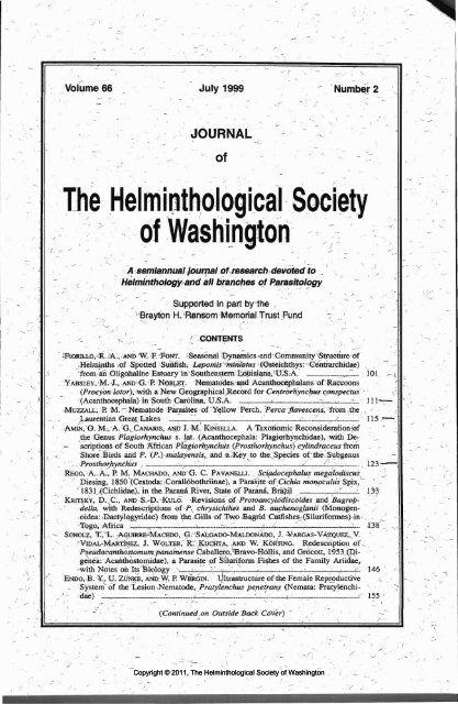
Figure 1. Mean monthly water temperature<br />
(°C) in Lake Pontchartrain-Lake Maurepas estuary<br />
(1992-1995). Vertical hars represent ±1 standard<br />
error <strong>of</strong> the mean.<br />
All stages<br />
NGR<br />
GRV<br />
HGR<br />
Helminth seasonal dynamics<br />
Prevalence <strong>of</strong> B. cupuloris differed significantly<br />
among time periods (x2 = 28.98, P <<br />
0.05). Thirty-six percent <strong>of</strong> hosts examined in<br />
May-August harbored at least 1 specimen. Prevalence<br />
increased to 40% in August-November<br />
and 41% in November-February before reaching<br />
82% in February-May. Irrespective <strong>of</strong> developmental<br />
stage, B. cupuloris was most abundant<br />
in February-May (8.8 ± 1.28, 0-37), displaying<br />
an 82% increase in abundance from the<br />
previous November-February time period (1.6<br />
± 0.47, 0-20) and a considerable decrease in the<br />
subsequent May-August period (1.1 ± 0.4, 0-<br />
15) (2-way ANCOVA, P < 0.05) (Fig. 2a).<br />
Abundance also differed with respect to developmental<br />
stage (2-way ANCOVA, P < 0.05),<br />
but no interaction effect was found (2-way AN-<br />
COVA, P > 0.05). Mature specimens were most<br />
abundant (1.5 ± 0.24, 0-21), followed by gravid<br />
specimens (1.4 ± 0.22, 0-18) and immature<br />
worms (0.6 ± 0.13, 0-14). In addition, each developmental<br />
stage <strong>of</strong> B. cupuloris showed a statistically<br />
significant seasonal cycle <strong>of</strong> abundance<br />
(ANCOVA, P < 0.05 for each stage). <strong>The</strong> abundance<br />
<strong>of</strong> each stage was lowest in May-August,<br />
remained low in the following August-November<br />
and November-February periods, and<br />
reached maximum abundance in February-May<br />
(Fig. 2a).<br />
Thirteen percent <strong>of</strong> hosts in May-August<br />
May-Aug Aug-Nov Nov-Feb Feb-May<br />
Figure 2. Seasonal abundances <strong>of</strong> (a) Barbulostomum<br />
cupuloris (all stages) and each developmental<br />
stage (IMM, immature; MAT, mature; GRV,<br />
gravid); (b) Genarchella sp. (all stages) and each<br />
developmental stage (NGR, nongravid; GRV, gravid;<br />
HGR, heavily gravid). Vertical bars represent<br />
± 1 standard error <strong>of</strong> the mean.<br />
were infected with Genarchella sp. Prevalence<br />
increased through August-November (34%) to<br />
reach a peak in November-February (49%) before<br />
decreasing in February-May (39%) (x2 =<br />
13.69, P < 0.05). Genarchella sp. was most<br />
abundant in November—February (6.9 ± 1.62,<br />
0-41), a 44% increase from the previous August—November<br />
time period (3.9 ± 1.39, 0—43)<br />
and showed its lowest abundance in May—August<br />
(1.4 ± 0.74, 0-23) (2-way ANOVA, P <<br />
0.05) (Fig. 2b). Overall, there was no difference<br />
in abundance among developmental stages and<br />
no interaction effect (2-way ANOVA, P ><br />
0.05). Both nongravid and gravid worms showed<br />
statistically significant seasonal cycles <strong>of</strong> abundance<br />
(ANOVA, P < 0.05 for each stage). Nongravid<br />
and gravid worms were most abundant in<br />
November-February (2.5 ± 0.89, 0-27) and Au-<br />
Copyright © 2011, <strong>The</strong> <strong>Helminthological</strong> <strong>Society</strong> <strong>of</strong> <strong>Washington</strong>
104 JOURNAL OF THE HELMINTHOLOGICAL SOCIETY OF WASHINGTON, 66(2), JULY 1999<br />
gust-November (2.2 ± 0.94, 0-41), respectively,<br />
and least abundant in May-August (nongravid:<br />
0.09 ± 0.09, 0-4; gravid: 0.1 ± 0.08, 0-3)<br />
(Fig. 2b).<br />
Camallanus oxycephalus was most prevalent<br />
and abundant in May-August (36%) (0.7 ±<br />
0.26, 0-11). Prevalence and abundance declined<br />
throughout the year and were lowest in February-May<br />
(10%) (0.1 ± 0.05, 0-2), (x2 = 12.888,<br />
P < 0.05) (ANOVA, P < 0.01), respectively<br />
(Table 1, Fig. 3). Prevalence <strong>of</strong> L. thecatus did<br />
not change seasonally (x2 = 5.278, P > 0.05)<br />
(Table 1), but its abundance did vary among<br />
time periods and peaked in May-August (0.6 ±<br />
0.19, 0-5) (ANOVA, P < 0.05) (Fig. 3). Crepidostomum<br />
cornutum, S. carolini, and N. cylindratus<br />
were uncommon (prevalence <strong>of</strong> each,<br />
< 7%), and too few individuals (abundance <strong>of</strong><br />
each, < 0.1) were recovered to determine seasonal<br />
patterns <strong>of</strong> prevalence and abundance (Table<br />
1).<br />
Parasite abundance (overall: 8.2 ± 0.8, 0—56)<br />
differed significantly among time periods (AN-<br />
COVA, P < 0.05). Abundance was lowest in<br />
May-August (4.0 ± 0.87, 0-26), increased during<br />
August-November (7.3 ± 1.66, 0-53) and<br />
November-February (8.5 ± 1.54, 0-45), and<br />
reached its highest value during February-May<br />
(12.8 ± 1.81, 0-56).<br />
Infracommunity analysis<br />
Overall, host body size was correlated with<br />
infracommunity diversity (r = 0.18, P < 0.05),<br />
but this relationship was not significant within<br />
time periods. <strong>The</strong> most diverse infracommunity<br />
was found in November-February (1.294 ±<br />
0.197, 0.0-4.14), whereas in February-May, infracommunity<br />
diversity was lowest (0.689 ±<br />
0.136, 0.0—3.35). <strong>The</strong> remaining 2 time periods,<br />
May—August and August-November, showed intermediate<br />
levels <strong>of</strong> infracommunity diversity<br />
(0.889 ± 0.151, 0.0-3.40 and 0.959 ± 0.185,<br />
0.0-4.01, respectively). However, infracommunity<br />
diversity did not differ significantly among<br />
time periods (ANCOVA, P > 0.05). Overall infracommunity<br />
diversity was 0.963 ± 0.087 (0.0-<br />
4.14).<br />
Both overall and within time periods, host<br />
body size did not influence infracommunity species<br />
richness. Helminth richness was lowest in<br />
May-August (1.244 ± 0.143, 0-4) and increased<br />
in August—November (1.302 ± 0.139,<br />
0-3) and November-February (1.353 ± 0.096,<br />
0-3). Infracommunity richness was greatest in<br />
February-May (1.549 ± 0.113, 0-3). However,<br />
infracommunity species richness did not differ<br />
significantly among time periods (ANOVA, P ><br />
0.05). Overall infracommunity species richness<br />
was 1.365 ± 0.062 (0-4). Infracommunity predictability<br />
differed significantly among time periods<br />
(ANOVA, P < 0.001). Infracommunity<br />
similarity was greatest in February-May (0.56<br />
± 0.01, 0-1) and lowest in May-August (0.22<br />
± 0.01, 0-1). In August-November and November-February,<br />
infracommunity similarity values<br />
were intermediate (0.31 ± 0.01, 0-1 and 0.34 ±<br />
0.01, 0-1, respectively). Overall infracommunity<br />
similarity was low (0.31 ± 0.003, 0-1).<br />
Component community analysis<br />
<strong>The</strong> trematodes B. cupuloris and Genarchella<br />
sp. were the most prevalent and abundant helminths<br />
and dominated the component community<br />
<strong>of</strong> Lepomis miniatus (Table 1). Barbulostomum<br />
cupuloris was most prevalent (50%), and<br />
although Genarchella sp. was recovered from<br />
fewer hosts (35%), its overall abundance (3.9 ±<br />
0.64, 0-43) did not differ significantly from that<br />
<strong>of</strong> B. cupuloris (3.5 ± 0.46, 0-37) (Mest, P ><br />
0.05). Although together these 2 trematodes accounted<br />
for 1,485 <strong>of</strong> the total 1,662 worms recovered<br />
during this study (89%), no significant<br />
association was found between them with respect<br />
to concurrent patterns <strong>of</strong> infection (x2 =<br />
0.032, P > 0.05). <strong>The</strong> nematode C. oxycephalus<br />
was recovered from 24% <strong>of</strong> hosts examined, but<br />
its abundance was low (0.4 ± 0.08, 0-11). <strong>The</strong><br />
remaining 4 helminth species showed low prevalence<br />
and abundance and, together with C. oxycephalus,<br />
represented only 11 % <strong>of</strong> the total helminth<br />
specimens recovered (Table 1).<br />
Component community diversity was low<br />
(0.47). It was greatest in February-May (1.16),<br />
progressively declined through May-August<br />
(0.63) and August-November (0.45), and was<br />
lowest in November-February (0.34). Component<br />
community species richness changed slightly<br />
over the year. Six helminth species were recovered<br />
during May—August, August—November,<br />
and November-February, whereas 7 helminth<br />
species were found in February-May<br />
(Table 1). <strong>The</strong> trematode C. cornutum and the<br />
acanthocephalan TV. cylindratus were the only<br />
helminths not found in all 4 time periods (Table<br />
1). Component community comparisons among<br />
time periods were made using Renkonen's co-<br />
Copyright © 2011, <strong>The</strong> <strong>Helminthological</strong> <strong>Society</strong> <strong>of</strong> <strong>Washington</strong>
FIORILLO AND FONT—SEASONAL DYNAMICS OF HELMINTH INFECTIONS 105<br />
Copyright © 2011, <strong>The</strong> <strong>Helminthological</strong> <strong>Society</strong> <strong>of</strong> <strong>Washington</strong>
106 JOURNAL OF THE HELMINTHOLOGICAL SOCIETY OF WASHINGTON, 66(2), JULY 1999<br />
Table 2. Renkonen's coefficients <strong>of</strong> component<br />
community similarity <strong>of</strong> helminths <strong>of</strong> Lepomis miniatus<br />
between paired time periods.<br />
Caox<br />
Leth<br />
May-Aug.<br />
Aug.—Nov.<br />
Nov.-Feb.<br />
Aug.-Nov. Nov.—Feb. Feb.—May<br />
0.46 0.41<br />
0.77<br />
0.46<br />
0.62<br />
0.45<br />
suit, the nature <strong>of</strong> the observed seasonal patterns<br />
<strong>of</strong> this and all other helminth species is biological<br />
and not a result <strong>of</strong> changes in host demographics.<br />
May-Aug Aug-Nov Nov-Feb Feb-May<br />
Figure 3. Seasonal abundance <strong>of</strong> Camallanus<br />
oxycephalus (Caox) and Leptorhynchoides thecatus<br />
(Leth). Vertical bars represent ±1 standard error<br />
<strong>of</strong> the mean.<br />
efficient <strong>of</strong> similarity. Mean seasonal component<br />
community similarity was 0.53 ± 0.058, 0.41-<br />
0.77. <strong>The</strong> helminth community <strong>of</strong> L. miniatus in<br />
August-November and November-February<br />
showed the greatest similarity (0.77), whereas<br />
the May-August and November-February communities<br />
were least similar (0.45). <strong>The</strong> remaining<br />
comparisons are shown in Table 2.<br />
Discussion<br />
<strong>The</strong> species composition, richness, and diversity<br />
<strong>of</strong> the helminth community <strong>of</strong> L. miniatus<br />
were similar throughout the year. Species-specific<br />
and overall abundance <strong>of</strong> these helminths,<br />
infracommunity similarity, and host body size<br />
did vary somewhat with season. <strong>The</strong> trematodes<br />
B. cupuloris, described from L. miniatus (as Lepomis<br />
punctatus (Valenciennes, 1831)) collected<br />
within our study site (Ramsey, 1965), and Genarchella<br />
sp. were the most prevalent and abundant<br />
helminths recovered in this study. <strong>The</strong>se<br />
parasites have been reported only in estuarine<br />
centrarchid fishes (Fiorillo and Font, 1996), in<br />
which they showed distinct seasonal cycles <strong>of</strong><br />
prevalence and abundance.<br />
Barbulostomum cupuloris was the only helminth<br />
to show, possibly as a result <strong>of</strong> an ontogenetic<br />
shift in diet, a significant relationship<br />
with host body size. However, the influence <strong>of</strong><br />
host size was removed statistically from the subsequent<br />
seasonal abundance analysis. As a re-<br />
Seasonal dynamics <strong>of</strong> helminth infections<br />
Conditions were optimal for the recruitment<br />
and maturation <strong>of</strong> B. cupuloris in February-<br />
May, when prevalence was greatest, and each<br />
developmental stage <strong>of</strong> this worm displayed<br />
maximum abundance. Following that period, the<br />
abundance <strong>of</strong> each developmental stage and the<br />
overall prevalence declined to their lowest values<br />
(Fig. 2a). Overall, most B. cupuloris specimens<br />
recovered were mature or gravid. <strong>The</strong>se<br />
data suggest that B. cupuloris matures quickly<br />
after recruitment. Immature, mature, and gravid<br />
specimens were recovered in all 4 collecting periods,<br />
indicating that recruitment and egg production<br />
occurred throughout the year, irrespective<br />
<strong>of</strong> water temperature. Because <strong>of</strong> south Louisiana's<br />
near-subtropical climate, seasonal<br />
changes in water temperature are not extreme<br />
(Fig. 1). Although water temperature does not<br />
affect egg production in B. cupuloris, temperature<br />
can influence timing and rate <strong>of</strong> cercarial<br />
production and dispersal (Chappell, 1969; review<br />
in Chubb, 1979) and the seasonal abundance<br />
<strong>of</strong> first or second intermediate hosts (Fernandez<br />
and Esch, 199la, b). It is likely that both<br />
factors interact to affect the seasonal abundance<br />
<strong>of</strong> B. cupuloris in its definitive host.<br />
Similarly, water temperature did not affect recruitment<br />
and maturation <strong>of</strong> Genarchella sp. in<br />
L. miniatus. As with B. cupuloris, all 3 developmental<br />
stages <strong>of</strong> Genarchella sp. were found<br />
throughout the year, but this helminth showed a<br />
more gradual increase in recruitment, which<br />
peaked in November-February (Fig. 2b). At that<br />
time <strong>of</strong> year, many gravid worms were also recovered.<br />
<strong>The</strong> seasonal cycles <strong>of</strong> Genarchella sp.<br />
and B. cupuloris were asynchronous. Unlike B.<br />
cupuloris, which showed maximum prevalence<br />
Copyright © 2011, <strong>The</strong> <strong>Helminthological</strong> <strong>Society</strong> <strong>of</strong> <strong>Washington</strong>
FIORILLO AND FONT—SEASONAL DYNAMICS OF HELMINTH INFECTIONS 107<br />
and abundance in February-May, Genarchella<br />
sp. was more common and numerous in November—February<br />
(Fig. 2a, b). However, as in B. cupuloris,<br />
the seasonal cycle <strong>of</strong> Genarchella sp. in<br />
L. rniniatus may be linked to the seasonal dynamics<br />
<strong>of</strong> cercarial production and dispersal and<br />
to the abundance <strong>of</strong> the intermediate host.<br />
<strong>The</strong> life cycles <strong>of</strong> B. cupuloris and Genarchella<br />
sp. are not known but are probably dependent<br />
on brackish water food webs for successful<br />
transmission (Ramsey, 1965; Fiorillo and<br />
Font, 1996). <strong>The</strong> lack <strong>of</strong> a concurrent pattern <strong>of</strong><br />
infection, as well as the apparent asynchrony in<br />
the seasonal cycles <strong>of</strong> these trematodes, suggest<br />
that they do not share the same intermediate<br />
hosts.<br />
A qualitative analysis <strong>of</strong> the gut contents <strong>of</strong><br />
L. miniatus in May-August showed that this<br />
centrarchid preyed primarily on amphipods<br />
(Fiorillo and Font, 1996). Similarly, Levine<br />
(1980) reported that amphipods made up 75% <strong>of</strong><br />
all prey items <strong>of</strong> L. miniatus in the Lake Pontchartrain-Lake<br />
Maurepas estuary. However,<br />
Fiorillo and Font (1996) showed that in May-<br />
August, both helminths were much more prevalent<br />
and abundant in redear sunfish Lepomis<br />
microlophus (Giinther, 1859), a host well known<br />
for its specialized diet <strong>of</strong> bivalves and other mollusks<br />
(Wilburn, 1969; Lauder, 1983). In this estuary,<br />
Levine (1980) reported that the diet <strong>of</strong> L.<br />
microlophus consisted primarily <strong>of</strong> molluscs, but<br />
some amphipods were also taken. A qualitative<br />
gut analysis in May-August showed that L. microlophus<br />
preyed on amphipods, isopods, and<br />
bivalves (Fiorillo and Font, 1996). <strong>The</strong>se data<br />
suggest that amphipods and bivalves may represent<br />
potential second intermediate hosts for<br />
these 2 trematodes.<br />
<strong>The</strong> nematode C. oxycephalus was most prevalent<br />
and abundant in May-August. Prevalence<br />
and abundance declined through the subsequent<br />
time periods and were lowest in February-May.<br />
It is likely that the seasonal dynamics <strong>of</strong> C. oxycephalus<br />
in this estuary are dependent on the<br />
seasonal abundance <strong>of</strong> its copepod intermediate<br />
host as shown by Stromberg and Crites (1975)<br />
in Lake Erie. Unfortunately, we have no data on<br />
the seasonal dynamics <strong>of</strong> copepod populations<br />
in the Lake Ponchartrain-Lake Maurepas estuary<br />
to support this assumption, but, generally, zooplankton<br />
populations in temperate climates increase<br />
in the summer months (Pennak, 1989).<br />
Although in our study C. oxycephalus abundance<br />
was low, we did recover gravid specimens,<br />
suggesting that L. miniatus is a suitable<br />
host for this nematode.<br />
As in C. oxycephalus, abundance <strong>of</strong> L. thecatus<br />
was low, but this acanthocephalan did<br />
show a seasonal cycle <strong>of</strong> abundance that peaked<br />
in May-August. Fiorillo and Font (1996)<br />
showed that, in this estuary, L. thecatus was<br />
much more prevalent and abundant in redear<br />
sunfish, L. microlophus, and C. oxycephalus occurred<br />
more frequently in bluegill, L. macrochirus<br />
Rafinesque, 1819, suggesting that L. miniatus<br />
is a suitable rather then a required host for<br />
these helminths. Leong and Holmes (1981) suggested<br />
that, within its environment, the seasonal<br />
cycle <strong>of</strong> a helminth is mostly determined by its<br />
seasonal dynamics within its most common host<br />
in which the parasite can become reproductive<br />
(required host). <strong>The</strong>refore, the seasonal cycles <strong>of</strong><br />
L. thecatus and C. oxycephalus in L. miniatus<br />
may not be indicative <strong>of</strong> the seasonal pattern<br />
found in L. microlophus and L. macrochirus, respectively.<br />
Too few specimens <strong>of</strong> the remaining<br />
3 helminth species were found to determine seasonal<br />
cycles <strong>of</strong> prevalence and abundance, but<br />
all are common parasites <strong>of</strong> centrarchids and<br />
other fishes from freshwater environments (see<br />
H<strong>of</strong>fman, 1967) (Table 1).<br />
Mostly because <strong>of</strong> increases in abundance <strong>of</strong><br />
B. cupuloris and Genarchella sp. (Fig. 2a, b),<br />
the overall parasite abundance was greatest in<br />
February-May. That time <strong>of</strong> year is generally<br />
associated with an increase in the feeding activity<br />
<strong>of</strong> fishes in Louisiana as water temperature<br />
begins to rise (Fig. 1) and many centrarchid species<br />
approach the reproductive season (Carlander,<br />
1977). Many invertebrate potential intermediate<br />
hosts also show seasonal changes in<br />
density, with abundance peaks in early spring<br />
(Heard, 1982). Seasonal dynamics <strong>of</strong> invertebrate<br />
intermediate hosts, coupled with seasonal<br />
variation in feeding rates and diet <strong>of</strong> L. miniatus,<br />
may play an important role in determining the<br />
seasonal cycles <strong>of</strong> abundance <strong>of</strong> these helminths.<br />
Infracommunity structure<br />
Kennedy (1990) characterized the helminth<br />
community <strong>of</strong> freshwater fishes as depauperate<br />
and isolationist. <strong>The</strong> infracommunity <strong>of</strong> L. miniatus<br />
displayed both characteristics. Infracommunities<br />
were characterized by a lack <strong>of</strong> helminth<br />
interactions, were species-poor, and included<br />
a small number <strong>of</strong> worms. Consequently,<br />
Copyright © 2011, <strong>The</strong> <strong>Helminthological</strong> <strong>Society</strong> <strong>of</strong> <strong>Washington</strong>
108 JOURNAL OF THE HELMINTHOLOGICAL SOCIETY OF WASHINGTON, 66(2), JULY 1999<br />
overall mean infracommunity diversity and species<br />
richness <strong>of</strong> L. miniatus were low, similar to<br />
other freshwater fishes (Kennedy et al., 1986).<br />
Most fish display indeterminate growth (Wooten,<br />
1990), so that body size is <strong>of</strong>ten highly correlated<br />
with age (Ricker, 1979; Swales, 1986).<br />
In the present study, larger hosts harbored a<br />
more diverse infracommunity. This was probably<br />
because <strong>of</strong> greater exposure time, which may<br />
increase the probability <strong>of</strong> these hosts being colonized<br />
by the less common helminth species.<br />
Cloutman (1975) noted a similar relationship between<br />
age and helminth diversity in largemouth<br />
bass, Micropterus salmoides (Lacepede, 1802).<br />
Seasonality did not affect infracommunity diversity<br />
and species richness. With the exception<br />
<strong>of</strong> C. cornutum and TV. cylindratus, all remaining<br />
helminths were recovered in all time periods,<br />
suggesting that the larval forms <strong>of</strong> the majority<br />
<strong>of</strong> these helminths are capable <strong>of</strong> colonizing L.<br />
miniatus year-round. However, the proportion <strong>of</strong><br />
infected intermediate hosts, as shown by Fernandez<br />
and Esch (199la, b), may have changed<br />
seasonally, resulting in the discrete cycles <strong>of</strong><br />
abundance shown by some <strong>of</strong> these helminths.<br />
Overall, the infracommunity structure <strong>of</strong> L.<br />
miniatus was not highly predictable, suggesting<br />
that each infracommunity represented a random<br />
subset <strong>of</strong> the parasites found in the component<br />
community <strong>of</strong> this host. Poulin (1997) noted that<br />
low infracommunity predictability is also a characteristic<br />
<strong>of</strong> isolationist communities, because<br />
helminth interactions, which <strong>of</strong>ten result in more<br />
predictably structured assemblages, are lacking.<br />
Infracommunity predictability did differ<br />
among time periods. Infracommunity structure<br />
was most and least predictable in February—May<br />
and May—August, respectively. In February-<br />
May, increases in prevalence and abundance <strong>of</strong><br />
B. cupuloris were largely responsible for the<br />
greatest degree <strong>of</strong> infracommunity similarity,<br />
whereas reductions in prevalence and abundance<br />
<strong>of</strong> this trematode, along with Genarchella sp.,<br />
may have contributed to low infracommunity<br />
predictability in the following season. <strong>The</strong> greater<br />
predictability in February-May suggests that<br />
larval helminths are more prevalent in their intermediate<br />
hosts during that time <strong>of</strong> year so that<br />
the probability <strong>of</strong> individual hosts acquiring a<br />
similar suite <strong>of</strong> parasites is greater.<br />
Component community structure<br />
<strong>The</strong> trematodes B. cupuloris and Genarchella<br />
sp. were the dominant species in the component<br />
community <strong>of</strong> L. punctatus and accounted for<br />
the majority <strong>of</strong> all worms recovered during this<br />
year-long study. <strong>The</strong>se helminths are not found<br />
in freshwater centrarchids but have been reported<br />
from other Lepomis spp. in the Lake Pontchartrain-Lake<br />
Maurepas estuary (Fiorillo and<br />
Font, 1996). Ramsey (1965) noted that B. cupuloris<br />
was replaced by the closely related Homalometron<br />
armatum (MacCallum, 1895) in centrarchid<br />
hosts collected in freshwater ponds located<br />
near this estuary. In this estuary, B. cupuloris<br />
and Genarchella sp. are more prevalent<br />
and abundant in L. microlophus (Fiorillo and<br />
Font, 1996), suggesting that L. miniatus is a suitable<br />
but not a required host for these trematodes<br />
(Leong and Holmes, 1981). However, the specificity<br />
<strong>of</strong> B. cupuloris and Genarchella sp. for<br />
estuarine hosts reaffirms the importance <strong>of</strong> ecological<br />
associations to the component community<br />
structure <strong>of</strong> L. miniatus.<br />
<strong>The</strong> remaining 5 helminths recovered from L.<br />
miniatus are common parasites <strong>of</strong> freshwater<br />
centrarchid fishes (see H<strong>of</strong>fman, 1967). Although<br />
mature forms were found in L. miniatus,<br />
these helminths showed low prevalence and<br />
abundance (Table 1). However, all 5 species<br />
were more prevalent and abundant in other Lepomis<br />
spp. from this estuary (Fiorillo and Font,<br />
1996). <strong>The</strong>se patterns suggest that L. miniatus is<br />
a suitable host for these helminths (Leong and<br />
Holmes, 1981) but that their occurrence in L.<br />
miniatus may represent accidental infections.<br />
Component community diversity was low and<br />
similar to that <strong>of</strong> other freshwater fishes (Kennedy<br />
et al., 1986). Qualitatively, component diversity<br />
varied seasonally and was greatest in<br />
February—May when B. cupuloris and Genarchella<br />
sp. occurred frequently and abundances<br />
were high. <strong>The</strong> component community <strong>of</strong> this<br />
host in August-November and November-February<br />
was most similar. In those time periods,<br />
most <strong>of</strong> the helminths recovered displayed similar<br />
measures <strong>of</strong> prevalence and abundance (Table<br />
1), resulting in a greater degree <strong>of</strong> similarity.<br />
Overall, the helminth species composition <strong>of</strong><br />
L. miniatus was similar to that <strong>of</strong> other centrarchid<br />
hosts in this estuary (see Fiorillo and Font,<br />
1996). All helminths found in the present study<br />
were also recovered in L. macrochirus and, with<br />
the exception <strong>of</strong> C. cornutum, in L. megalotis.<br />
However, compared to L. miniatus, species richness<br />
was much lower in L. microlophus. Dietary<br />
differences between and among hosts may ac-<br />
Copyright © 2011, <strong>The</strong> <strong>Helminthological</strong> <strong>Society</strong> <strong>of</strong> <strong>Washington</strong>
FIORILLO AND FONT—SEASONAL DYNAMICS OF HELMINTH INFECTIONS 109<br />
count for this result (Bell and Burt, 1991), but<br />
unequal sampling effort may have biased this<br />
pattern (see Levine, 1980; Fiorillo and Font,<br />
1996, for diet analyses).<br />
Further studies are necessary to determine the<br />
life cycles <strong>of</strong> B. cupuloris and Genarchella sp.<br />
Knowledge <strong>of</strong> the intermediate hosts <strong>of</strong> these<br />
trematodes and their seasonal patterns <strong>of</strong> abundance,<br />
as well as <strong>of</strong> temporal changes in the trophic<br />
interactions <strong>of</strong> intermediate hosts and fish,<br />
is essential to our understanding <strong>of</strong> the mechanisms<br />
that determine the seasonal dynamics <strong>of</strong><br />
these helminths and the parasite community<br />
structure <strong>of</strong> this centrarchid host. In addition, a<br />
better understanding <strong>of</strong> these life cycles and seasonal<br />
patterns <strong>of</strong> incidence and abundance<br />
would further elucidate the importance <strong>of</strong> L.<br />
miniatus to the circulation <strong>of</strong> these helminths in<br />
this estuarine ecosystem.<br />
Acknowledgments<br />
We thank G. W. Childers, R. A. Seigel, R. W.<br />
Hastings, and G. P. Shaffer for their counsel and<br />
support. We thank Bill Lutterschmidt, Becky<br />
Fiorillo, John Chelchowski, Scott Thompson,<br />
and Kelley Smith for their help in the field. We<br />
are especially grateful to Tom Blanchard for his<br />
relentless help in the field and support throughout<br />
the duration <strong>of</strong> this study and to an anonymous<br />
reviewer for a critical and perceptive review<br />
<strong>of</strong> the manuscript and recommendations<br />
for its improvement.<br />
Literature Cited<br />
Anderson, R. M. 1974. Population dynamics <strong>of</strong> the<br />
cestode Caryophyllaeus laticeps (Fellow, 1781) in<br />
the bream (Abramus brama L.). Journal <strong>of</strong> Animal<br />
Ecology 43:305-321.<br />
. 1976. Seasonal variation in the population dynamics<br />
<strong>of</strong> Caryophyllaeus laticeps. Parasitology<br />
72:281-305.<br />
Bell, G., and A. Burt. 1991. <strong>The</strong> comparative biology<br />
<strong>of</strong> parasite species diversity: internal helminths <strong>of</strong><br />
freshwater fish. Journal <strong>of</strong> Animal Ecology 60:<br />
1047-1064.<br />
Bush, A. O., K. D. Lafferty, J. M. Lotz, and A. W.<br />
Shostak. 1997. Parasitology meets ecology on its<br />
own terms: Margolis et al. revisited. Journal <strong>of</strong><br />
Parasitology 83:575-583.<br />
Carlander, K. D. 1977. Handbook <strong>of</strong> Freshwater<br />
Fishery Biology. Life History Data on Centrarchid<br />
Fishes <strong>of</strong> the United <strong>State</strong>s and Canada. Iowa<br />
<strong>State</strong> University Press, Ames, Iowa. 431 pp.<br />
Chappell, L. H. 1969. <strong>The</strong> parasites <strong>of</strong> the threespined<br />
stickleback Gasterosteus aculeatus L. from<br />
a Yorkshire pond. I. Seasonal variation <strong>of</strong> parasite<br />
fauna. Journal <strong>of</strong> Fish Biology 1:137-152.<br />
Chubb, J. S. 1979. Seasonal occurrence <strong>of</strong> helminths<br />
in freshwater fishes. Part II. Trematoda. Pages<br />
141-313 in W. H. R. Lumsden, R. Muller, and J.<br />
R. Baker, eds. Advances in Parasitology. Vol. 17.<br />
Academic Press, London. 415 pp.<br />
Cloutman, D. G. 1975. Parasite community structure<br />
<strong>of</strong> largemouth bass, warmouth, and bluegill in<br />
Lake Fort Smith, Arkansas. Transactions <strong>of</strong> the<br />
American Fisheries <strong>Society</strong> 104:277-283.<br />
Eure, H. 1976. Seasonal abundance <strong>of</strong> Neoechinorhyncus<br />
cylindratus from the largemouth bass<br />
(Micropterus salmoides) in a heated reservoir.<br />
Parasitology 73:355-370.<br />
Fernandez, J., and G. W. Esch. 199la. Guild structure<br />
<strong>of</strong> larval trematodes in the snail Helisoma<br />
anceps and its impact on fecundity <strong>of</strong> the snail<br />
host. Journal <strong>of</strong> Parasitology 77:528-539.<br />
, and . 1991b. <strong>The</strong> component community<br />
structure <strong>of</strong> larval trematodes in the pulmonate<br />
snail Helisoma anceps. Journal <strong>of</strong> Parasitology<br />
77:540-550.<br />
Fiorillo, R. A., and W. F. Font. 1996. Helminth community<br />
structure <strong>of</strong> four species <strong>of</strong> Lepomis (Osteichthyes:<br />
Centrarchidae) from an oligohaline estuary<br />
in southeastern Louisiana. Journal <strong>of</strong> the<br />
<strong>Helminthological</strong> <strong>Society</strong> <strong>of</strong> <strong>Washington</strong> 64:24-<br />
30.<br />
Granath, W. O., and G. W. Esch. 1983a. Temperature<br />
and other factors that regulate the composition<br />
and infrapopulation densities <strong>of</strong> Bothriocephalus<br />
acheilognathi (Cestoda) in Gambusia affinis<br />
(Pisces). Journal <strong>of</strong> Parasitology 69:1 1 16-1124.<br />
, and . 1983b. Survivorship and parasite-induced<br />
mortality among mosquito fish in a<br />
predator-free, North Carolina cooling reservoir.<br />
American Midland Naturalist 110:314-323.<br />
, and . 1983c. Seasonal dynamics <strong>of</strong><br />
Bothriocephalus acheilognathi in ambient and<br />
thermally altered areas <strong>of</strong> a North Carolina cooling<br />
reservoir. Proceedings <strong>of</strong> the <strong>Helminthological</strong><br />
<strong>Society</strong> <strong>of</strong> <strong>Washington</strong> 50:205-218.<br />
Heard, R. W. 1982. Guide to common tidal marsh<br />
invertebrates <strong>of</strong> the northeastern Gulf <strong>of</strong> Mexico.<br />
Mississippi Alabama Sea Grant Consortium Publication<br />
79-004. 82 pp.<br />
H<strong>of</strong>fman, G. L. 1967. Parasites <strong>of</strong> North American<br />
Freshwater Fish. University <strong>of</strong> California Press,<br />
Berkeley. 486 pp.<br />
Kennedy, C. R. 1990. Helminth communities in freshwater<br />
fish: structured communities or stochastic<br />
assemblages? Pages 131-156 in G. W. Esch, O.<br />
Bush, and J. M. Aho, eds. Parasite Communities:<br />
Patterns and Process. Chapman and Hall, London.<br />
335 pp.<br />
, A. O. Bush, and J. M. Aho. 1986. Patterns<br />
in helminth communities: why are birds and fish<br />
different? Parasitology 93:205-215.<br />
Lauder, G. V. 1983. Functional and morphological<br />
bases <strong>of</strong> trophic specialization in sunfishes (Teleostei,<br />
Centrarchidae). Journal <strong>of</strong> Morphology<br />
178:1-21.<br />
Leong, T. S., and J. C. Holmes. 1981. Communities<br />
<strong>of</strong> metazoan parasites in open water fishes <strong>of</strong> Cold<br />
Copyright © 2011, <strong>The</strong> <strong>Helminthological</strong> <strong>Society</strong> <strong>of</strong> <strong>Washington</strong>
110 JOURNAL OF THE HELMINTHOLOGICAL SOCIETY OF WASHINGTON, 66(2), JULY 1999<br />
Lake, Alberta. Journal <strong>of</strong> Fish Biology 18:693-<br />
713.<br />
Levine, S. J. 1980. Gut contents <strong>of</strong> 44 Lake Pontchartrain<br />
fish species. Pages 973-1029 in J. H.<br />
Stone, ed. Environmental Analysis <strong>of</strong> Lake Pontchartrain,<br />
Louisiana, Its Surrounding Wetlands<br />
and Selected Land Uses. Vol. 2. Coastal Ecology<br />
Laboratory, Center for Wetland Resources, Louisiana<br />
<strong>State</strong> University, Baton Rouge, Louisiana.<br />
Technical Report 899-1030. 1219 pp.<br />
McDaniel, J. S., and H. H. Bailey. 1974. Seasonal<br />
population dynamics <strong>of</strong> some helminth parasites<br />
<strong>of</strong> centrarchid fishes. Southwestern Naturalist 18:<br />
403-416.<br />
Pennak, R. W. 1989. Fresh-water Invertebrates <strong>of</strong> the<br />
United <strong>State</strong>s: Protozoa to Mollusca, 3rd ed. John<br />
Wiley & Sons, New York. 628 pp.<br />
Pielou, E. C. 1977. Mathematical Ecology. John Wiley<br />
& Sons, New York. 385 pp.<br />
Poulin, R. 1997. Species richness <strong>of</strong> parasite assemblages:<br />
evolution and patterns. Annual Review <strong>of</strong><br />
Ecology and Systematics 28:341-358.<br />
Ramsey, J. S. 1965. Barbulostomiim cupuloris gen. et<br />
sp. n. (Trematoda: Lepocreadiidae) from sunfishes<br />
(Lepomis spp.) in Lake Pontchartrain, Louisiana.<br />
Journal <strong>of</strong> Parasitology 51:777-780.<br />
Ricker, W. E. 1979. Growth rates and models. Pages<br />
677-743 in W. S. Hoar, D. J. Randall, and J. R.<br />
Brett, eds. Fish Physiology. Vol. 8. Academic<br />
Press, London. 792 pp.<br />
Stromberg, P. C., and J. L. Crites. 1975. An analysis<br />
<strong>of</strong> the changes in the prevalence <strong>of</strong> Camallanus<br />
oxycephalus (Nematoda: Camallanidae) in western<br />
Lake Erie. Ohio Journal <strong>of</strong> Science 75:1-6.<br />
Swales, S. 1986. Population dynamics, production and<br />
angling catch <strong>of</strong> brown trout, Salmo trutta, in a<br />
mature upland reservoir in Mid-Wales. Environmental<br />
Biology <strong>of</strong> Fishes 16:279-293.<br />
Wilbur, R. L. 1969. <strong>The</strong> Redear Sunfish in Florida.<br />
Florida Game and Fish Commission Fisheries<br />
Bulletin 5. 64 pp.<br />
Wootton, R. J. 1990. An Ecology <strong>of</strong> Teleost Fishes.<br />
Chapman and Hall, New York. 404 pp.<br />
Editors' Acknowledgments<br />
In addition to the members <strong>of</strong> the Editorial Board, we would like to acknowledge, with thanks,<br />
the following persons for providing their valuable help and insights in reviewing manuscripts for<br />
the Journal: Jasem Abdul-Salam, Martin Adamson, Omar Amin, James Baldwin, Diane Barton,<br />
George Benz, Ian Beveridge, Walter Boeger, Burton Bogitsh, Daniel Brooks, Charles Bursey, Albert<br />
Bush, Gilbert Castro, Hilda Ching, Rebecca Cole, Bruce Conn, John Crites, John Cross, Murray<br />
Dailey, Tommy Dunagan, Donald Forrester, Stephen Goldberg, David Hall, Hideo Hasegawa, Richard<br />
Heard, Gary Hendrickson, Sherman Hendrix, Russell Hobbs, Jane Huffman, Hugh Jones,<br />
James Joy, Michael Kinsella, Thomas Klei, Delane Kritsky, Ralph Lichtenfels, Donald Linzey,<br />
Jeffrey Lotz, Eugene Lyons, David Marcogliese, Gary MacCallister, Donald McAlpine, Lena Measures,<br />
Frantisek Moravec, Darwin Murrell, Patrick Muzzall, Brent Nickol, Thomas Nolan, Paul<br />
Nollen, David Oetinger, Robin Overstreet, Raphael Payne, Danny Pence, Thomas Platt, Mark Pokras,<br />
Dennis Richardson, Guillermo Salgado-Maldonado, Gerhard Schad, Mark Siddall, Donald<br />
Smith, Marilyn Spalding, George Stewart, Michael Sukhdeo, Dennis Thoney, John Ubelaker.<br />
Copyright © 2011, <strong>The</strong> <strong>Helminthological</strong> <strong>Society</strong> <strong>of</strong> <strong>Washington</strong>
J. Helminthol. Soc. Wash.<br />
66(2), 1999 pp. 11 1-114<br />
Nematodes and Acanthocephalans <strong>of</strong> Raccoons (Procyon lotor}, with<br />
a New Geographical Record for Centrorhynchus conspectus<br />
(Acanthocephala) in South Carolina, U.S.A.<br />
MICHAEL J. YABSLEY AND GAYLE PITTMAN NoBLET1<br />
Department <strong>of</strong> Biological Sciences, Clemson University, Clemson, South Carolina 29634-1903, U.S.A. (e--<br />
mail: myabsle@clemson.cdu; gnoblet@clemson.edu)<br />
ABSTRACT: From April 1997 through April 1998, 128 raccoons (Procyon lotor (Linnaeus)) collected from 7<br />
sites representing 4 physiographic areas in South Carolina were examined for gastrointestinal helminth parasites.<br />
Four species <strong>of</strong> nematodes (Gnathostoma procyonis (Chandler), Physaloptera rara Hall and Wigdor, Arthrocephahis<br />
lotoris (Schwartz), and Molineus barhatus Chandler) and 2 species <strong>of</strong> acanthocephalans (Macracanthorhynchus<br />
ingens (von Linstow) and Centrorhynchus conspectus Van Cleave and Pratt) were collected. <strong>The</strong><br />
finding <strong>of</strong> 11 immature C. conspectus in 3 South Carolina raccoons represents a new geographical record for<br />
this species.<br />
KEY WORDS: Centrorhynchus conspectus, raccoon, Procyon lotor, Nematoda, Acanthocephala, helminths,<br />
Gnathostoma procyonis, Physaloptera rara, Arthrocephalus lotoris, Molineus barbatus, Macracanthorhynchus<br />
ingens, South Carolina, U.S.A.<br />
<strong>The</strong> raccoon (Procyon lotor (Linnaeus, 1758))<br />
is an omnivore that ranges over most <strong>of</strong> North<br />
America and occurs in both rural and urban settings.<br />
Consequently, the range <strong>of</strong> zoonoses for<br />
raccoons is important in assessing risk to humans<br />
and domestic animals. In South Carolina,<br />
only limited studies on helminth parasites <strong>of</strong> raccoons<br />
have been reported previously (Harkema<br />
and Miller, 1964; Stansell, 1974). More recent<br />
reports <strong>of</strong> serious human illnesses from the<br />
northern and midwestern United <strong>State</strong>s, such as<br />
cerebrospinal nematodiasis because <strong>of</strong> infection<br />
with the gastrointestinal nematode Baylisascaris<br />
procyonis (Stefanski and Zarnowski, 1951), led<br />
to the current study, which includes raccoons<br />
collected statewide from a wide variety <strong>of</strong> habitats<br />
(e.g., mountains, farms, urban areas, beaches,<br />
swamps, and barrier islands), allowing for a<br />
comparison <strong>of</strong> parasite burdens and consideration<br />
<strong>of</strong> human health risks associated with these<br />
parasites (Williams et al., 1997; Boschetti and<br />
Kasznica, 1995).<br />
Materials and Materials<br />
Raccoons (n = 128) were collected between April<br />
1997 and April 1998 with foot-hold traps or wire livetraps.<br />
Traps were set at 7 sites that included 4 <strong>of</strong> the<br />
5 physiographic areas <strong>of</strong> South Carolina. Site 1 included<br />
both urban and waterfowl management areas<br />
(WMA) in Pickens County (Foothills); Site 2 was a<br />
WMA in Union County (Piedmont); Site 3 was inland<br />
1 Corresponding author.<br />
111<br />
farm areas <strong>of</strong> Horry County (Lower Coastal Plains<br />
North, LCPN); Site 4 included both beach and wooded<br />
habitats in the tourist area <strong>of</strong> Myrtle Beach, Horry<br />
County (LCPN); Site 5 was a swamp located on the<br />
Savannah River in Hampton County (Lower Coastal<br />
Plains South, LCPS); and Sites 6 and 7 were both on<br />
barrier islands located in Charleston County (LCPS).<br />
John's Island (Site 6), next to and continuous with the<br />
mainland at times <strong>of</strong> low tide, is primarily forest and<br />
farmland with many freshwater ponds, whereas Seabrook<br />
Island (Site 7) is a small residential island about<br />
1.5 km <strong>of</strong>fshore, which lacks freshwater habitats. Each<br />
raccoon was subjected to multiple evaluations, which<br />
included not only our study <strong>of</strong> gastrointestinal helminth<br />
parasites, but also seroprevalence, culture and<br />
DNA studies for Trypanosoma cruzi, and museum<br />
study specimens. In addition, most animals were included<br />
in a trap-type capture effectiveness study conducted<br />
by the South Carolina Department <strong>of</strong> Natural<br />
Resources (SCDNR).<br />
Raccoons were either euthanized by intramuscular<br />
injection <strong>of</strong> 0.2 ml/kg ketamine/xylazine followed by<br />
intraperitoneal injection <strong>of</strong> 1 ml/kg sodium pentobarbital,<br />
or were hunter-shot. Stomach and intestines from each<br />
animal were examined as soon as possible after death<br />
(within 1-2 hr). However, animals from 2 <strong>of</strong> the physiographic<br />
regions (Sites 3-7) were frozen at — 4°C for<br />
1-3 mo prior to examination for helminths because <strong>of</strong><br />
the use <strong>of</strong> the animals for a trap-type study conducted<br />
by the International Association <strong>of</strong> Fish and Wildlife<br />
Agencies. <strong>The</strong>refore, trematodes and cestodes were excluded<br />
from the overall analyses because freezing <strong>of</strong> a<br />
large number <strong>of</strong> hosts resulted in difficult collection<br />
and unreliable identification <strong>of</strong> flatworms.<br />
All nematodes collected from the stomachs and<br />
small intestines <strong>of</strong> raccoons were preserved and stored<br />
in a 70% ethanol-5% glycerine solution. Representative<br />
specimens <strong>of</strong> each nematode were mounted in glycerine<br />
jelly. Acanthocephalans collected from the small<br />
Copyright © 2011, <strong>The</strong> <strong>Helminthological</strong> <strong>Society</strong> <strong>of</strong> <strong>Washington</strong>
M<br />
*<br />
12 JOURNAL OF THE HELMINTHOLOGICAL SOCIETY OF WASHINGTON, 66(2), JULY 1999<br />
intestine were placed in water until the proboscis everted,<br />
preserved in acetic acid-formalin-alcohol (AFA),<br />
and stored in 70% ethanol. Temporary wet mounts and<br />
permanent Mayer's acid carmine-stained mounts in<br />
Canada balsam were made for identification. Voucher<br />
specimens deposited at the U.S. National Parasite Collection<br />
in Beltsville, Maryland, have been assigned<br />
USNPC accession numbers 87838-87843. Fisher's exact<br />
test was used to detect significant differences (P <<br />
0.05) in helminth prevalence (%) between study sites.<br />
Two-thirds <strong>of</strong> the animals caught were male, and 85%<br />
<strong>of</strong> all animals were mature. Because <strong>of</strong> the large bias<br />
toward males and adults, no statistical analyses were<br />
performed.<br />
Results and Discussion<br />
Of the 128 raccoons examined, 103 (80%)<br />
were infected with 1 or more <strong>of</strong> the 4 nematodes<br />
and 2 acanthocephalans listed in Table 1. Gnathostoma<br />
procyonis Chandler, 1942, and Physaloptera<br />
rara Hall and Wigdor, 1918, were recovered<br />
primarily from the stomach. Arthrocephalus<br />
(=Placoconus) lotoris (Schwartz, 1925)<br />
and Molineus barbatus Chandler, 1942, were<br />
collected from the posterior and anterior ends <strong>of</strong><br />
the small intestine, respectively. Both Macracanthorhynchus<br />
ingens (von Linstow, 1879) and<br />
Centrorhynchus conspectus Van Cleave and<br />
Pratt, 1940, were recovered exclusively from the<br />
small intestine. Interestingly, 96.1% <strong>of</strong> raccoons<br />
examined from Sites 1-6 were infected with at<br />
least 1 helminth species, whereas only 5 <strong>of</strong> 26<br />
(19.2%) raccoons examined from Seabrook Island<br />
(Site 7) were infected.<br />
Gnathostoma procyonis, a stomach nematode<br />
that forms large nodules in the mucosa, was<br />
found at Sites 3 and 5 in significantly larger<br />
numbers than at other sites (Table 1). Extensive<br />
freshwater habitats were present at both sites,<br />
providing a favorable environment for the required<br />
first intermediate host, which is 1 <strong>of</strong> several<br />
species <strong>of</strong> cyclopoid copepods (Miyazaki,<br />
1960). In contrast, no infections <strong>of</strong> G. procyonis<br />
were observed at 2 coastal locations (Sites 4 and<br />
7), which lacked permanent freshwater habitats.<br />
Physaloptera rara, a spirurid nematode recovered<br />
from both the stomach and small intestine<br />
<strong>of</strong> hosts, does not require the presence <strong>of</strong> freshwater<br />
habitats, because raccoons become infected<br />
by ingestion <strong>of</strong> various terrestrial arthropods<br />
(e.g., Gryllus pennsylvanicus Burmeister, 1838,<br />
Pennsylvania field cricket; Blattella germanica<br />
(Linnaeus, 1767), German cockroach; and Centophiles<br />
spp., camel crickets) (Lincoln and Anderson,<br />
1973). Compared to all other sites, a sig-<br />
"E a<br />
^N<br />
OJj<br />
3<br />
o<br />
5<br />
t^<br />
sON<br />
iH<br />
9 c a<br />
E o£a<br />
j30<br />
«<br />
U<br />
—*j<br />
o<br />
JS<br />
i<br />
1"o 0<br />
c ,g<br />
~<br />
|><br />
1<br />
Qq<br />
e<br />
o<br />
u<br />
u<br />
03<br />
£<br />
00<br />
E<br />
0<br />
tM<br />
C<br />
JS<br />
a<br />
u<br />
o<br />
1<br />
s<br />
•o ea<br />
VI<br />
0<br />
a u<br />
X s<br />
*o<br />
" CN<br />
1)<br />
.S II<br />
00 ^<br />
c .<br />
fll ^<br />
.« o<br />
oo J-<br />
OJ<br />
a<br />
D.<br />
O<br />
X<br />
tL)<br />
u<br />
a.<br />
00<br />
NO<br />
>O C — ^J" ^j" CO<br />
— — oo ON •* ON<br />
J. J. J. J. ' '<br />
__<br />
V") t~-; NO ON CO P~;<br />
O V~l CO NO •— CN<br />
+1 +1 +1 +1 +1 +1<br />
CN r- ON NO r- t-<br />
•^t oo ON CO ON CO<br />
CO — CN<br />
— 00 00 CO CO ^^<br />
^t CO CN -^<br />
V~) CN<br />
CN ON NO NO OO CO<br />
IO ^1" CO • — ' NO<br />
^^ >-^ ^-~. ^^<br />
o o ** c<br />
C O — O<br />
r-~ ^^<br />
— o<br />
TJ- C<br />
!o oo ^ ^ in ^<br />
V) — ^ ^t ON i/~) ON<br />
NO CN >/1 — NO —<br />
-^ ^<br />
f^ C S o ^-.<br />
oo r- CN — oo o<br />
ON >O t —• "^" 00 O<br />
CN CN N<br />
C 00 CN — NO C<br />
O — CO CN OO O<br />
^ ^ ^^ ^ ^ ^<br />
r — r^- CN t — NO —<br />
— — CO — ON (N<br />
0 ^ O ^ O ^<br />
•* O •* O CN C<br />
CN O CN O<br />
— O<br />
. — X , — s , * ^ — s<br />
— CO O CO -3" C<br />
*t O >/~> OO CN O<br />
CN —<br />
•—<br />
__<br />
tjo<br />
^<br />
oo oo" oo So<br />
>•<br />
oo oo<br />
c^<br />
I; 5<br />
-Ii ^<br />
'S -^2 ^ Ci.<br />
K c -^ K<br />
G<br />
s *»<br />
s ~?<br />
S<br />
"B "5<br />
S "*-<br />
^ "5<br />
3*<br />
•S X *^- ^ Q" S ^<br />
S^"^J"-* CJ "^"^<br />
^<br />
"O ^; *^. O ^ O C ^<br />
2*5i?h^ -^ P § ^ t: £ ^^§<br />
£
YABSLEY AND NOBLET—HELMINTHS OF RACCOONS 13<br />
nificantly higher prevalence <strong>of</strong> P. ram was observed<br />
in raccoons trapped at Site 1, with urbancaptured<br />
animals dominating the number <strong>of</strong><br />
infected animals. Because broad host specificity<br />
has been documented for physalopteroids, domesticated<br />
animals could accumulate large numbers<br />
<strong>of</strong> these worms by ingesting an infected intermediate<br />
host that commonly occurs in urban<br />
settings (Morgan, 1941).<br />
High prevalences <strong>of</strong> both the raccoon hookworm,<br />
A. lotoris, and the trichostrongylid, M.<br />
barbatus, frequently have been reported in previous<br />
surveys (Harkema and Miller, 1964; Cole<br />
and Shoop, 1987). In the present study, however,<br />
overall prevalence <strong>of</strong> A. lotoris and M. barbatus<br />
was 28.1 and 12.5%, respectively (Table 1).<br />
Data from Seabrook Island are consistent with<br />
those <strong>of</strong> Harkema and Miller (1962), who previously<br />
reported an almost complete absence <strong>of</strong><br />
A. lotoris and M. barbatus from Cape Island,<br />
South Carolina. <strong>The</strong>se investigators suggested<br />
that the low prevalence <strong>of</strong> these 2 nematodes on<br />
coastal islands was due possibly to the detrimental<br />
effect <strong>of</strong> high tides, salinity <strong>of</strong> soil, and<br />
dry habitats on the free-living larval stages <strong>of</strong><br />
these helminths. Additionally, seasonal variations<br />
have been documented, with lower prevalence<br />
during winter months (Smith et al., 1985),<br />
which could have contributed to the lower numbers<br />
observed in the current survey.<br />
Although B. procyonis was not collected from<br />
any raccoon examined in this study, surveillance<br />
for this parasite should be continued because <strong>of</strong><br />
its medical and veterinary significance and its<br />
reported widespread distribution in the United<br />
<strong>State</strong>s (Kazacos and Boyce, 1989). Based on reports<br />
from southeastern states, Jones and Mc-<br />
Ginnes (1983) suggested that B. procyonis was<br />
found primarily in the more mountainous regions.<br />
<strong>The</strong> northwestern range <strong>of</strong> our study site,<br />
although classified as "Foothills," is not truly<br />
mountainous, which might account for the lack<br />
<strong>of</strong> B. procyonis. In contrast, however, B. procyonis<br />
recently was found in 70% <strong>of</strong> 33 raccoons<br />
examined in southern coastal Texas (Kerr<br />
et al., 1997). <strong>The</strong>se investigators suggested that<br />
the nematode could have been acquired by ingestion<br />
<strong>of</strong> larvae in migratory wild birds, introduced<br />
from infected translocated raccoons, or<br />
the result <strong>of</strong> a northern expansion from Latin<br />
America. This recent finding <strong>of</strong> B. procyonis in<br />
southern Texas and limited reports from the adjacent<br />
state <strong>of</strong> Georgia (only 2 reports, each <strong>of</strong><br />
a single infected raccoon [Babero and Shepperson,<br />
1958; Kazacos and Boyce, 1989]) suggests<br />
the possibility <strong>of</strong> introduction <strong>of</strong> this nematode<br />
into South Carolina.<br />
<strong>The</strong> most prevalent parasite collected was the<br />
acanthocephalan M. ingens. Infections occurred<br />
in raccoons from all study sites, with an overall<br />
prevalence <strong>of</strong> 53%. Although not considered a<br />
threat to public health, M. ingens infection in<br />
humans has been reported (Dingley and Beaver,<br />
1985).<br />
Six species <strong>of</strong> the acanthocephalan genus<br />
Centrorhynchus have been reported from North<br />
American birds <strong>of</strong> prey; however, little is known<br />
about the life cycle, geographical distribution, or<br />
prevalence <strong>of</strong> these acanthocephalan species.<br />
Read (1950) demonstrated experimentally that<br />
Centrorhynchus spinosus (Kaiser, 1893) was capable<br />
<strong>of</strong> developing to adults in laboratory rats,<br />
suggesting that members <strong>of</strong> this genus have the<br />
ability to complete development not only in bird<br />
definitive hosts, but also in mammalian hosts.<br />
One raccoon from John's Island (Site 6) and 2<br />
raccoons from the Horry County inland site (Site<br />
3) were infected with immature C. conspectus.<br />
Prior to the current study, immature forms <strong>of</strong> C.<br />
conspectus had been reported from 26 mammals<br />
representing 6 host species (3 Didelphis virginiana<br />
Kerr, 1792, Virginia opossum; 3 P. lotor,<br />
raccoon; 17 Mustela vison Schreber, 1775, mink;<br />
1 Spilogale putorius (Linnaeus, 1758), spotted<br />
skunk; 1 Blarina brevicauda Gray, 1838, shorttailed<br />
shrew; and 1 Urocyon cinereoargenteus<br />
Schreber, 1775, gray fox) from 5 states (Virginia,<br />
Arkansas, North Carolina, Ohio, and Florida)<br />
(Nickol, 1969; see Richardson and Nickol,<br />
1995). <strong>The</strong> largest number <strong>of</strong> worms previously<br />
reported from any individual mammalian host<br />
was 2 worms, whereas 1 raccoon in the current<br />
survey from the inland Horry County site (Site<br />
3) was infected with 9 C. conspectus (see Richardson<br />
and Nickol, 1995). Several owl species,<br />
including Bubo virginianus (Gmelin, 1788), the<br />
great horned owl; Otus asio (Linnaeus, 1758),<br />
the eastern screech owl; and Strix varia Barton,<br />
1799, the barred owl, have been reported as definitive<br />
hosts for C. conspectus (Richardson and<br />
Nickol, 1995). No intermediate host has been<br />
identified, although cystacanths <strong>of</strong> C. conspectus<br />
have been found in paratenic hosts (Nerodia sipedon<br />
(Linnaeus, 1758), the water snake, from<br />
North Carolina; Rana clamitans Latreille, 1801,<br />
the aquatic green frog, from Virginia; and Des-<br />
Copyright © 2011, <strong>The</strong> <strong>Helminthological</strong> <strong>Society</strong> <strong>of</strong> <strong>Washington</strong>
114 JOURNAL OF THE HELMINTHOLOGICAL SOCIETY OF WASHINGTON, 66(2), JULY 1999<br />
inognathus fuscus (Green, 1818), the northern<br />
dusky salamander and Plethodon glutinosus<br />
(Green, 1818), the slimy salamander from Louisiana)<br />
(Nickol, 1969; see Richardson and Nickol,<br />
1995). <strong>The</strong> finding <strong>of</strong> 11 immature C. conspectus<br />
in 3 South Carolina raccoons represents<br />
a new geographical record for this species. <strong>The</strong><br />
collection only <strong>of</strong> immature C. conspectus from<br />
raccoons in this study supports earlier reports<br />
that suggested that wild mammals are aberrant<br />
hosts for this parasite (Richardson, 1993).<br />
Acknowledgments<br />
<strong>The</strong> authors thank Osborne E. (Buddy) Baker<br />
III, SCDNR, for aid in procuring coastal raccoon<br />
specimens, and Dr. William C. Bridges, Jr., Department<br />
<strong>of</strong> Experimental Statistics, Clemson<br />
University, for assistance with statistical methods.<br />
Literature Cited<br />
Babero, B. B., and J. R. Shepperson. 1958. Some<br />
helminths <strong>of</strong> raccoons in Georgia. Journal <strong>of</strong> Parasitology<br />
44:519.<br />
Boschetti, A., and J. Kasznica. 1995. Visceral larva<br />
migrans induced eosinophilic cardiac pseudotumor:<br />
a cause <strong>of</strong> sudden death in a child. Journal<br />
<strong>of</strong> Forensic Sciences 40:1097-1099.<br />
Cole, R. A., and W. L. Shoop. 1987. Helminths <strong>of</strong><br />
the raccoon (Procyon lotor) in western Kentucky.<br />
Journal <strong>of</strong> Parasitology 73:762-768.<br />
Dingley, D., and P. C. Beaver. 1985. Macracanthorhynchus<br />
ingens from a child in Texas. American<br />
Journal <strong>of</strong> Tropical Medicine and Hygiene 34:<br />
918-920.<br />
Harkema, R., and G. C. Miller. 1962. Helminths <strong>of</strong><br />
Procyon lotor solutus from Cape Island, South<br />
Carolina. Journal <strong>of</strong> Parasitology 48:333-335.<br />
, and . 1964. Helminth parasites <strong>of</strong> the<br />
raccoon, Procyon lotor, in the southeastern United<br />
<strong>State</strong>s. Journal <strong>of</strong> Parasitology 50:60-66.<br />
Jones, E. J., and B. S. McGinnes. 1983. Distribution<br />
<strong>of</strong> adult Baylisascaris procyonis in raccoons from<br />
Virginia. Journal <strong>of</strong> Parasitology 69:653.<br />
Kazacos, K. R., and W. M. Boyce. 1989. Baylisascaris<br />
larva migrans. Journal <strong>of</strong> the American Veterinary<br />
Medical Association 195:894-903.<br />
Kerr, C. L., S. C. Henke, and D. B. Pence. 1997<br />
Baylisascariasis in raccoons from southern coastal<br />
Texas. Journal <strong>of</strong> Wildlife Diseases 33:653-655.<br />
Lincoln, R. C., and R. C. Anderson. 1973. <strong>The</strong> relationship<br />
<strong>of</strong> Physaloptera maxillaris (Nematoda:<br />
Physalopteroidea) to skunk (Mephitis mephitis).<br />
Canadian Journal <strong>of</strong> Zoology 51:437-441.<br />
Miyazaki, I. 1960. On the genus Gnathostoma and<br />
human gnathostomiasis, with special reference to<br />
Japan. Experimental Parasitology 9:338-370.<br />
Morgan, B. B. 1941. A summary <strong>of</strong> Physalopterinae<br />
(Nematoda) <strong>of</strong> North America. Proceedings <strong>of</strong> the<br />
<strong>Helminthological</strong> <strong>Society</strong> <strong>of</strong> <strong>Washington</strong> 8:28-30.<br />
Nickol, B. B. 1969. Acanthocephala <strong>of</strong> Louisiana Caudata<br />
with notes on the life history <strong>of</strong> Centrorhynchus<br />
conspectus. American Midland Naturalist 81:<br />
262-265.<br />
Read, C. P. 1950. <strong>The</strong> rat as an experimental host <strong>of</strong><br />
Centrorhynchus spinosus (Kaiser, 1893), with remarks<br />
on host specificity <strong>of</strong> the Acanthocephala.<br />
Transactions <strong>of</strong> the American Microscopical <strong>Society</strong><br />
69:179-182.<br />
Richardson, D. J. 1993. Acanthocephala <strong>of</strong> the Virginia<br />
opossum (Didelphis virginiand) in Arkansas,<br />
with a note on the life history <strong>of</strong> Centrorhynchus<br />
wardae (Centrorhynchidae). Journal <strong>of</strong> the <strong>Helminthological</strong><br />
<strong>Society</strong> <strong>of</strong> <strong>Washington</strong> 60:128-130.<br />
, and B. B. Nickol. 1995. <strong>The</strong> genus Centrorhynchus<br />
(Acanthocephala) in North America<br />
with description <strong>of</strong> Centrorhynchus robustus n.<br />
sp., redescription <strong>of</strong> Centrorhynchus conspectus,<br />
and a key to species. Journal <strong>of</strong> Parasitology 81:<br />
737-772.<br />
Smith, R. A., M. L. Kennedy, and W. E. Wilhelm.<br />
1985. Helminth parasites <strong>of</strong> the raccoon (Procyon<br />
lotor) from Tennessee and Kentucky. Journal <strong>of</strong><br />
Parasitology 71:599-601.<br />
Stansell, K. B. 1974. Internal parasites <strong>of</strong> coastal raccoons<br />
with notes on changes in parasite burdens<br />
after transportation and release in the upper Piedmont<br />
section <strong>of</strong> South Carolina. <strong>State</strong>wide Wildlife<br />
Research Project, South Carolina Wildlife and<br />
Marine Resources Department, 112 pp.<br />
Williams, C. K., R. D. McKown, J. K. Veatch, and<br />
R. D. Applegate. 1997. Baylisascaris sp. found<br />
in a wild northern bobwhite (Colinus virginianus).<br />
Journal <strong>of</strong> Wildlife Diseases 33:158-160.<br />
Copyright © 2011, <strong>The</strong> <strong>Helminthological</strong> <strong>Society</strong> <strong>of</strong> <strong>Washington</strong>
J. Helminthol. Soc. Wash.<br />
66(2), 1999 pp. 115-122<br />
Nematode Parasites <strong>of</strong> Yellow Perch, Perca flavescens, from the<br />
Laurentian Great Lakes<br />
PATRICK M. MUZZALL<br />
Department <strong>of</strong> Zoology, Natural Science Building, Michigan <strong>State</strong> University, East Lansing, Michigan 48824<br />
U.S.A. (e-mail: muzzall@pilot.msu.edu)<br />
ABSTRACT: Yellow perch, Perca flavescens (Mitchill), from 4 localities in the Laurentian (North American)<br />
Great Lakes were examined for nematodes: from eastern Lake Michigan in 1990; from southern Lake Michigan<br />
in 1991; from <strong>The</strong> Black Hole, Saginaw Bay, Lake Huron in 1991; and from Oak Point, Saginaw Bay, Lake<br />
Huron in 1996. Dichelyne cotylophora (Ward and Magath) infected perch from each location and had the highest<br />
prevalence, mean intensity, and mean abundance at Oak Point. Eustrongylides tubifex (Nitzsch) Jagerskiold was<br />
a common parasite <strong>of</strong> perch from Saginaw Bay, but it infrequently infected Lake Michigan perch. Philometra<br />
cylindracea (Ward and Magath) Van Cleave and Mueller was found in perch only from Saginaw Bay. Contracaecum<br />
sp. infrequently infected perch from Lake Michigan and <strong>The</strong> Black Hole. A comparative summary <strong>of</strong><br />
the literature on nematodes infecting yellow perch from the Great Lakes is presented, listing 27 studies published<br />
since 1917. Four nematode genera utilize perch as intermediate hosts, and 5 genera utilize them as definitive<br />
hosts. Information on the life cycles and pathology caused by nematodes infecting yellow perch is presented.<br />
KEY WORDS: Yellow perch, Perca flavescens, Percidae, Pisces, parasites, nematodes, Laurentian Great Lakes,<br />
Lake Michigan, Lake Huron, Saginaw Bay.<br />
Several nematodes have been reported from<br />
yellow perch, Perca flavescens (Mitchill, 1814)<br />
(Percidae), in the Laurentian (North American)<br />
Great Lakes. In recent years, federal and state<br />
fisheries personnel, aquaculturists, and anglers<br />
have asked me to identify nematodes infecting<br />
yellow perch from the Great Lakes and to answer<br />
questions about them. Declines in the catch<br />
rates <strong>of</strong> perch have been reported in southern<br />
Lake Michigan; Saginaw Bay, Lake Huron; and<br />
western Lake Erie (Francis et al., 1996). <strong>The</strong><br />
present study reports on the occurrence <strong>of</strong> Dichelyne<br />
cotylophora (Ward and Magath, 1917);<br />
Eustrongylides tubifex (Nitzsch, 1819) Jagerskiold,<br />
1909; Philometra cylindracea (Ward and<br />
Magath, 1917) Van Cleave and Mueller, 1934;<br />
and Contracaecum sp. in yellow perch from<br />
Lake Michigan and Saginaw Bay, Lake Huron.<br />
A summary <strong>of</strong> the nematodes infecting yellow<br />
perch from the Great Lakes is presented, with<br />
accompanying information on their life cycles<br />
and pathology. <strong>The</strong> possible relationship between<br />
the decline <strong>of</strong> yellow perch populations<br />
in some areas <strong>of</strong> the Great Lakes and the occurrence<br />
<strong>of</strong> parasitic nematodes is also discussed.<br />
Materials and Methods<br />
A total <strong>of</strong> 364 yellow perch was collected by beach<br />
seine and trawl from southern Lake Michigan (Michigan<br />
City, Indiana) in 1991; eastern Lake Michigan<br />
(Ludington, Michigan) in 1990; Saginaw Bay, Lake<br />
Huron (<strong>The</strong> Black Hole) in 1991; and Saginaw Bay,<br />
Lake Huron (Oak Point) in 1996. Ludington is approximately<br />
247 km north <strong>of</strong> Michigan City. Fish were<br />
sampled from the open water in Lake Michigan and<br />
also along the shore at Ludington. Saginaw Bay, a<br />
large shallow eutrophic bay divided into inner and outer<br />
areas, is the southwestern extension <strong>of</strong> Lake Huron<br />
located in east central Michigan. <strong>The</strong> inner area is shallower<br />
and warmer than the outer area, and is enriched<br />
with domestic, agricultural, and industrial inputs from<br />
the Saginaw River. <strong>The</strong> Black Hole in the Inner Saginaw<br />
Area and Oak Point in the Outer Saginaw Area<br />
are approximately 50 km apart.<br />
Perch were put on ice in the field, frozen at the<br />
laboratory, and measured and sexed at necropsy when<br />
the abdominal cavity, viscera, muscle, gastrointestinal<br />
tract, and head were examined. Dichelyne cotylophora,<br />
Eustrongylides tubifex, and Contracaecum sp. were<br />
preserved in 70% alcohol and later cleared in glycerin<br />
for identification. Philometra cylindracea were broken<br />
during necropsy and pieces were placed in glycerin on<br />
a glass slide, allowed to clear, and examined with a<br />
light microscope; specimens were not kept. Prevalence<br />
is the percentage <strong>of</strong> fish infected in each sample, mean<br />
intensity is the mean number <strong>of</strong> nematodes <strong>of</strong> a species<br />
per infected fish, and mean abundance is the mean<br />
number <strong>of</strong> worms per examined fish. Voucher specimens<br />
have been deposited in the United <strong>State</strong>s National<br />
Parasite Collection (USNPC), Beltsville, Maryland<br />
20705: Dichelyne cotylophora (USNPC 88506)<br />
and Eustrongylides tubifex (USNPC 88507).<br />
Results<br />
Yellow perch from 2 locations in Lake Michigan<br />
and 2 locations in Saginaw Bay, Lake Huron,<br />
were examined for nematodes (Table 1).<br />
115<br />
Copyright © 2011, <strong>The</strong> <strong>Helminthological</strong> <strong>Society</strong> <strong>of</strong> <strong>Washington</strong>
116 JOURNAL OF THE HELMINTHOLOGICAL SOCIETY OF WASHINGTON, 66(2), JULY 1999<br />
Table 1. Number, collection time, and mean total length (mm) <strong>of</strong> Perca flavescens examined from Lake<br />
Michigan and Saginaw Bay, Lake Huron.<br />
Location<br />
Month(s), year<br />
Mean total length<br />
± SD (range, 95%<br />
confidence interval)<br />
Michigan City, Indiana, southern Lake Michigan (100)* August 1991<br />
Ludington, Michigan, eastern Lake Michigan (64)* May-September 1990<br />
<strong>The</strong> Black Hole, inner Saginaw Bay, Lake Huron, Michigan September 1991<br />
(100)*<br />
Oak Point, outer Saginaw Bay, Lake Huron, Michigan (100)* August 1996<br />
154 ± 67(50-280, 141-168)<br />
136 ± 23(105-177, 131-142)<br />
172 ± 36(110-278, 164-178)<br />
202 ± 23(170-287,197-206)<br />
* (Number <strong>of</strong> yellow perch examined.)<br />
<strong>The</strong>re was a significant difference in the lengths<br />
<strong>of</strong> perch between locations (analysis <strong>of</strong> variance,<br />
F = 35.9, P < 0.0001) with those from Oak<br />
Point being larger. Forty-eight percent (48) <strong>of</strong><br />
yellow perch from Michigan City in 1991, 26%<br />
(17) from Ludington in 1990, 96% (96) from<br />
<strong>The</strong> Black Hole in 1991, and 98% (98) from<br />
Oak Point in 1996 were infected with 1 or more<br />
nematodes.<br />
Gravid Dichelyne cotylophora infected the intestines<br />
<strong>of</strong> yellow perch from each location (Table<br />
2). It was significantly more prevalent in<br />
perch from Michigan City than from Ludington,<br />
Michigan (chi-square, x2 = 26.6, P < 0.005);<br />
intensities were not significantly different, but<br />
abundances were (Mann-Whitney test, U =<br />
9,187, P < 0.0001). In Saginaw Bay, prevalence<br />
(chi-square, x2 = 128.6, P < 0.005) and abundance<br />
(Mann-Whitney test, U = 6,064, P <<br />
0.0001) <strong>of</strong> D. cotylophora were significantly<br />
higher in perch from Oak Point than from <strong>The</strong><br />
Black Hole. Contracaecum sp. infrequently occurred<br />
encysted on the surface <strong>of</strong> the heart, in<br />
the liver, and associated with the mesentery <strong>of</strong><br />
perch from Lake Michigan and <strong>The</strong> Black Hole.<br />
Eustrongylides tubifex was most common in<br />
perch from Saginaw Bay. <strong>The</strong> intensity (Mann-<br />
Whitney test, U = 9,773, P < 0.0001) and abundance<br />
(Mann-Whitney test, U = 12,798, P <<br />
0.0001) <strong>of</strong> E. tubifex were significantly higher<br />
in perch from <strong>The</strong> Black Hole than from Oak<br />
Point. Larvae occurred in capsules associated<br />
with the mesentery on the surface <strong>of</strong> the ovaries,<br />
testes, liver, spleen, and gastrointestinal tract and<br />
free in the body cavity, viscera, and muscle. Of<br />
the 303 E. tubifex found in perch from Oak Point<br />
in 1996, 92% <strong>of</strong> the worms or capsules with<br />
worms were seen with the unaided eye, whereas<br />
8% were detected only with a dissecting microscope.<br />
Small and large E. tubifex were found in<br />
perch from both Saginaw Bay locations.<br />
Philometra cylindracea, some <strong>of</strong> which were<br />
larvigerous, occurred free in the body cavity <strong>of</strong><br />
perch only from Saginaw Bay, and was most<br />
common at Oak Point. Remains <strong>of</strong> crenulated<br />
and hardened masses <strong>of</strong> nematodes, probably<br />
dead P. cylindracea from past infections, were<br />
found in the body cavities and viscera <strong>of</strong> yellow<br />
perch from <strong>The</strong> Black Hole and Oak Point. All<br />
perch from Saginaw Bay in 1991 and 69% <strong>of</strong><br />
them in 1996 that were infected with P. cylindracea<br />
were concurrently infected with E. tubifex.<br />
Ninety-six percent <strong>of</strong> Oak Point perch harbored<br />
at least 1 E. tubifex or P. cylindracea or<br />
remains <strong>of</strong> dead P. cylindracea.<br />
<strong>The</strong>re were no significant differences in the<br />
prevalence (chi-square analysis, P > 0.05) and<br />
intensity or abundance (Mann-Whitney test, P<br />
> 0.05) <strong>of</strong> D. cotylophora, E. tubifex, P. cylindracea,<br />
and Contracaecum sp. between female<br />
and male perch at any location. <strong>The</strong>re were no<br />
significant correlations between the intensities <strong>of</strong><br />
each nematode species and host length.<br />
Discussion<br />
At least 27 studies mentioning the nematode<br />
parasites <strong>of</strong> yellow perch from the Great Lakes<br />
have been published since 1917. <strong>The</strong> number <strong>of</strong><br />
studies (in parentheses) performed in each Great<br />
Lake and associated connecting waters are: Lake<br />
Michigan (5), Lake Superior (1), St. Marys River<br />
(1), Lake Huron (7), Lake St. Clair (1), Lake<br />
Erie (12), and Lake Ontario (3) (Table 3). Many<br />
<strong>of</strong> these investigations did not report the number,<br />
length, and age <strong>of</strong> perch. Rosinski et al. (1997)<br />
reported that the nematode fauna <strong>of</strong> yellow<br />
perch in Saginaw Bay, Lake Huron, and Lake<br />
Huron proper are similar.<br />
Copyright © 2011, <strong>The</strong> <strong>Helminthological</strong> <strong>Society</strong> <strong>of</strong> <strong>Washington</strong>
MUZZALL—NEMATODES OF YELLOW PERCH 17<br />
S3<br />
E<br />
o<br />
CM<br />
55<br />
^U<br />
1<br />
4<br />
AS<br />
M<br />
U<br />
cS<br />
*aa 0)<br />
1<br />
cd<br />
e<br />
'o<br />
I<br />
4><br />
U<br />
1<br />
T3 C<br />
S<br />
e<br />
E<br />
•a<br />
3-<br />
^n<br />
0)<br />
A<br />
3<br />
S<br />
E<br />
3<br />
•g<br />
03<br />
*<br />
HH<br />
^ •4J OB<br />
11<br />
I.S<br />
c «<br />
1 £<br />
g^" fe, £<br />
rti<br />
C3<br />
u C<br />
It cs cfl<br />
£"§<br />
CL, a<br />
e<br />
as<br />
.C u<br />
cc 12<br />
H fi<br />
N §0<br />
^<br />
2<br />
^<br />
><<br />
1<br />
Q<br />
CO<br />
+1<br />
§<br />
0.<br />
u<br />
S •*<br />
oo<br />
•*' (^ d<br />
+1 +1 +1<br />
*o ^ NO<br />
(N C<br />
^t r*"<br />
..<br />
^ ^<br />
(N —<br />
(N<br />
d<br />
, £<br />
+ 1 +1 +1<br />
O O O<br />
in ^t _;<br />
Q CO<br />
+1<br />
-^<br />
^<br />
,— s<br />
o<br />
c ,—,<br />
•^ X<br />
a<br />
0. _§<br />
O<br />
Q<br />
CO<br />
03"<br />
CO<br />
+1<br />
S<br />
D-<br />
Q<br />
CO<br />
fe 1<br />
^<br />
X<br />
a<br />
§<br />
I<br />
03<br />
Q<br />
CO<br />
03"<br />
00 +1<br />
S<br />
0.<br />
Q<br />
CO<br />
+1<br />
<<br />
•1—<br />
2<br />
ff ^_^<br />
S- X d<br />
J<br />
S Q<br />
CO<br />
+1<br />
§<br />
Cu<br />
Q<br />
CO<br />
+1<br />
*<br />
-1-<br />
2<br />
x-^<br />
•:•:•<br />
U<br />
§<br />
_J<br />
rt r<br />
d<br />
d d r-<br />
+1 +1 +1 +1<br />
•3- >n o O O vO —<br />
d d 06 d<br />
O O<br />
ON —<br />
NO<br />
NO<br />
d ri<br />
+ 1 +1<br />
in oo<br />
— r-<br />
d d<br />
^ 00<br />
CM r-<br />
+1 +1<br />
S o (N<br />
(N •*<br />
OO<br />
ON<br />
CN OO r<br />
d m —<br />
+1 +1 +1<br />
co in c*<br />
O — C i<br />
d IN d<br />
ON*<br />
+ 1<br />
O<br />
- **' -<br />
m r- rn<br />
><br />
d|||<br />
|<br />
«<br />
| |-5 "^<br />
U<br />
a ~<br />
g "f'<br />
If<br />
5 •-« » '1^<br />
(J Q t<br />
3 a,<br />
H<br />
'1<br />
a<br />
O<br />
^«<br />
03<br />
CS<br />
C<br />
'5b<br />
«<br />
CO<br />
II<br />
OH<br />
0<br />
n<br />
CO<br />
r<br />
u<br />
"o<br />
X<br />
5<br />
u<br />
x<br />
H<br />
>><br />
1<br />
B<br />
'oo CS<br />
CO<br />
X<br />
03<br />
PQ<br />
CO<br />
B<br />
0<br />
00<br />
•a<br />
J<br />
c<br />
_M<br />
o<br />
S<br />
0)<br />
3<br />
j<br />
-<br />
j<br />
u<br />
c<br />
Cd '<br />
•<br />
00 -a<br />
•g B<br />
is 1 x<br />
- x ^n c D<br />
.*« -g ^<br />
•S i e<br />
§ r-J<br />
QJ O TD<br />
y — ] tu<br />
II "o J<br />
u s S<br />
s.li<br />
Si!<br />
•v- ^^ .{.}.<br />
A total <strong>of</strong> 15 nematode taxa has been reported<br />
from yellow perch in the Great Lakes (Table 3).<br />
Of these, Agamonema sp., Contracaecum sp., E.<br />
tubifex, Eustrongylides sp., Hysterothylacium<br />
brachyurum (Ward and Magath, 1917) Van<br />
Cleave and Mueller, 1934, Raphidascaris acus<br />
(Bloch, 1779) Railliet and Henry, 1915, and Raphidascaris<br />
sp. are represented by larval or immature<br />
stages. Of the 10 nematodes identified to<br />
species, 6 mature in the intestine <strong>of</strong> perch. Prevalence<br />
data from the literature indicate that D.<br />
cotylophora is the most common nematode infecting<br />
perch from Lake Michigan, R. acus is<br />
most common in Lake Superior perch, and D.<br />
cotylophora and E. tubifex are most common in<br />
perch from Lakes Huron and Erie. <strong>The</strong> nematodes<br />
<strong>of</strong> perch from Lake Ontario have prevalences<br />
<strong>of</strong> 8% or less. <strong>The</strong> report <strong>of</strong> Bangham and<br />
Hunter (1939) <strong>of</strong> Agamonema sp. from perch in<br />
Lake Erie refers to an unidentified larval form,<br />
an immature nematode (J. Crites, pers. comm.),<br />
and will not be considered further.<br />
In the present study, Contracaecum sp. is reported<br />
for the first time from perch in Lakes<br />
Michigan and Huron; all other nematodes found<br />
have been reported infecting perch from these<br />
lakes. Four nematode taxa were found in perch<br />
in the present study compared to the 15 nematode<br />
taxa reported in the literature. <strong>The</strong>re are<br />
several possible reasons for this, including 1) I<br />
only examined perch from 2 Lake Michigan locations<br />
and Saginaw Bay, 2) more parasitological<br />
studies have been done on perch in Lakes<br />
Erie and Huron, and 3) it is difficult to determine<br />
if fish were collected from different habitats. It<br />
is pointless to discuss whether some nematodes<br />
<strong>of</strong> yellow perch have disappeared in the Great<br />
Lakes, because so few studies have been done<br />
in the past to which I can compare this study.<br />
Dichelyne cotylophora infects yellow perch<br />
from all the Great Lakes and is commonly found<br />
in the anterior intestine. Visible lesions were not<br />
observed at the sites <strong>of</strong> adult attachment. I have<br />
found worms up to 8 mm in length. Based on<br />
experimental evidence, Baker (1984b) suggested<br />
that prey fish (cyprinid minnows) are intermediate<br />
hosts for D. cotylophora. This parasite is<br />
not host-specific to perch, since it has been reported<br />
from several fish species in Lake Michigan,<br />
St. Marys River, Lake Huron, Lake St.<br />
Clair, Lake Erie, and Lake Ontario (Ward and<br />
Magath, 1917; Pearse, 1924; Bangham, 1933,<br />
1955; Bangham and Hunter, 1939; Muzzall,<br />
Copyright © 2011, <strong>The</strong> <strong>Helminthological</strong> <strong>Society</strong> <strong>of</strong> <strong>Washington</strong>
118 JOURNAL OF THE HELMINTHOLOGICAL SOCIETY OF WASHINGTON, 66(2), JULY 1999<br />
Table 3. Reported nematodes <strong>of</strong> Perca flavescens from the Laurentian Great Lakes.<br />
Species<br />
Lake*<br />
Prevalencet<br />
Locality<br />
Reference<br />
Agamonema sp.±<br />
E<br />
2(2/128)<br />
OH<br />
Bangham and Hunter, 1939<br />
Contracaecum sp.i<br />
M<br />
H<br />
3 (3/100)<br />
8 (5/640)<br />
4 (4/100)<br />
IN<br />
MI<br />
MI<br />
Michigan City, this study<br />
Ludington, this study<br />
<strong>The</strong> Black Hole, this study<br />
Camallanus oxycephalus<br />
H<br />
E<br />
— §<br />
2(1/45)<br />
5 (5/93)<br />
— S<br />
6 (45/735)<br />
7 (27/408)<br />
MI<br />
OH, ONT<br />
OH<br />
OH<br />
OH<br />
ONT<br />
Rosinski et al., 1997<br />
Bangham and Hunter, 1939<br />
Bangham, 1972<br />
Stromberg and Crites, 1972<br />
Cooper et al., 1977<br />
Dechtiar and Nepszy, 1988<br />
Dichelync cotylophora<br />
Eustrongylid.es tubifex'j.<br />
Eustrongylides sp . ±<br />
Hysterothylacium brachyiintm^<br />
Philometra cylindracea<br />
M<br />
S<br />
SMR<br />
H<br />
LSC<br />
E<br />
0<br />
M<br />
H<br />
E<br />
E<br />
0<br />
E<br />
S<br />
E<br />
O<br />
H<br />
—8<br />
9(1/11)<br />
47 (47/100)<br />
19(12/64)<br />
42(10/24)<br />
33 (24/73)<br />
— §<br />
55(110/201)<br />
2(3/134)<br />
— §<br />
4(4/100)<br />
68 (68/100)<br />
— §<br />
— S<br />
65 (45/69)<br />
10 (76/735)<br />
— §<br />
50(6/12)||<br />
6 (25/408)<br />
— §<br />
— S<br />
5 (7/150)<br />
2 (4/374)<br />
3 (3/100)<br />
35(293/831)<br />
2(3/134)<br />
80(193/240)<br />
95 (95/100)<br />
74 (74/100)<br />
38 (19/50)<br />
41 (304/735)<br />
—5<br />
— §<br />
50 (204/408)<br />
8(5/150)<br />
8 (8/98)<br />
33 (8/24)<br />
4(16/408)<br />
8(5/150)<br />
1 (2/201)<br />
4(5/134)<br />
— §<br />
24 (57/240)<br />
10(10/100)<br />
16(16/100)<br />
WI<br />
WI, IL<br />
IN<br />
MI<br />
ONT<br />
MI<br />
ONT<br />
ONT<br />
ONT<br />
MI<br />
MI<br />
MI<br />
—<br />
ONT<br />
OH, ONT,<br />
NY, PA<br />
OH<br />
ONT<br />
ONT<br />
ONT<br />
ONT<br />
ONT<br />
ONT<br />
MI<br />
IN<br />
MI<br />
ONT<br />
MI<br />
MI<br />
MI<br />
OH<br />
OH<br />
OH<br />
OH<br />
ONT<br />
ONT<br />
OH<br />
ONT<br />
ONT<br />
ONT<br />
ONT<br />
ONT<br />
MI<br />
MI<br />
MI<br />
MI<br />
Pearse, 1924<br />
Amin, 1977<br />
Michigan City, this study<br />
Ludington, this study<br />
Dechtiar and Lawrie, 1988<br />
Muz/.all, 1984<br />
Smedley, 1934<br />
Bangham, 1955<br />
Dechtiar et al., 1988<br />
Rosinski et al., 1997<br />
<strong>The</strong> Black Hole, this study<br />
Oak Point, this study<br />
Ward and Magath, 1917<br />
Smedley, 1934<br />
Bangham and Hunter, 1939<br />
Cooper et al., 1977<br />
Baker, 1984a<br />
Baker, 1984b<br />
Dechtiar and Nepszy, 1988<br />
Tedla and Fernando, 1 969<br />
Tedla and Fernando, 1970<br />
Dechtiar and Christie, 1988<br />
Allison, 1966<br />
Michigan City, this study<br />
Allison, 1966<br />
Dechtiar et al., 1988<br />
Rosinski et al., 1997<br />
<strong>The</strong> Black Hole, this study<br />
Oak Point, this study<br />
Measures, 1988b<br />
Cooper et al., 1977<br />
Cooper et al., 1978<br />
Crites, 1982<br />
Dechtiar and Nepszy, 1988<br />
Dechtiar and Christie, 1988<br />
Bangham, 1972<br />
Dechtiar and Lawrie, 1988<br />
Dechtiar and Nepszy, 1988<br />
Dechtiar and Christie, 1988<br />
Bangham, 1955<br />
Dechtiar et al., 1988<br />
Salz, 1989<br />
Rosinski et al., 1997<br />
<strong>The</strong> Black Hole, this study<br />
Oak Point, this study<br />
Copyright © 2011, <strong>The</strong> <strong>Helminthological</strong> <strong>Society</strong> <strong>of</strong> <strong>Washington</strong>
MUZZALL—NEMATODES OF YELLOW PERCH 19<br />
Table 3. Continued.<br />
Species<br />
Lake*<br />
Prevalencet<br />
Locality<br />
Reference<br />
Raphidascaris acus$<br />
Raphidascaris sp.t<br />
Rhabdochona canadensis<br />
Moravec and Aral, 1971<br />
Rhabdochona ovtfikanenta<br />
Weller, 1938<br />
Spinitectux carolini Roll,<br />
1928<br />
E<br />
0<br />
S<br />
11<br />
E<br />
M<br />
S<br />
E<br />
S<br />
1 (1/128)<br />
8 (62/735)<br />
— §<br />
10(40/408)<br />
5(8/150)<br />
63(15/24)<br />
2(3/134)<br />
— §<br />
8 (32/408)<br />
1 (1/136)<br />
8 (2/24)<br />
9 (6/69)<br />
8 (2/24)<br />
OH, ONT<br />
OH<br />
OH<br />
ONT<br />
ONT<br />
ONT<br />
ONT<br />
Spinitectus gracilis Ward 0<br />
1 (2/150) ONT<br />
Dechtiar and Christie, 1988<br />
and Magath, 1917<br />
Spinitectus sp. 5 (5/98) OH Bangham, 1972<br />
* E, Lake Erie; M, Lake Michigan; H, Lake Huron; S, Lake Superior;<br />
Ontario.<br />
t Percent infected (number <strong>of</strong> fish infected/number <strong>of</strong> fish examined).<br />
± Larval stage.<br />
S Parasite present but prevalence not given.<br />
|| Prevalence calculated from winter 1984 sample.<br />
MI<br />
ONT<br />
MI<br />
ONT<br />
OH<br />
ONT<br />
Bangham and Hunter, 1939<br />
Cooper et al., 1977<br />
Crites, 1982<br />
Dechtiar and Nepszy, 1988<br />
Dechtiar and Christie, 1988<br />
Dechtiar and Lawrie, 1988<br />
Dechtiar et al., 1988<br />
Rosinski et al., 1997<br />
Dechtiar and Nepszy, 1988<br />
Weller, 1938<br />
Dechtiar and Lawrie, 1988<br />
Jilek and Crites, 1981<br />
Dechtiar and Lawrie, 1988<br />
SMR, St. Marys River; LSC, Lake St. Clair; O, Lake<br />
1984; Dechtiar and Christie, 1988). Cooper et<br />
al. (1977) demonstrated that the prevalence <strong>of</strong><br />
D. cotylophora in perch in the western basin <strong>of</strong><br />
Lake Erie decreased from 1927-1929, to 1957,<br />
to 1974.<br />
Rhabdochona spp. and Spinitectus spp. infrequently<br />
occur in yellow perch from Lakes Michigan,<br />
Superior, and Erie, and Lakes Superior,<br />
Erie, and Ontario, respectively. Both genera are<br />
found in the intestine <strong>of</strong> several fish species and<br />
do little or no damage to their hosts. <strong>The</strong>y utilize<br />
mayfly larvae and other arthropods as intermediate<br />
hosts.<br />
Species not found as adults in the intestine <strong>of</strong><br />
yellow perch are: H. brachyurum, R. acus, Raphidascaris<br />
sp., Contracaecum sp., P. cylindracea,<br />
E. tubifex, and Eustrongylides sp. Larval H.<br />
brachyurum have been reported in perch from 3<br />
<strong>of</strong> the Great Lakes. Dechtiar and Lawrie (1988)<br />
found H. brachyurum and R. acus larvae in the<br />
liver <strong>of</strong> perch from Lake Superior and suggested<br />
that moderate to heavy liver damage occurred<br />
with iibrosis. Similarly, encysted H. brachyurum<br />
caused liver damage to perch in Lake Ontario<br />
(Dechtiar and Christie, 1988). Piscivorous fishes<br />
serve as definitive hosts for H. brachyurum and<br />
Raphidascaris spp. Contracaecum spp. mature<br />
in piscivorous birds and mammals.<br />
<strong>The</strong> redworm nematode complex <strong>of</strong> yellow<br />
perch in the Great Lakes is composed <strong>of</strong> Camallanus<br />
oxycephalus, P. cylindracea, and E. tubifex.<br />
<strong>The</strong> term "redworm" was coined by anglers<br />
asking the question, "What are these red<br />
worms in my fish?" (J. Crites, pers. comm.). Camallanus<br />
oxycephalus has been reported from<br />
yellow perch in 2 <strong>of</strong> the Great Lakes. Stromberg<br />
and Crites (1974) found that during July and August<br />
in Lake Erie, female C. oxycephalus protrude<br />
from the anus <strong>of</strong> white bass, Morone chrysops,<br />
and rupture, releasing infective larvae that<br />
are ingested by copepods. <strong>The</strong> life cycle is completed<br />
when infected copepods or small paratenic<br />
forage fish hosts are eaten by larger fish. Cooper<br />
et al. (1977) found that the prevalence <strong>of</strong> C.<br />
oxycephalus in yellow perch in western Lake<br />
Erie increased from 1927-1929, to 1957, to<br />
1974.<br />
In the present study, P. cylindracea only occurred<br />
in yellow perch from Saginaw Bay. Copepods<br />
are intermediate hosts for P. cylindracea<br />
(see Molnar and Fernando, 1975; Crites, 1982).<br />
It is not known if P. cylindracea utilizes a trans-<br />
Copyright © 2011, <strong>The</strong> <strong>Helminthological</strong> <strong>Society</strong> <strong>of</strong> <strong>Washington</strong>
120 JOURNAL OF THE HELMINTHOLOGICAL SOCIETY OF WASHINGTON, 66(2), JULY 1999<br />
port host in its life cycle. Philometra cylindracea<br />
has been found in perch from 3 <strong>of</strong> the Great<br />
Lakes, occurring unencysted in the body cavity.<br />
This nematode matures in, and is host-specific<br />
to, yellow perch, since it has been reported from<br />
no other fish species. Mature females are about<br />
the same length as males (4 mm) or longer. Larvigerous<br />
females, which are delicate and have a<br />
thin transparent cuticle, may exceed 100 mm in<br />
length and are easily broken during host necropsy.<br />
Eustrongylides tubifex had significantly higher<br />
prevalences, mean intensities, and mean abundances<br />
in yellow perch from Saginaw Bay than<br />
in those from Lake Michigan. Karmanova<br />
(1968) and Measures (1988a, b) reported that<br />
tubificid oligochaetes serve as intermediate hosts<br />
for E. tubifex. Although Brinkhurst (1967) and<br />
Schneider et al. (1969) found large numbers <strong>of</strong><br />
tubificids in Saginaw Bay, Haas and Schaeffer<br />
(1992) did not find tubificids in perch stomachs<br />
in Saginaw Bay, and Rosinski et al. (1997)<br />
found them to be infrequent. <strong>The</strong> lack <strong>of</strong> tubificids<br />
in stomachs is surprising, since perch from<br />
Saginaw Bay and other areas <strong>of</strong> Lake Huron are<br />
heavily infected. Tubificids have been found in<br />
the stomachs <strong>of</strong> yellow perch from Lake Erie (J.<br />
Crites, pers. comm.), another lake where E. tubifex<br />
commonly occurs. <strong>The</strong> difference in infection<br />
values <strong>of</strong> E. tubifex between Saginaw Bay<br />
and Lake Michigan may be explained by the<br />
large number <strong>of</strong> tubificids in the bay and by the<br />
small numbers <strong>of</strong> them in Lake Michigan. Piscivorous<br />
birds (e.g., mergansers, Mergus merganser<br />
Linnaeus, 1758; see Measures, 1988c)<br />
serve as definitive hosts for E. tubifex, and differences<br />
in their numbers between these locations<br />
may also play a role in this difference.<br />
Eustrongylides tubifex has been reported from<br />
yellow perch in 4 <strong>of</strong> the Great Lakes. It is infrequent<br />
in Lake Michigan, and the small number<br />
<strong>of</strong> perch examined from Lake Superior may<br />
not reflect its absence. Allison (1966) reported<br />
perch from the Detroit River infected with E.<br />
tubifex. Dechtiar and Christie (1988) found E.<br />
tubifex in several fish species from Lake Ontario<br />
and suggested that it caused damage to perch.<br />
This nematode is very common in yellow perch<br />
from Lake Erie. Interestingly, Bangham and<br />
Hunter (1939) did not report E. tubifex in an<br />
extensive survey <strong>of</strong> parasites <strong>of</strong> Lake Erie fishes,<br />
including 128 yellow perch. Bangham (1972)<br />
was the first to report the occurrence <strong>of</strong> E. tubifex<br />
in yellow perch collected in 1957 from<br />
Lake Erie.<br />
Eustrongylides tubifex is pink to red in color<br />
and thicker than P. cylindracea. Larval E. tubifex<br />
in fish intermediate hosts can reach 10 cm in<br />
length. Cooper et al. (1978) and Crites (1982)<br />
demonstrated experimentally that E. tubifex can<br />
be transferred when a small infected fish is eaten<br />
by a larger one. Crites (1982) reported that E.<br />
tubifex can live in capsules <strong>of</strong> host origin for at<br />
least 1.5 yr and demonstrated that the walls <strong>of</strong><br />
the capsule have several different tissues and are<br />
furnished with capillaries. <strong>The</strong> larvae are nourished<br />
during their development and growth in<br />
these capsules. Measures (1988b) reported on<br />
the pathology <strong>of</strong> E. tubifex in fishes, including<br />
the yellow perch. It appears that E. tubifex infections<br />
in perch do not give rise to immunity,<br />
since larvae <strong>of</strong> different lengths were found in<br />
the same perch in the present study.<br />
Crites (1982) showed that E. tubifex and P.<br />
cylindracea were associated with weight loss in<br />
yellow perch. It is not known if this weight loss<br />
affected fecundity. In addition, P. cylindracea<br />
sometimes infected the ovaries <strong>of</strong> perch, but<br />
whether this impairs reproductive capacity was<br />
not determined. Allison (1966) and Salz (1989)<br />
suggested these E. tubifex and P. cylindracea<br />
play a role in reduced perch growth and high<br />
mortality.<br />
Excluding the Salmoniformes, percids are<br />
probably the most important group <strong>of</strong> fishes in<br />
the Great Lakes. Based on a review <strong>of</strong> the literature<br />
and the present study, it appears that<br />
nematodes do not greatly harm yellow perch, except<br />
for E. tubifex and P. cylindracea, which<br />
commonly infect perch in Saginaw Bay, other<br />
areas <strong>of</strong> Lake Huron, and Lake Erie (Allison,<br />
1966; Crites, 1982; Salz, 1989; Rosinski et al.,<br />
1997). <strong>The</strong>se are Great Lakes areas where the<br />
catch rates <strong>of</strong> perch have declined (Francis et al.,<br />
1996), but the direct effects <strong>of</strong> E. tubifex and P.<br />
cylindracea on reducing the numbers <strong>of</strong> perch<br />
in these areas are not known.<br />
Acknowledgments<br />
I thank Dan Brazo, Indiana Department <strong>of</strong><br />
Natural Resources, Michigan City, Indiana; Tom<br />
McComish, Ball <strong>State</strong> University, Muncie, Indiana;<br />
Rob Elliott and Doug Peterson, Michigan<br />
<strong>State</strong> University, East Lansing, Michigan; and<br />
Bob Haas, Jack Hodge, Dave Fielder, and Larry<br />
Shubel, Michigan Department <strong>of</strong> Natural Re-<br />
Copyright © 2011, <strong>The</strong> <strong>Helminthological</strong> <strong>Society</strong> <strong>of</strong> <strong>Washington</strong>
MUZZALL—NEMATODES OF YELLOW PERCH 121<br />
sources, Mt. Clemens and Alpena, Michigan, for<br />
providing the yellow perch; Bernadette Hermann<br />
for technical assistance in 1996; and John<br />
Crites for reviewing an early draft <strong>of</strong> the manuscript<br />
and sharing information on the nematodes<br />
<strong>of</strong> yellow perch in the Great Lakes.<br />
Literature Cited<br />
Allison, L. N. 1966. <strong>The</strong> redworm (Philometra cylindraced)<br />
<strong>of</strong> yellow perch (ferca flavescens) in<br />
Michigan waters <strong>of</strong> the Great Lakes. Michigan<br />
Department <strong>of</strong> Conservation Research and Development<br />
Report 53, Institute for Fisheries Report<br />
1712. 8 pp.<br />
Amin, O. M. 1977. Helminth parasites <strong>of</strong> some southwestern<br />
Lake Michigan fishes. Proceedings <strong>of</strong> the<br />
<strong>Helminthological</strong> <strong>Society</strong> <strong>of</strong> <strong>Washington</strong> 44:210—<br />
217.<br />
Baker, M. R. 1984a. Redescription <strong>of</strong> Dichelyne (Cucullanellus)<br />
cotylophora (Ward and Magath,<br />
1917) (Nematoda: Cucullanidae) parasitic in<br />
freshwater fishes <strong>of</strong> eastern North America. Canadian<br />
Journal <strong>of</strong> Zoology 62:2053—2061.<br />
. 1984b. On the biology <strong>of</strong> Dichelyne (Cucullanellus)<br />
cotylophora (Ward and Magath, 1917)<br />
(Nematoda: Cucullanidae) in perch (Perca flavescens)<br />
from Lake Erie, Ontario. Canadian Journal<br />
<strong>of</strong> Zoology 62:2062-2073.<br />
Bangham, R. V. 1933. Parasites <strong>of</strong> the spotted bass,<br />
Micropterus pseudoalplites Hubbs, and summary<br />
<strong>of</strong> parasites <strong>of</strong> smallmouth and largemouth bass<br />
from Ohio streams. Transactions <strong>of</strong> the American<br />
Fisheries <strong>Society</strong> 63:220-227.<br />
. 1955. Studies on fish parasites <strong>of</strong> Lake Huron<br />
and Manitoulin Island. American Midland Naturalist<br />
53:184-194.<br />
. 1972. A resurvey <strong>of</strong> the fish parasites <strong>of</strong> western<br />
Lake Erie. Bulletin <strong>of</strong> the Ohio Biological<br />
Survey 4:1-23.<br />
-, and G. W. Hunter III. 1939. Studies on fish<br />
parasites <strong>of</strong> Lake Erie. Distribution studies. Zoologica<br />
24:385-448.<br />
Brinkhurst, R. O. 1967. <strong>The</strong> distribution <strong>of</strong> aquatic<br />
oligochaetes in Saginaw Bay, Lake Huron. Limnology<br />
and Oceanography 12:137-143.<br />
Cooper, C. L., R. R. Ashmead, and J. L. Crites.<br />
1977. Prevalence <strong>of</strong> certain endoparasitic helminths<br />
<strong>of</strong> the yellow perch from western Lake<br />
Erie. Proceedings <strong>of</strong> the <strong>Helminthological</strong> <strong>Society</strong><br />
<strong>of</strong> <strong>Washington</strong> 44:96.<br />
, J. L. Crites, and D. J. Sprinkle-Fastkie.<br />
1978. Population biology and behavior <strong>of</strong> larval<br />
Eiistrongylides tubifex (Nematoda: Dioctophymatida)<br />
in poikilothermous hosts. Journal <strong>of</strong> Parasitology<br />
64:102-107.<br />
Crites, J. L. 1982. Impact <strong>of</strong> the nematode parasite<br />
Eiistrongylides tubifex on yellow perch in Lake<br />
Erie. U.S. Department <strong>of</strong> Commerce Commercial<br />
Fisheries Research and Developmental Project. 3-<br />
298-D. Clear Technical Report 258. 84 pp.<br />
Dechtiar, A. O., and W. J. Christie. 1988. Survey <strong>of</strong><br />
the parasite fauna <strong>of</strong> Lake Ontario fishes, 1961-<br />
1971. Great Lakes Fisheries Commission, Technical<br />
Report 51:66-106.<br />
, J. J. Collins, and J. A. Reckahn. 1988. Survey<br />
<strong>of</strong> the parasite fauna <strong>of</strong> Lake Huron fishes,<br />
1961-1971. Great Lakes Fisheries Commission,<br />
Technical Report 51:19-48.<br />
, and A. H. Lawrie. 1988. Survey <strong>of</strong> the parasite<br />
fauna <strong>of</strong> Lake Superior fishes, 1969-1975.<br />
Great Lakes Fisheries Commission, Technical Report<br />
51:1-18.<br />
, and S. J. Nepszy. 1988. Survey <strong>of</strong> the parasite<br />
fauna <strong>of</strong> selected fish species from Lake Erie,<br />
1970-1975. Great Lakes Fisheries Commission,<br />
Technical Report 51:49-65.<br />
Francis, J. T., S. R. Robillard, and J. E. Marsden.<br />
1996. Yellow perch management in Lake Michigan:<br />
a multi-jurisdictional challenge. Fisheries 21:<br />
18-20.<br />
Haas, R. C., and J. S. Schaeffer. 1992. Predator-prey<br />
and competitive interactions among walleye, yellow<br />
perch, and other forage fishes in Saginaw<br />
Bay, Lake Huron. Michigan Department <strong>of</strong> Natural<br />
Resources, Fisheries Division, Research Report<br />
1984. 135 pp.<br />
Jilek, R., and J. L. Crites. 1981. Prevalence <strong>of</strong> Spinitectus<br />
carolini Holl, 1928, and Spinitectus gracilis<br />
Ward and Magath, 1916 (Spirurida: Nematoda)<br />
in fishes from Lake Erie. Canadian Journal <strong>of</strong><br />
Zoology 59:141-142.<br />
Karmanova, E. M. 1968. Dioctophymidea <strong>of</strong> Animals<br />
and Man and Diseases Caused by <strong>The</strong>m. Fundamentals<br />
<strong>of</strong> Nematology. Vol. 20. Academy <strong>of</strong> Science<br />
<strong>of</strong> the USSR. Translated and published for<br />
U.S. Department <strong>of</strong> Agriculture. Amerind Publishing,<br />
New Delhi, 1985. 383 pp.<br />
Measures, L. N. 1988a. <strong>The</strong> development <strong>of</strong> Eiistrongylides<br />
tubifex (Nematoda: Dioctophymatoidea) in<br />
oligochaetes. Journal <strong>of</strong> Parasitology 74:296-304.<br />
. 1988b. Epizootiology, pathology, and description<br />
<strong>of</strong> Eustrongylides tubifex (Nematoda: Dioctophymatoidea)<br />
in fish. Canadian Journal <strong>of</strong> Zoology<br />
68:2212-2222.<br />
. 1988c. <strong>The</strong> development and pathogenesis <strong>of</strong><br />
Eustrongylides tubifex (Nematoda: Dioctophymatoidea)<br />
in piscivorous birds. Canadian Journal <strong>of</strong><br />
Zoology 66:2223-2232.<br />
Molnar, K., and C. H. Fernando. 1975. Morphology<br />
and development <strong>of</strong> Philometra cylindracea<br />
(Ward and Magath, 1916) (Nematoda: Philometridae).<br />
Journal <strong>of</strong> Helminthology 49:19-24.<br />
Muzzall, P. M. 1984. Helminths <strong>of</strong> fishes from the St.<br />
Marys River, Michigan. Canadian Journal <strong>of</strong> Zoology<br />
62:516-519.<br />
Pearse, A. S. 1924. <strong>The</strong> parasites <strong>of</strong> lake fishes. Transactions<br />
<strong>of</strong> the Wisconsin Academy <strong>of</strong> Sciences,<br />
Arts, and Letters 21:161-194.<br />
Rosinski, J. L., P. M. Muzzall, and R. C. Haas.<br />
1997. Nematodes <strong>of</strong> yellow perch from Saginaw<br />
Bay, Lake Huron with emphasis on Eustrongylides<br />
tubifex (Dioctophymatidae) and Philometra<br />
cylindracea (Philometridae). Journal <strong>of</strong> the <strong>Helminthological</strong><br />
<strong>Society</strong> <strong>of</strong> <strong>Washington</strong> 64:96-101.<br />
Salz, R. J. 1989. Factors influencing growth and survival<br />
<strong>of</strong> yellow perch from Saginaw Bay, Lake<br />
Copyright © 2011, <strong>The</strong> <strong>Helminthological</strong> <strong>Society</strong> <strong>of</strong> <strong>Washington</strong>
JOURNAL OF THE HELMINTHOLOGICAL SOCIETY OF WASHINGTON, 66(2), JULY 1999<br />
Huron. Michigan Department <strong>of</strong> Natural Resourc- Magath, 1916 (Nematoda: Camallanidae). Journal<br />
es, Fisheries Division, Fisheries Research Report <strong>of</strong> Parasitology 60:117-124.<br />
1964. 80 pp. Tedla, S., and C. H. Fernando. 1969. Observations<br />
Schneider, J. C., F. C. Hooper, and A. M. Beeton. on the seasonal changes <strong>of</strong> the parasite fauna <strong>of</strong><br />
1969. <strong>The</strong> distribution and abundance <strong>of</strong> benthic yellow perch (Perca flavescens) from the Bay <strong>of</strong><br />
fauna in Saginaw Bay, Lake Huron. Proceedings Quinte, Lake Ontario. Journal <strong>of</strong> the Fisheries Re<strong>of</strong><br />
the 12th Conference <strong>of</strong> Great Lakes Research search Board <strong>of</strong> Canada 26:833-843.<br />
1969:80-90. , and . 1970. On the characterization <strong>of</strong><br />
Smedley, E. M. 1934. Some parasitic nematodes from the parasite fauna <strong>of</strong> the yellow perch (Perca flu-<br />
Canadian fishes. Journal <strong>of</strong> Helminthology 12: viatilis L.) in five lakes in southern Ontario, Can-<br />
205-220. ada. Helminthologia 11:23-33.<br />
Stromberg, P. C., and J. L. Crites. 1972. A new Ward, H. B., and T. B. Magath. 1917 (dated 1916).<br />
nematode Dichelyne bullocki sp. n. (Cucullanidae) Notes on some nematodes from freshwater fishes,<br />
from Fundulus heteroclitus (Linnaeus). Proceed- Journal <strong>of</strong> Parasitology 3:57-64.<br />
ings <strong>of</strong> the <strong>Helminthological</strong> <strong>Society</strong> <strong>of</strong> Washing- Weller, T. H. 1938. Description <strong>of</strong> Rhabdochona oviton<br />
39:131-134. filamenta n. sp. (Nematoda: <strong>The</strong>laziidae) with a<br />
, and . 1974. <strong>The</strong> life cycle and devel- note on the life history. Journal <strong>of</strong> Parasitology<br />
opment <strong>of</strong> Camallanus oxycephalus Ward and 24:403-408.<br />
NOTICE<br />
EFFECTIVE WITH THE JANUARY, 2000 (VOL. 67, NO. 1) ISSUE, THE<br />
JOURNAL OF THE HELMINTHOLOGICAL SOCIETY OF WASHINGTON<br />
WILL BE RENAMED<br />
COMPARATIVE PARASITOLOGY<br />
THE FIRST VOLUME NUMBER WILL BE "VOLUME 67",<br />
INDICATING THAT<br />
COMPARATIVE PARASITOLOGY<br />
WILL BE<br />
"CONTINUING THE JOURNAL OF THE HELMINTHOLOGICAL SOCIETY OF WASHINGTON'<br />
YOUR MEMBERSHIP IN THE SOCIETY WILL CONTINUE TO BRING YOUR SEMIANNUAL<br />
ISSUES OF COMPARATIVE PARASITOLOGY<br />
Copyright © 2011, <strong>The</strong> <strong>Helminthological</strong> <strong>Society</strong> <strong>of</strong> <strong>Washington</strong>
J. Helminthol. Soc. Wash.<br />
66(2), 1999 pp. 123-132<br />
A Taxonomic Reconsideration <strong>of</strong> the Genus Plagiorhynchus s. lat.<br />
(Acanthocephala: Plagiorhynchidae), with Descriptions <strong>of</strong> South<br />
African Plagiorhynchus (Prosthorhynchus) cylindraceus from Shore<br />
Birds and P. (P.) malayensis, and a Key to the Species <strong>of</strong> the<br />
Subgenus Prosthorhynchus<br />
OMAR M. AMIN,M ALBERT G. CANARis,2 AND J. MICHAEL KINSELLAS<br />
1 Institute <strong>of</strong> Parasitic Diseases, P.O. Box 28372, Tempe, Arizona 85285 and Department <strong>of</strong> Zoology, Arizona<br />
<strong>State</strong> University, Tempe, Arizona 85287 U.S.A. (e-mail: omaramin@aol.com),<br />
2 P.O. Box 1479, Hamilton, Montana 59840-1479 U.S.A. (e-mail: acanaris@bitterroot.net), and<br />
3 2108 Hilda Avenue, Missoula, Montana 59801 U.S.A. (e-mail: wormdwb@aol.com)<br />
ABSTRACT: A population <strong>of</strong> Plagiorhynchus (Prosthorhynchus) cylindraceus (Goeze) Schmidt and Kuntz is<br />
described from 4 species <strong>of</strong> shore birds in South Africa. Specimens <strong>of</strong> 3 supposed synonyms <strong>of</strong> P. (P.) cylindraceus,<br />
namely P. (P.) formosus Van Cleave, P. (P.) taiwanensis Schmidt and Kuntz, and P. (P.) transversus<br />
(Rudolphi) Travassos, were studied and this synonymy was verified. <strong>The</strong> taxonomic status <strong>of</strong> Plagiorhynchus s.<br />
str. and <strong>of</strong> Prosthorhynchus was reconsidered, and both were retained as subgenera. Females <strong>of</strong> Plagiorhynchus<br />
(Prosthorhynchus) malayensis (Tubangui) Schmidt and Kuntz (nee malayense) are described for the first time;<br />
males are redescribed. A key to species <strong>of</strong> the subgenus Prosthorhynchus is provided.<br />
KEY WORDS: Acanthocephala, Plagiorhynchus (Prosthorhynchus) cylindraceus, description, South Africa,<br />
shore birds, Aves, subgenera Plagiorhynchus s. str. and Prosthorhynchus, Plagiorhynchus (Prosthorhynchus)<br />
malayensis, taxonomic key.<br />
A collection <strong>of</strong> acanthocephalans was made<br />
by one <strong>of</strong> us (A.G.C.) from 7 species <strong>of</strong> shore<br />
birds in South Africa in 1981. All 7 species<br />
yielded a new centrorhynchid acanthocephalan,<br />
Neolacunisoma geraldschmidti Amin and Canaris,<br />
1997. Additionally, 5 <strong>of</strong> these 7 host species<br />
harbored 2 species <strong>of</strong> plagiorhynchid acanthocephalans.<br />
One unidentified species <strong>of</strong> Plagiorhynchus<br />
infected 1 host species, and the other 4 host species<br />
were infected with Plagiorhynchus (Prosthorhynchus)<br />
cylindraceus (Goeze, 1782)<br />
Schmidt and Kuntz, 1966. <strong>The</strong> study <strong>of</strong> the latter<br />
species, a number <strong>of</strong> its synonyms, and various<br />
plagiorhynchid species prompted reconsideration<br />
<strong>of</strong> the generic-subgeneric status <strong>of</strong> Plagiorhynchus<br />
and Prosthorhynchus and the construction<br />
<strong>of</strong> a key to species <strong>of</strong> the latter subgenus.<br />
Among the acanthocephalans borrowed for this<br />
study were a few specimens <strong>of</strong> Plagiorhynchus<br />
(Prosthorhynchus) malayensis (Tubangui, 1935)<br />
Schmidt and Kuntz, 1966 (nee malayense), that<br />
were sufficiently informative to describe females<br />
for the first time and redescribe males. This paper<br />
reports on these findings.<br />
4 Corresponding author.<br />
Materials and Methods<br />
Twenty-eight individuals (12 males and 16 females)<br />
<strong>of</strong> P. (P.) cylindraceus were recovered from 4 species<br />
<strong>of</strong> shore birds (Charadriiformes) collected by one <strong>of</strong><br />
us (A.G.C.) from the Berg River, Cape Province, South<br />
Africa, between 24 May and 31 July 1981. <strong>The</strong> host<br />
species were the curlew sandpiper (Calidris ferruginea<br />
(Pontoppidan, 1763), 1 individual infected with 25<br />
acanthocephalans); Kittlitz' plover (Charadrius pecuarius<br />
(Temminck, 1823), 1 <strong>of</strong> 4 individuals infected<br />
with 1 acanthocephalan); triple-banded plover (Charadrius<br />
tricollaris (Vieillot, 1818), 1 <strong>of</strong> 5 individuals<br />
infected with 1 acanthocephalan); and blacksmith plover<br />
(Holopterus armatus (Burchell, 1822), 1 <strong>of</strong> 7 individuals<br />
infected with 1 acanthocephalan). In addition,<br />
26 unidentifiable plagiorhynchid acanthocephalans<br />
were collected by A.G.C. from 2 white-fronted<br />
sand plovers (Charadrius marginatus Vieillot, 1818)<br />
and 10 uninformative plagiorhynchid acanthocephalans<br />
from the stilt (Himantopus hirnantopus (Linnaeus,<br />
1758)), H. armatus, Charadrius pallidus Strickland,<br />
1852, and C. pecuarius. <strong>The</strong>se unidentified specimens<br />
are in the collection <strong>of</strong> M. Kinsella, Missoula, Montana.<br />
Specimens were processed by the late Gerald D.<br />
Schmidt. We do not know the processing method used.<br />
Measurements, made using an ocular micrometer and<br />
conversion table, are in micrometers unless otherwise<br />
stated. Width measurements refer to maximum width.<br />
Most specimens were deposited in the United <strong>State</strong>s<br />
National Parasite Collection (USNPC), Beltsville,<br />
Maryland, and a few were retained in the collection <strong>of</strong><br />
the first author (O.M.A.). A few study specimens were<br />
123<br />
Copyright © 2011, <strong>The</strong> <strong>Helminthological</strong> <strong>Society</strong> <strong>of</strong> <strong>Washington</strong>
124 JOURNAL OF THE HELMINTHOLOGICAL SOCIETY OF WASHINGTON, 66(2), JULY 1999<br />
loaned from USNPC, but most were from the Harold<br />
W. Manter Laboratory Collection (HWMLC), University<br />
<strong>of</strong> Nebraska <strong>State</strong> Museum, Lincoln, Nebraska.<br />
We report the results <strong>of</strong> examination <strong>of</strong> the specimens<br />
collected from the South African shore birds.<br />
Results and Discussion<br />
Plagiorhynchus (Plagiorhynchus} sp.<br />
<strong>The</strong> 26 specimens <strong>of</strong> Plagiorhynchus (Plagiorhynchus)<br />
sp. collected from C. marginatus<br />
were slender, with the proboscis wider near its<br />
middle, long lemnisci and uterus, a near-terminal<br />
female gonopore, elliptical eggs with polar prolongation<br />
<strong>of</strong> the fertilization membrane, and cement<br />
glands <strong>of</strong> unequal length and altogether<br />
about as long as the 2 testes. <strong>The</strong> specimens<br />
were not sufficiently informative to make a specific<br />
designation.<br />
Plagiorhynchus (Prosthorhynchus)<br />
cylindraceus (Goeze, 1782) Schmidt and<br />
Kuntz, 1966<br />
Except for 1 female in the ovarian ball stage,<br />
all 13 other female and 11 male P. (P.) cylindraceus<br />
collected from the single curlew sandpiper<br />
examined were sexually mature adults<br />
with ripe eggs and sperm, respectively. Of the<br />
other 3 host species examined, 1 individual <strong>of</strong><br />
each was infected with 1 immature female. <strong>The</strong><br />
curlew sandpiper appears to be the natural host<br />
<strong>of</strong> P. (P.) cylindraceus in South Africa.<br />
Our South African specimens were diagnosed<br />
as P. (P.) cylindraceus based on their close similarities<br />
with that species and taxa now synonymized<br />
with it, as listed in Amin (1985) and<br />
compared herein (Table 1). Measurements <strong>of</strong> the<br />
1 available female Plagiorynchus (Prosthorhynchus)<br />
transversus (Rudolphi, 1819) Travassos,<br />
1926, the other supposed synonym (USNPC<br />
#65269) agreed with those listed in the table.<br />
Some <strong>of</strong> the specimens examined, and particularly<br />
European P. (P.) cylindraceus, however,<br />
appeared less robust and more slender, and females<br />
as long as 40 mm were reported (Golvan,<br />
1956, Fig. 1). Another difference was related to<br />
the roots <strong>of</strong> the middle proboscis hooks, which<br />
were longer than the blades in European P. (P.)<br />
cylindraceus (see Golvan, 1956, pi. 1A). This<br />
was also observed in some but not all P. (P.)<br />
cylindraceus from Long Island, New York, and<br />
New Hampshire, U.S.A. (HWMLC 33444-<br />
33452), but not in specimens from Israel<br />
(HWMLC 34871). Golvan's specimens reached<br />
lengths <strong>of</strong> 15 mm in males and 40 mm in females<br />
and had as many as 24 longitudinal rows<br />
<strong>of</strong> proboscis hooks. In all other respects, the synonymy<br />
<strong>of</strong> P. (P.) cylindraceus, P. (P.) transversus,<br />
Plagiorhynchus (Prosthorhynchus) formosus<br />
Van Cleave, 1918, and Plagiorhynchus<br />
(Prosthorhynchus) taiwanensis Schmidt and<br />
Kuntz, 1966, was upheld.<br />
Description <strong>of</strong> South African Plagiorhynchus<br />
(Prosthorhynchus) cylindraceus<br />
GENERAL: Specimens robust and bluntly<br />
pointed, females not much longer but more<br />
plump than males. Subdermal nuclei discoidal,<br />
in shallow ameboid branched interconnected<br />
vesicles, appearing rod-shaped in pr<strong>of</strong>ile, with<br />
vertical orientation at almost regular intervals<br />
from anterior end <strong>of</strong> trunk to short distance from<br />
posterior end. Secondary lacunar vessels transverse<br />
throughout trunk. Proboscis hooks in<br />
straight longitudinal rows, without dorsoventral<br />
or any other differentiation. Blades generally<br />
similar in length, but becoming slightly shorter<br />
abruptly anteriorly and more gradually posteriorly<br />
(Table 1). Hook roots simple, posteriorly<br />
directed, and usually about as long as or slightly<br />
shorter than blades. Posterior 2 hooks <strong>of</strong> each<br />
row spiniform, second to last hook with short<br />
root which may be further reduced to large<br />
knob; last hook rootless and invariably with<br />
small knob instead. Lemnisci long and slender,<br />
much longer than proboscis receptacle, nucleated,<br />
subequal, sometimes branched or multiple,<br />
may extend past posterior end <strong>of</strong> posterior testis.<br />
Testes ovoid, contiguous, usually in anterior half<br />
<strong>of</strong> trunk. Four cement glands in 2 sets <strong>of</strong> 2 each,<br />
originating at various levels beginning anteriorly<br />
near posterior end <strong>of</strong> posterior testis. Four separate<br />
cement gland ducts originating anteriorly<br />
at level <strong>of</strong> anterior end <strong>of</strong> Saefttigen's pouch and<br />
joining pouch at its posterior end. Gonopore<br />
near-terminal in adult males but distinctly subterminal<br />
in adult females, vagina usually curved<br />
anteriad in a 90 degree angle. Ripe eggs mostly<br />
elliptical with concentric shell and no polar prolongation<br />
<strong>of</strong> fertilization membrane. Fertilization<br />
membrane <strong>of</strong> a few eggs in gravid females (5—<br />
15%) may exhibit unipolar or, less frequently,<br />
bipolar prolongation.<br />
SPECIMENS DEPOSITED: USNPC 88031 (10<br />
males and 10 females on 10 slides from Calidris<br />
ferruginea in the Berg River, Cape Province,<br />
South Africa).<br />
Copyright © 2011, <strong>The</strong> <strong>Helminthological</strong> <strong>Society</strong> <strong>of</strong> <strong>Washington</strong>
Table 1. Comparison between the South African Plagiorhynchus (Prosthorhynchus) cylindraceus and<br />
paper) in selected diagnostic characteristics.<br />
South Africa,<br />
this paper<br />
(n = 23)<br />
P. cylindraceus<br />
Golvan,<br />
1956<br />
(n = ?)<br />
This paper<br />
(n = 20)<br />
P. formosus<br />
Van Cleave, 1918, 1942;<br />
Schmidt and Olsen,<br />
1964 (n = ?)<br />
This pape<br />
(n = 20)<br />
Trunk (mm)<br />
Males<br />
Females<br />
7.79-0.15<br />
8.97-11.06<br />
X 1.67-1.88<br />
X 2.06-2.55<br />
Proboscis (mm)<br />
Males 1.15-1.21 X 0.21-0.24<br />
Females 1.24-1.39 X 0.24-0.27<br />
Proboscis hooks<br />
Rows (no.) 14-17<br />
10-15 X<br />
20-40 X<br />
14-20, up<br />
to 24<br />
Hooks/row 15-18 10-18<br />
Proboscis hooks (mean length from anterior)<br />
M(ll)* 59 62 64 68 68 71 70 71 69 72 71 69 68 62 59 56 53 Eggs 64-78 F(12)* 56 66 69 69 69 73 75 76 76 73 73 73 69 66 62 60 60 X 25-28 M<br />
NGt<br />
NG<br />
NG<br />
NG<br />
NG<br />
NG<br />
NG<br />
NG<br />
NG<br />
NG<br />
NG<br />
NG<br />
NG<br />
NG<br />
NG<br />
NG<br />
NG<br />
80 X<br />
F<br />
NG<br />
NG<br />
NG<br />
NG<br />
NG<br />
NG<br />
NG<br />
NG<br />
NG<br />
NG<br />
NG<br />
NG<br />
NG<br />
NG<br />
NG<br />
NG<br />
NG<br />
30<br />
4.545-10.45<br />
5.15-12.12<br />
X 0.61-1.82<br />
X 0.45-2.818<br />
0.88-1.03 X 0.24-0.33<br />
0.97-1.15 x 0.27-0.33<br />
M(8)<br />
53<br />
66<br />
67<br />
66<br />
70<br />
70<br />
69<br />
74<br />
71<br />
67<br />
71<br />
71<br />
66<br />
66<br />
56<br />
—<br />
—<br />
42-70 X<br />
16-17<br />
13-15<br />
F(12)<br />
55<br />
67<br />
66<br />
72<br />
73<br />
79<br />
79<br />
78<br />
82<br />
80<br />
75<br />
72<br />
64<br />
63<br />
61<br />
—<br />
—<br />
12-34<br />
8-13 X 1.5-2.5<br />
9-15 X 2-3<br />
0.80-1.10 X 0.25-0.33<br />
0.80-1.10 X 0.25-0.33<br />
V.C.<br />
71<br />
77<br />
83<br />
83<br />
83<br />
83<br />
83<br />
83<br />
83<br />
77<br />
77<br />
77<br />
65<br />
—<br />
—<br />
—<br />
—<br />
40-75+<br />
15-18<br />
11-15<br />
S.&O.<br />
60<br />
79<br />
79<br />
79<br />
79<br />
79<br />
79<br />
79<br />
79<br />
79<br />
79<br />
79<br />
79<br />
60<br />
60<br />
—<br />
—<br />
X 18-30+<br />
5.61-12.42<br />
8.03-13.32<br />
X 1.2<br />
X 1.3<br />
0.94-1.12 X 0.2<br />
1.00-1.21 X 0.<br />
M(10)<br />
56<br />
56<br />
62<br />
70<br />
72<br />
73<br />
77<br />
77<br />
75<br />
73<br />
75<br />
69<br />
66<br />
63<br />
60<br />
—<br />
—<br />
56-73<br />
13-17<br />
13-15<br />
F<br />
X 25-<br />
* Numbers in parentheses indicate the numbers <strong>of</strong> specimens used for determination.<br />
t ?, Number <strong>of</strong> specimens examined not given.<br />
± NG, not given.<br />
Copyright © 2011, <strong>The</strong> <strong>Helminthological</strong> <strong>Society</strong> <strong>of</strong> <strong>Washington</strong>
126 JOURNAL OF THE HELMINTHOLOGICAL SOCIETY OF WASHINGTON, 66(2), JULY 1999<br />
Figures 1-8. Species <strong>of</strong> Plagiorhynchus (Prosthorhynchus) and /*. (Plagiorhynchus). 1-5. Plagiorhynchus<br />
(Prosthorhynchus) inalayensis, female. 1. Lateral view <strong>of</strong> whole specimen. 2. Posterior end and reproductive<br />
system. 3. Egg from the body cavity. 4. Proboscis. 5. Proboscis hook numbers 1, 4, 8, 13, 18, 20 <strong>of</strong> 1 row.<br />
6. Plagiorhynchus (Prosthorhynchus) bullocki, egg from the body cavity <strong>of</strong> a gravid female. 7, 8. Plagiorhynchus<br />
(Plagiorhynchus) paulus. 7. Egg from the body cavity <strong>of</strong> a gravid female. 8. Posterior end and<br />
reproductive system <strong>of</strong> a female, showing the subterminal position <strong>of</strong> the gonopore.<br />
SPECIMENS EXAMINED: P. (P.) cylindraceus<br />
adults: HWMLC 33443-33449, 33451, 35658,<br />
36785 (Nebraska, New Hampshire, New York,<br />
U.S.A.); 34871, 34882 (Israel). P. (P.)formosus<br />
adults: USNPC 4598 (syntypes), 60023;<br />
HWMLC 30539, 30978, 30983, 30987, 31037,<br />
33877, 33938-33941, 34480, 34652, 35005<br />
(Colorado, Oregon, and Kansas, U.S.A.), many<br />
slides <strong>of</strong> larvae from various intermediate hosts.<br />
HWMLC 30975, 30978, 30983, 30987, 31037,<br />
31037, 31061 labeled "Plagiorhynchus formosus<br />
ex. Sturnus vulgaris, intestine; Kansas"<br />
Copyright © 2011, <strong>The</strong> <strong>Helminthological</strong> <strong>Society</strong> <strong>of</strong> <strong>Washington</strong>
AMIN ET AL.—PLAGIORHYNCHUS IN SOUTH AFRICA AND REVIEW 127<br />
were clearly misidentified and placed in the<br />
wrong genus as judged by their thin body form<br />
and small size, proboscis size and armature, and<br />
eggs; some had spiny trunks. P. (P.) taiwanensis<br />
adults: USNPC 60718 (paratypes). HWMLC<br />
34124-34126 (paratypes). P. (P.) transversus<br />
adult: USNPC 65269.<br />
<strong>The</strong> examined specimens provided additional<br />
data that are not included in Table 1: 1 P. (P.)<br />
transversus female (USNPC 65269) had hook<br />
roots that were considerably longer than the<br />
blades and eggs with concentric membranes,<br />
with no more than 5% having polar prolongation<br />
<strong>of</strong> the fertilization membrane. <strong>The</strong> position <strong>of</strong><br />
the gonopore was obscured. <strong>The</strong> P. (P.) formosus<br />
specimens had proboscides with only up to<br />
15 hooks per row. <strong>The</strong> roots <strong>of</strong> the middle proboscis<br />
hooks were longer than the blades in<br />
some specimens. Gravid females had up to 10%<br />
<strong>of</strong> their ripe eggs showing some polar prolongation<br />
<strong>of</strong> the fertilization membrane. <strong>The</strong> female<br />
gonopore was invariably and definitively subterminal.<br />
<strong>The</strong> P. (P.) taiwanensis specimens were<br />
robust and almost identical to P. (P.) formosus.<br />
Distinct differences in lemniscal length, which<br />
were used to justify the designation <strong>of</strong> P. (P.)<br />
taiwanensis as a separate species (Schmidt and<br />
Kuntz, 1966), were not observed in this study,<br />
in agreement with later observations by Schmidt<br />
(1981). <strong>The</strong> proboscis had only up to 15 hooks<br />
per row. <strong>The</strong> roots <strong>of</strong> the middle proboscis<br />
hooks were invariably slightly shorter than the<br />
blades. Up to 15% <strong>of</strong> the ripe eggs had some<br />
polar prolongation <strong>of</strong> the fertilization membrane.<br />
<strong>The</strong> female gonopore was definitively subterminal.<br />
Plagiorhynchus (Prosthorhynchus) malayensis<br />
(Tubangui, 1935) Schmidt and Kuntz, 1966<br />
(Figs. 1-5)<br />
GENERAL: Tubangui (1935) originally described<br />
this species from 1 male specimen obtained<br />
from the gruiform bird, the banded landrail<br />
Gallirallus (=Hypotaenidia) philippensis<br />
Linnaeus, 1766, in Luzon, Philippines, as Oligoterorhynchus<br />
malayensis. It was later transferred<br />
to the genus Prosthorhynchus by Yamaguti<br />
(1963) because <strong>of</strong> its cylindrical proboscis.<br />
Schmidt and Kuntz (1966) redescribed the males<br />
based on 2 new specimens (USNPC 60730) collected<br />
from 2 other species <strong>of</strong> gruiform birds<br />
from Taiwan (the white-breasted water hen,<br />
Amauromis phoenicurus chinensis (Boddaert,<br />
1783) and the banded crake, Rallina eurozonoides<br />
formosana Seebohm, 1894) and on the original<br />
description. <strong>The</strong> female remained unknown.<br />
Eleven specimens (6 males and 5 females on 8<br />
slides) <strong>of</strong> the same species, all from the G. D.<br />
Schmidt collection, became available for this<br />
study (10 specimens from HWMLC, 1 from<br />
USNPC). Seven <strong>of</strong> the 8 slides were dated 1965;<br />
the remaining slide (1 male specimen) was dated<br />
1972. One <strong>of</strong> the 2 males described by Schmidt<br />
and Kuntz (1966) (USNPC 60730) was also dated<br />
1965. <strong>The</strong> 5 female specimens in this collection<br />
were adequate for description. <strong>The</strong> 6 male<br />
specimens in the same collection also provided<br />
additional new information.<br />
FEMALE: Trunk elongate, slender, cylindrical<br />
(Fig. 1), 11.5-18.2 (Jc = 15.1) mm long by 1.12-<br />
1.37 (1.23) mm wide. Proboscis cylindrical,<br />
rounded anteriorly 1.06-1.30 (1.18) mm long by<br />
0.26-0.30 (0.28) mm wide (Fig. 4), with 19<br />
hook rows, each with 20-21 hooks. All hooks<br />
similar in shape, except basal hooks spiniform.<br />
Hooks increasing in size posteriorly to hooks 4-<br />
8, then gradually decreasing to hooks 20, 21,<br />
reaching size <strong>of</strong> anterior hooks. Lengths <strong>of</strong> 1<br />
row <strong>of</strong> hooks <strong>of</strong> 1 female (Figs. 1, 4, 5) from<br />
anterior 48, 53, 56, 56, 62, 62, 62, 64, 62, 62,<br />
62, 59, 56, 56, 56, 56, 56, 53, 53, 50, 50. Roots<br />
<strong>of</strong> posterior 4 hooks in each row greatly and<br />
more progressively reduced posteriorly, well developed<br />
in all other hooks, and with anterior manubria<br />
in anterior 4-6 hooks; manubria most developed<br />
anteriorly (Fig. 5). Neck <strong>of</strong> same female<br />
303 long by 333 wide. Proboscis receptacle<br />
1.97-2.03 (2.00) mm long by 0.27-0.48 (0.37)<br />
mm wide. Lemnisci narrow and much longer<br />
than proboscis receptacle, 4.30-5.45 (4.74) mm<br />
long by 0.12 mm wide. Reproductive system<br />
short, robust with well-developed vagina, very<br />
short uterus, and comparatively large uterine<br />
bell, 757 long (5% <strong>of</strong> trunk length). Gonopore<br />
decidedly subterminal (Fig. 2). Eggs elongate<br />
ovoid, 53-84 (64) long by 22-31 (28) wide; external<br />
shell sculptured with elevated ridges and<br />
grooves particularly at poles, all shells concentric<br />
(Fig. 3) with less than 5% <strong>of</strong> ripe eggs showing<br />
mild to moderate polar prolongation <strong>of</strong> fertilization<br />
membrane.<br />
FEMALE (Fig. 1): HWMLC 36329.<br />
OTHER FEMALES: HWMLC 33878, 36327,<br />
36328.<br />
HOST: Amauromis phoenicurus (Boddaert,<br />
1783).<br />
Copyright © 2011, <strong>The</strong> <strong>Helminthological</strong> <strong>Society</strong> <strong>of</strong> <strong>Washington</strong>
JOURNAL OF THE HELMINTHOLOGICAL SOCIETY OF WASHINGTON, 66(2), JULY 1999<br />
SITE OF INFECTION: Intestine.<br />
17, 16) and most ripe eggs (at least 80%)<br />
LOCALITY: Borneo, Indonesia; Taiwan. showed mild to strong polar prolongation <strong>of</strong> the<br />
MALE: Trunk slender, cylindrical, 10.0-13.0 fertilization membrane (Fig. 6). Schmidt and<br />
(11.5) mm long by 0.82-1.42 (1.09) mm wide.<br />
Proboscis cylindrical with rounded anterior end,<br />
1.00-1.21 (1.11) mm long by 0.20-0.24 (0.23)<br />
mm wide. Proboscis with 16-21 longitudinal<br />
rows <strong>of</strong> 20-22 hooks each. Differences between<br />
anterior, middle, and posterior hook sizes and<br />
shape and size <strong>of</strong> roots comparable to females.<br />
Lengths <strong>of</strong> hooks from anterior 42 (42), 48-56<br />
(52), 53-56 (54), 53-59 (56), 50-59 (54), 50-<br />
62 (57), 50-56 (54), 48-59 (54), 48-64 (57),<br />
Kuntz (1966) did not refer to a polar prolongation<br />
<strong>of</strong> the fertilization membrane, and their figure<br />
11 shows none. <strong>The</strong> gonopore <strong>of</strong> both sexes<br />
is decidedly subterminal. <strong>The</strong> above 2 traits are<br />
in conflict with the traditional criteria for the<br />
subgenus Prosthorhynchus (females with subterminal<br />
gonopore and eggs with concentric shells)<br />
or the subgenus Plagiorhynchus (females with<br />
terminal gonopore and eggs with polar prolongation<br />
<strong>of</strong> fertilization membrane). See Remarks<br />
48-56 (53), 45-56 (52), 45-59 (53), 48-56 (53), following.<br />
48-56 (53), 48-56 (53), 45-56 (51), 45-56 (50), SPECIMENS EXAMINED: HWMLC 34074,<br />
45-56 (51), 45-53 (49), 45-50 (47), 42-48 (45). 34133.<br />
Neck 151-242 (181) long by 212-333 (273)<br />
wide. Proboscis receptacle 1.88-2.12 (1.98) mm<br />
long by 0.30-0.42 (0.34) mm wide. Lemnisci<br />
narrow and markedly longer than proboscis receptacle,<br />
2.36-3.33 (2.82) mm long. Testes<br />
ovoid, contiguous, at middle <strong>of</strong> trunk. Anterior<br />
testis 0.94-1.15 (1.05) mm long by 0.45-0.70<br />
(0.52) mm wide. Posterior testis 0.91-1.88<br />
(1.14) mm long by 0.45-0.73 (0.55) mm wide.<br />
Four tubular cement glands, 2.12-4.24 (3.14)<br />
mm long by 0.09-0.30 (0.18) mm wide; cement<br />
glands begin at posterior end <strong>of</strong> posterior testis<br />
and join into 2 cement ducts posteriorly at level<br />
<strong>of</strong> anterior end <strong>of</strong> Saefftigen's pouch, which they<br />
join at its posterior end. Saefftigen's pouch<br />
1.21-1.36 (1.29) mm long by 0.45-0.48 (0.47)<br />
mm wide. Bursa 0.94—1.36 (1.15) mm long by<br />
0.97-1.21 (1.09) mm wide.<br />
SPECIMENS EXAMINED: USNPC 60730;<br />
HWMLC 33878, 36327, 36328, 36329.<br />
Other species <strong>of</strong> the 2 Plagiorhynchus subgenera<br />
were studied to help with the construction<br />
<strong>of</strong> the following key. This study produced<br />
the following unexpected information, which<br />
demonstrated the wide variability within the genus<br />
Plagiorhynchus and provided a context<br />
against which its taxonomic complexity could be<br />
evaluated.<br />
Plagiorhynchus (Prosthorhynchus) bullocki<br />
Schmidt and Kuntz, 1966<br />
<strong>The</strong> specimens (5 males and 4 females from<br />
the Formosan hill partridge, Arborophilia crudigularis<br />
(Swinhoe, 1864) from Taiwan) were in<br />
general agreement with the original description,<br />
except that proboscis hooks numbered 17—18 in<br />
each <strong>of</strong> 14-16 longitudinal rows (instead <strong>of</strong> 16-<br />
Plagiorhynchus (Prosthorhynchus) gracilis<br />
(Petrochenko, 1958) Schmidt and Kuntz, 1966<br />
One male from the intestine <strong>of</strong> the masked<br />
lapwing, Vanellus miles (Boddaert, 1783), in<br />
Tasmania was slender and somewhat robust anteriorly,<br />
with lemnisci about as long as the proboscis<br />
receptacle. <strong>The</strong> proboscis had 21 rows <strong>of</strong><br />
more than 15 hooks each and 6 tubular cement<br />
glands. All <strong>of</strong> Petrochenko's (1958) male specimens<br />
were "wrinkled," and the resulting "corrugation"<br />
affected the "subsequent distribution<br />
<strong>of</strong> internal organs." His males had 20 proboscis<br />
hook rows, each with 16 hooks and only 3 tubular<br />
cement glands (Petrochenko, 1958, p.<br />
182).<br />
SPECIMEN EXAMINED: HWMLC 39385.<br />
Plagiorhynchus (Prosthorhynchus) golvani<br />
Schmidt and Kuntz, 1966<br />
Observations on 1 male from the intestine <strong>of</strong><br />
a collared bush-robin, Tarsiger (=Erithacus)<br />
johnstoniae (Ogilvie-Grant, 1906) (Turdidae), in<br />
Taiwan were in agreement with the original description.<br />
SPECIMEN EXAMINED: HWMLC 34299.<br />
Plagiorhynchus (Plagiorhynchus) charadrii<br />
(Yamaguti, 1939) Van Cleave, 1951<br />
<strong>The</strong> specimens (9 males and 12 females on 10<br />
slides), dated 1965 to 1978 and collected from<br />
shore birds in Taiwan, Hawaii, and Tasmania,<br />
generally agreed with the descriptions <strong>of</strong> Yamaguti<br />
(1939) and Schmidt and Kuntz (1966).<br />
<strong>The</strong> proboscides had 17-18 rows <strong>of</strong> 14-15<br />
hooks each. <strong>The</strong> gonopore was terminal in both<br />
sexes, but eggs varied considerably in size and<br />
Copyright © 2011, <strong>The</strong> <strong>Helminthological</strong> <strong>Society</strong> <strong>of</strong> <strong>Washington</strong>
AMIN ET M_.—PLAGIORHYNCHUS IN SOUTH AFRICA AND REVIEW 129<br />
degree <strong>of</strong> polar prolongation <strong>of</strong> the middle membrane,<br />
if any. For example, females collected<br />
from the Kentish plover, Charadrius alexandrinus<br />
Deignan, 1941, and the golden plover Pluvialis<br />
dominica Gmelin, 1789, in Taiwan and<br />
Hawaii had eggs up to 85 X 28 and 132 X 50,<br />
respectively. <strong>The</strong>se eggs mostly had a polar prolongation<br />
<strong>of</strong> the middle membrane as described<br />
by Yamaguti (1939) and Schmidt and Kuntz<br />
(1966), whose specimens' eggs measured 105-<br />
120 X 30-45. Some females from the redcapped<br />
plover, Charadrius (Alexandrinus) ruficapillus<br />
Temminck, 1822, in Tasmania had larger<br />
eggs, up to 168 X 67, that mostly had no<br />
visible prolongation <strong>of</strong> the fertilization membrane.<br />
In most other females examined, however,<br />
about 80% <strong>of</strong> the eggs normally had no<br />
polar prolongation. This extreme variation in the<br />
polar swelling <strong>of</strong> the fertilization membrane poses<br />
taxonomic problems and is clearly not related<br />
to egg size or maturity. It may be associated<br />
with host species or with unknown geographical<br />
factors.<br />
SPECIMENS EXAMINED: HWMLC 34128, 34747,<br />
39347, 39374.<br />
Plagiorhynchus (Plagiorhynchus) paulus Van<br />
Cleave and Williams, 1951<br />
Measurements <strong>of</strong> 2 males and 2 females from<br />
the varied thrush, Zoothera (=Ixoreus) naevius<br />
(Gmelin, 1784), in the <strong>State</strong> <strong>of</strong> <strong>Washington</strong>,<br />
U.S.A., did not agree with the original description.<br />
For example, testes were longer (anterior<br />
0.848 X 0.364 mm, posterior 0.666 X 0.364<br />
mm), proboscis receptacle 1.060 X 0.212 mm in<br />
1 male and 1.394 X 0.273 mm in 1 female, cement<br />
glands 0.697 X 0.106 mm to 1.515 X<br />
0.121 mm and eggs 50-76 (66) X 14-28 (19)<br />
(n = 8). A few (5-10%) <strong>of</strong> the eggs showed no<br />
polar prolongation <strong>of</strong> the fertilization membrane,<br />
but most did (Fig. 7). <strong>The</strong> female gonopore was,<br />
however, not terminal as would be expected in<br />
a species placed in Plagiorhynchus. <strong>The</strong> female<br />
gonopore was actually subterminal (Fig. 8). No<br />
reference to the position <strong>of</strong> the female gonopore<br />
was made in the original description (Van<br />
Cleave and Williams, 1951) or in subsequent accounts<br />
by other authors (e.g., Petrochenko,<br />
1958). Based on this character alone, this species<br />
would be assigned to Prosthorhynchus.<br />
However, the polar prolongation <strong>of</strong> the egg fertilization<br />
membrane, among other factors discussed<br />
below, further complicates the issue. No<br />
reassignment is made at this time.<br />
SPECIMEN EXAMINED: HWMLC 34333.<br />
Inclusion <strong>of</strong> species in the key<br />
Amin (1985) listed 19 species in the subgenus<br />
Prosthorhynchus, and Golvan (1994) listed 27,<br />
while Hoklova (1986) listed 11 species from<br />
land vertebrates. Part <strong>of</strong> this discrepancy is because<br />
<strong>of</strong> synonyms not acknowledged by Golvan<br />
(1994) or Hoklova (1986) and hence not included<br />
in the key. <strong>The</strong> following species are not recognized<br />
as valid: P. (P.) formosus, P. (P.) taiwanensis,<br />
and P. (P.) transversus (synonyms <strong>of</strong><br />
P. (P.) cylindraceus, see this paper; Schmidt,<br />
1981; Amin, 1985). Other synonyms <strong>of</strong> P. (P.)<br />
cylindraceus noted by Golvan (1994) are P. (P.)<br />
rosai (Porta, 1910) Meyer, 1932, and P. (P.)<br />
upupae Lopez-Neyra, 1946. Rhadinorhynchus<br />
asturi Gupta and Lata, 1967, was erroneously<br />
named Prosthorhynchus asturi by Golvan<br />
(1994); this species, with a spinose trunk, is<br />
clearly a rhadinorhynchid. Golvan (1956) proposed<br />
other synonymies that he later retracted<br />
(Golvan, 1994). Plagiorhynchus (Prosthorhynchus}<br />
pupa (von Linstow, 1905) Meyer, 1931, is<br />
a synonym <strong>of</strong> Polymorphic pupa (von Linstow,<br />
1905) Kostylev, 1922 (see Amin, 1992). Golvan<br />
(1994) removed Prosthorhynchus (Prosthorhynchus}<br />
limnobaeni Tubangui, 1933, to the subgenus<br />
Plagiorhynchus despite the fact that this<br />
species is known from only 2 males. This reassignment<br />
to Plagiorhynchus is unjustified, and<br />
the species is retained in the subgenus Prosthorhynchus.<br />
It is not, however, included in the<br />
key because <strong>of</strong> controversy regarding the only<br />
usable diagnostic trait, the proboscis armature.<br />
Tubangui (1933) indicated that proboscis hooks<br />
are "in forty-three alternating anteroposterior<br />
rows <strong>of</strong> eight hooks each," but his Plate 5, Figure<br />
1 shows a proboscis with about 18-20 longitudinal<br />
rows, each with 30 hooks. Golvan<br />
(1956) accepted the 43 X 8 formula and Petrochenko<br />
(1958, after Meyer, 1932-1933) indicated<br />
16 longitudinal rows <strong>of</strong> 17 hooks each. Yamaguti<br />
(1963) quoted both figures, 43 X 8 and<br />
16 x 17. Both Petrochenko (1958) and Yamaguti<br />
(1963) retained the species in Prosthorhynchus<br />
as originally described. Golvan (1956,<br />
1994) synonymized P. (Prosthorhynchus) rectus<br />
Sphern, 1942 nee Linton, 1892, with "Prosthorhynchus<br />
schmidti nom. nov." This entity,<br />
originally described as Echinorhynchus rectus<br />
Copyright © 2011, <strong>The</strong> <strong>Helminthological</strong> <strong>Society</strong> <strong>of</strong> <strong>Washington</strong>
130 JOURNAL OF THE HELMINTHOLOGICAL SOCIETY OF WASHINGTON, 66(2), JULY 1999<br />
Linton, 1892, was declared incertae sedis by Posterior 1-8 hooks smaller, spine-like, with<br />
Schmidt<br />
o i- •_! b»"ock! Sc^1 d Kunt7' 196J<br />
Proboscis with 1-4 spine-like hooks 6<br />
(1994) also listed "Prosthorhynchus luehei Tra- 6 Proboscis with 3_4 spine-like hooks having<br />
vassos, 1916" (=Echinorhynchus spirilla Ru- greatly reduced but definite roots<br />
dolphi, 1819; E. spirilla Linstow, 1878, 1897;<br />
P- (P.) malayensis<br />
Gigantorhynchus spirula Porta, 1908, 1909; (Tubangui, 1935) Schmidt and Kuntz, 1966<br />
_ , , . , , . „ ir,nr n ; Proboscis with 1-3 spine-like hooks having<br />
Prosthenorchis luhei Travassos, 1916; Prosthor- underdeveloped, rudimentary, or no roots 7<br />
hynchus spiralis (Rudolphi, 1809) Schmidt and 7 Adults very long (males 45 mm, females 60<br />
Kuntz, 1966). <strong>The</strong> species is considered incertae mm) P. (P.) scolopacidis<br />
sedis (Schmidt and Kuntz, 1966) and is not in- (Kostylev, 1915) Schmidt and Kuntz, 1966<br />
eluded in the key because its inadequate descrip- Adults sj°rter (alef UP to 30 mm' females o<br />
,, . , . . , , up to 40 mm long) 8<br />
tion does not allow its placement in either <strong>of</strong> the g Eggs large> 125_i3o x 45-50 ..... P. (P.) pittamm<br />
2 subgenera <strong>of</strong> Plagiorhynchus. Another spe- (Tubangui, 1935) Schmidt and Kuntz, 1966<br />
cies, Plagiorhynchus kuntzi Gupta and Fatma, Eggs smaller than 125-130 x 45-50 9<br />
1988, is not included in the key because it is not 9- Proboscis with 8 rows <strong>of</strong> hooks, posterior<br />
assignable to either subgenus. <strong>The</strong> position <strong>of</strong> ho°ks with very short roots; most eggs with<br />
; , . , , , , . , polar prolongation or fertilization memthe<br />
female gonopore was described as terminal brane<br />
or subterminal"; the description was based on .... p. (/>.) ,-usselli (Tadros, 1970) Golvan, 1994<br />
only 1 female and 1 male (Gupta and Fatma, Proboscis with 14 or more rows <strong>of</strong> hooks, pos-<br />
1988). Petrochenko (1958) and Yamaguti (1963) terior hooks with greatly reduced, rudimenlisted<br />
22 and 21 species <strong>of</strong> Prosthorhynchus, re- ^ or no roots; most e§§s with Concentric<br />
, shells<br />
spectively, but the taxonomic status and assign- 1Q Vaginal sphincter strongly developed on 1 side<br />
10<br />
ment <strong>of</strong> many <strong>of</strong> these species also has been p. (/>.) asymmetricus Belopolskaja, 1983<br />
changed since. Vaginal sphincter symmetrical 11<br />
Based on the above account, 21 species are 1L Lemnisci considerably shorter than proboscis<br />
considered valid and are included in the follow- receptacle; proboscis with 18 rows, each<br />
. _ _ , , , /ir> each with 10-22 hooks 12<br />
cies from the former U.S.S.R., some <strong>of</strong> which 12' Ventral surface <strong>of</strong> female gonopore with elevated<br />
papilla<br />
are synonyms. p (/)) genitopapillosus Lundstrom, 1942<br />
No papilla at female gonopore 13<br />
Key to Species <strong>of</strong> the Subgenus 13. Proboscis small, 640-770 x 190-230, with 18<br />
Prosthorhynchus rows <strong>of</strong> hooks P. (P.) ogati (Fukui<br />
and Morisita, 1936) Schmidt and Kuntz, 1966<br />
1. Proboscis with 30 rows <strong>of</strong> hooks; eggs small Proboscis larger, with 14-20 rows <strong>of</strong> hooks 14<br />
(40 X 20); trunk pigmented 14. Proboscis less than 1.0 mm long 15<br />
P. (P.) pigmentatus Proboscis 1.0 mm long, or longer 16<br />
(Marval, 1902) Meyer, 1932 15. Proboscis 800-900 X 200 with 16-18 rows <strong>of</strong><br />
Proboscis with 8-21 rows <strong>of</strong> hooks; eggs larg-<br />
15-18 hooks each, hooks very small, middle<br />
er; trunk not pigmented 2 and posterior hooks 23 and 4 long; females<br />
2. All proboscis hooks <strong>of</strong> almost uniform size 17 mm long; eggs 70 X 10 P. (P.) rheae<br />
(50-54 long), with rectangular well-devel- (Marval, 1902) Schmidt and Kuntz, 1966<br />
oped roots P. (P.) limnobaeni Proboscis 957 X 65 with 16—18 rows <strong>of</strong> 20—<br />
(Tubangui, 1933) Golvan, 1956 22 hooks each, middle and posterior hooks<br />
Copyright © 2011, <strong>The</strong> <strong>Helminthological</strong> <strong>Society</strong> <strong>of</strong> <strong>Washington</strong>
AMIN ET AL.—PLAGIORHYNCHUS IN SOUTH AFRICA AND REVIEW 131<br />
39 and 13 long; females 4.6 mm long; eggs<br />
44-46 X 26-28 P. (P.) rossicus<br />
(Kostylev, 1915) Schmidt and Kuntz, 1966<br />
16. Proboscis consistently longer than 1.0 mm ___ 17<br />
Proboscis length averaging about 1.0 mm ..... 20<br />
17. Proboscis with 18-20 rows <strong>of</strong> hooks 18<br />
Proboscis with 14-16 rows <strong>of</strong> hooks 19<br />
18. Proboscis 1.25-1.44 X 0.33 mm with 18-20<br />
rows <strong>of</strong> 15 hooks each, middle hooks 58-59<br />
long, posterior 3 hooks rootless<br />
- P. (P.) gallinagi (Schachtachtinskaia, 1953)<br />
Schmidt and Kuntz, 1966<br />
Proboscis 1.18 X 0.260-0.033 mm with 20<br />
rows <strong>of</strong> 16 hooks each, middle hooks 71-77<br />
long, posterior 3 hooks with underdeveloped<br />
but definite roots<br />
P. (P.) gracilis<br />
(Petrochenko, 1958) Schmidt and Kuntz, 1966<br />
19. Proboscis 1.0-1.3 mm long with 16-17 hooks<br />
per row, 1—3 basal hooks with broadened<br />
base but no definite root; females 12-15 mm<br />
long; eggs 80 X 40 P. (P.) reticulatus<br />
(Westrumb, 1821) Golvan, 1956<br />
Proboscis 1.1 X 0.3 mm with 14-15 hooks per<br />
row, posterior hooks spiniform and rootless;<br />
females 7.0-8.5 mm long; eggs 70 X 35<br />
P. (P.) nicobarensis (Soota and Kansal, 1970)<br />
Zafar and Farooqi, 1981<br />
20. Proboscis 0.96-1.1 X 0.19-0.22 mm with 20<br />
rows <strong>of</strong> 19-20 hooks each, most posterior<br />
hooks rootless; proboscis receptacle 1.8 mm<br />
long; males 7 X 1.1 mm, females 8X1.1<br />
mm<br />
P. (P.) longirostris<br />
(Travassos, 1927) Amin, 1985<br />
Proboscis 0.8-1.3 X 0.2-0.36 mm with 14-20<br />
(usually 14-18) rows <strong>of</strong> 10-18 (usually 13-<br />
18) hooks each, posterior 1-3 spiniform<br />
hooks with greatly reduced or no roots; proboscis<br />
receptacle 2.0-2.5 mm long; males<br />
4.5-30 X 0.6-2.4 mm, females 5-40 X 0.4-<br />
3.2 mm P. (P.) cylindraceus<br />
(Goeze, 1782) Schmidt and Kuntz, 1966<br />
Remarks<br />
Plagiorhynchinae was established by Meyer<br />
(1931) as a subfamily <strong>of</strong> Polymorphidae, within<br />
which he included the genera Plagiorhynchus<br />
Liihe, 1911, and Prosthorhynchus Kostylew<br />
1915, as well as Sphaerechinorhynchus Johnston<br />
and Deland, 1929, and Porrorchis Fukui, 1929.<br />
Golvan (1956, 1960) erected 2 new subfamilies,<br />
Porrorchinae and Sphaerechinorhynchinae, to<br />
accommodate forms with short spheroid proboscides.<br />
This left only 2 genera, Plagiorhynchus<br />
and Prosthorhynchus, in the Plagiorhynchinae.<br />
Petrochenko (1956) established the family Prosthorhynchidae<br />
to contain Prosthorhynchus,<br />
among other genera, that infect terrestrial vertebrates<br />
as adults and terrestrial insects as larvae<br />
and that have eggs with concentric shells and no<br />
polar prolongations. Yamaguti (1963) placed<br />
Plagiorhynchidae Golvan, 1960 emend, in Echinorhynchidea<br />
Southwell and Macfie, 1925, in<br />
which adult and larval worms infected aquatic<br />
vertebrates and crustaceans, respectively, and<br />
eggs had a polar prolongation <strong>of</strong> the middle<br />
membrane. Schmidt and Kuntz (1966) synonymized<br />
Prosthorhynchus with Plagiorhynchus<br />
and reduced the 2 genera to subgenera <strong>of</strong> the<br />
genus Plagiorhynchus s. lat. Schmidt and Kuntz<br />
(1966) observed that the only 2 consistent morphological<br />
differences between the 2 taxa, the<br />
position <strong>of</strong> the female genital pore and the presence<br />
or absence <strong>of</strong> polar swelling in the egg fertilization<br />
membrane, were "not invariable."<br />
Amin (1982, 1985) accepted Schmidt and<br />
Kuntz's (1966) classification, and additional<br />
documentation was produced by this study. Hoklova<br />
(1986) and Golvan (1994), however, preferred<br />
to retain the original independent status<br />
<strong>of</strong> the 2 genera in Polymorphidae.<br />
In the present work, an examination <strong>of</strong> many<br />
specimens and a review <strong>of</strong> relevant literature<br />
provided additional documentation and justification<br />
<strong>of</strong> Schmidt and Kuntz's (1966) decision<br />
to reduce Plagiorhynchus s. str. and Prosthorhynchus<br />
to subgenera <strong>of</strong> the genus Plagiorhynchus<br />
s. lat. All characteristics examined were<br />
found to vary considerably within each taxon,<br />
and to overlap between the 2 taxa. Characters<br />
found with some degree <strong>of</strong> variation and with<br />
very little but evident overlap include hosts, egg<br />
membranes, and female gonopore. Species <strong>of</strong><br />
the subgenus Plagiorhynchus s. str. normally infect<br />
shore and aquatic arthropods (crustaceans<br />
and insects) as larvae, have a terminal gonopore<br />
in the female, and have eggs with polar prolongation<br />
<strong>of</strong> the fertilization membrane. Species <strong>of</strong><br />
subgenus Prosthorhynchus normally infect terrestrial<br />
birds and occasionally mammals as<br />
adults and terrestrial arthropods as larvae, have<br />
a subterminal gonopore in the female, and have<br />
eggs with concentric shells showing no prolongation<br />
<strong>of</strong> any membrane. Despite Golvan's<br />
(1994) assertions and Hoklova's (1986) reservations,<br />
we have found exceptions to each <strong>of</strong><br />
these 3 more stable characteristics, constituting<br />
an overlap between the concept <strong>of</strong> Plagiorhynchus<br />
s. str. and that <strong>of</strong> Prosthorhynchus. Our P.<br />
(Prosthorhynchus) cylindraceus specimens from<br />
South Africa were collected from 5 species <strong>of</strong><br />
shore birds, suggesting an aquatic life cycle in<br />
the definitive and intermediate hosts. <strong>The</strong> same<br />
specimens and many others reported as syno-<br />
Copyright © 2011, <strong>The</strong> <strong>Helminthological</strong> <strong>Society</strong> <strong>of</strong> <strong>Washington</strong>
132 JOURNAL OF THE HELMINTHOLOGICAL SOCIETY OF WASHINGTON, 66(2), JULY 1999<br />
nyms <strong>of</strong> the same species included females having<br />
up to 15% <strong>of</strong> their eggs with polar prolongation<br />
<strong>of</strong> the fertilization membrane. Most eggs<br />
(at least 80%) <strong>of</strong> the P. (Prosthorhynchus) bullocki<br />
female specimens examined also had polar<br />
prolongation <strong>of</strong> the fertilization membrane. Females<br />
<strong>of</strong> P. (Prosthorhynchus) bullocki have a<br />
definite subterminal gonopore; thus, this taxon<br />
remains in limbo between the 2 subgenera. Similarly,<br />
females <strong>of</strong> P. {Plagiorhynchus) paulus<br />
with eggs mostly having prolongation <strong>of</strong> the fertilization<br />
membrane have a subterminal gonopore.<br />
Because the eggs vary in size, shape, and<br />
the presence and degree <strong>of</strong> polar prolongation<br />
and because host ecological parameters are not<br />
consistent within each subgenus, the position <strong>of</strong><br />
the female gonopore becomes the only remaining<br />
reliable trait distinguishing the 2 subgenera.<br />
Examples <strong>of</strong> the limitations to sole use <strong>of</strong> this<br />
characteristic include that <strong>of</strong> P. (P.) paulus and<br />
the fact that males cannot be keyed out. Variability<br />
within and between the 2 subgenera in all<br />
3 characteristics (host, female gonopore, eggs)<br />
should be considered in toto while considering<br />
the limitations inherent in each.<br />
Despite the above documented variations and<br />
limitations, no new subgeneric diagnoses are<br />
given or believed necessary; those provided by<br />
Schmidt and Kuntz (1966) are considered adequate.<br />
Literature Cited<br />
Amin, O. M. 1982. Acanthocephala. Pages 933-941<br />
in S. P. Parker, ed. Synopsis and Classification <strong>of</strong><br />
Living Organisms. McGraw-Hill Book Company,<br />
New York.<br />
. 1985. Classification. Pages 27-72 in D. W. T.<br />
Crompton and B. B. Nickol, eds. Biology <strong>of</strong> the<br />
Acanthocephala. Cambridge University Press,<br />
London.<br />
-. 1992. Review <strong>of</strong> the genus Polymorphus<br />
Liihe, 1911 (Acanthocephala: Polymorphidae),<br />
with the synonymization <strong>of</strong> Hexaglandula Petrochenko,<br />
1950, and Subcorynozoma Hoklova,<br />
1967, and a key to the species. Qatar University<br />
Science Journal 12:115-123.<br />
Golvan, Y. J. 1956. Acanthocephales d'oiseaux. Troisieme<br />
note. Revision des especes Europeennes de<br />
la sous-famille de Plagiorhynchinae A. Meyer<br />
1931 (Polymorphidae). Annales de Parasitologie<br />
Humaine et Comparee 31:351-384.<br />
. 1960. Le phylum des Acanthocephala. Troisieme<br />
note. La classe de Palaeacanthocephala<br />
(Meyer 1931). Annales de Parasitologie Humaine<br />
et Comparee 35:350-386.<br />
-. 1994. Nomenclature <strong>of</strong> the Acanthocephala.<br />
Research and Reviews in Parasitology 54:35-205.<br />
Gupta, V. and S. Fatma. 1988. On four acanthocephalan<br />
parasites <strong>of</strong> vertebrates from Uttar Pradesh<br />
and Tamil Nadu. Indian Journal <strong>of</strong> Helminthology<br />
(1987) 39:128-142.<br />
Hoklova, I. G. 1986. <strong>The</strong> Acanthocephalan Fauna <strong>of</strong><br />
Terrestial Vertebrates <strong>of</strong> S.S.S.R. Nauka Press,<br />
Moscow. 276 pp.<br />
Meyer, A. 1931. Neue Acanthocephalen aus dem Berliner<br />
Museum. Begrundung eines neuen Acanthocephalensystems<br />
auf Grund einer Untersuchung<br />
der Berliner Sammlung. Zoologische Jahrbucher,<br />
Abteilung fiir Systematik, Okologie und Geographic<br />
der Tiere 62:53-108.<br />
. 1932-1933. Acanthocephala. Pages 1-582 in<br />
Dr. H. G. Bronn's Klassen und Ordnungen des<br />
Tier-Reichs. Vol. 4. Akademisch Verlagsgesellschaft<br />
MBH, Leipzig.<br />
Petrochenko, V. I. 1956. Acanthocephala <strong>of</strong> Domestic<br />
and Wild Animals. Vol. 1. Izdatel'stvo Academii<br />
Nauk S.S.S.R., Moscow. 465 pp. (English translation<br />
by Israel Program for Scientific Translations<br />
Ltd., 1971)<br />
. 1958. Acanthocephala <strong>of</strong> Domestic and Wild<br />
Animals. Vol. 2. Izdatel'stvo Akademii Nauk<br />
S.S.S.R., Moscow. 478 pp. (English translation by<br />
Israel Program for Scientific Translations Ltd.,<br />
1971)<br />
Schmidt, G. D. 1981. Plagiorhynchus formosus Van<br />
Cleave, 1918, a synonym <strong>of</strong> Plagiorhynchus cylindraceus<br />
(Goeze, 1782) Schmidt and Kuntz,<br />
1966. Journal <strong>of</strong> Parasitology 67:597-598.<br />
, and R. E. Kuntz. 1966. New and little-known<br />
plagiorhynchid Acanthocephala from Taiwan and<br />
the Pescadores Islands. Journal <strong>of</strong> Parasitology 52:<br />
520-527.<br />
-, and O. W. Olsen. 1964. Life cycle and development<br />
<strong>of</strong> Prosthorhynchus formosus (Van<br />
Cleave, 1918) Travassos, 1926, an acanthocephalan<br />
parasite <strong>of</strong> birds. Journal <strong>of</strong> Parasitology 50:<br />
721-730.<br />
Tubangui, M. A. 1933. Notes on Acanthocephala in<br />
the Philippines. Philippine Journal <strong>of</strong> Science 50:<br />
115-128, 6 plates.<br />
. 1935. Additional notes on Philippine Acanthocephala.<br />
Philippine Journal <strong>of</strong> Science 56:13-<br />
17, 2 plates.<br />
Van Cleave, H. J. 1918. <strong>The</strong> Acanthocephala <strong>of</strong> North<br />
American birds. Transactions <strong>of</strong> the American Microscopical<br />
<strong>Society</strong> 37:19-47.<br />
. 1942. A reconsideration <strong>of</strong> Plagiorhynchus<br />
formosus and observations <strong>of</strong> Acanthocephala<br />
with atypical lemnisci. Transactions <strong>of</strong> the American<br />
Microscopical <strong>Society</strong> 61:206-210.<br />
Yamaguti, S. 1939. Studies on the helminth fauna <strong>of</strong><br />
Japan. Part 29. Acanthocephala, II. Japanese Journal<br />
<strong>of</strong> Zoology 13:317-351.<br />
. 1963. Systema Helminthum. Vol. 5. Acanthocephala.<br />
Interscience Publishers, New York. 423<br />
pp.<br />
Copyright © 2011, <strong>The</strong> <strong>Helminthological</strong> <strong>Society</strong> <strong>of</strong> <strong>Washington</strong>
J. Helminthol. Soc. Wash.<br />
66(2), 1999 pp. 133-137<br />
Sciadocephalus megalodiscus Diesing, 1850 (Cestoda:<br />
Corallobothriinae), a Parasite <strong>of</strong> Cichla monoculus Spix, 1831<br />
(Cichlidae), in the Parana River, <strong>State</strong> <strong>of</strong> Parana, Brazil<br />
AMILCAR ARANDAS REGO,' PATRICIA MIYUKI MACHADO,2 AND GILBERTO CEZAR<br />
PAVANELLI2-3<br />
1 Department <strong>of</strong> Helminthology, Fundacao Institute Oswaldo Cruz (FIOCRUZ), Rua Marques de Valen9a, 25,<br />
Apt. 1004, 20550-030, Rio de Janeiro, Rio de Janeiro, Brazil (e-mail: arego@openlink.com.br)<br />
2 Center for Research in Limnology, Ichthyology and Aquaculture (NUPELIA), <strong>State</strong> University <strong>of</strong> Maringa,<br />
87020-900 Maringa, Parana, Brazil (e-mail: gcpavanelli@ppg.uem.br)<br />
ABSTRACT: Sciadocephalus megalodiscus Diesing, gen. et sp. inquirenda, is redescribed from the tucunare,<br />
Cichla monoculus Spix, collected in the Parana River, Brazil. <strong>The</strong> position <strong>of</strong> the reproductive system <strong>of</strong> the<br />
parasite is clarified, thus revalidating the genus and species. Sciadocephalus megalodiscus is recorded from the<br />
Parana River for the first time.<br />
KEY WORDS: Cestoda, Proteocephalidae, Sciadocephalus megalodiscus, Cichla monoculus, Cichlidae, Teleostei,<br />
Parana River, Brazil.<br />
<strong>The</strong> basis <strong>of</strong> the taxonomy <strong>of</strong> the South American<br />
cestodes <strong>of</strong> the Order Proteocephalidea<br />
Mola, 1928, parasitizing freshwater fishes was<br />
established by W. N. F. Woodland, who, in a<br />
series <strong>of</strong> studies published in the 1930's, described<br />
numerous proteocephalid parasites <strong>of</strong><br />
fishes <strong>of</strong> the Amazon basin. Some older species<br />
were described by Diesing (1850, 1855). Interest<br />
in these helminths has increased recently, and<br />
new cestodes are frequently being added to the<br />
South American species list (Rego et al., 1999).<br />
Some <strong>of</strong> the older species were placed as species<br />
inquirenda, as is the case with Sciadocephalus<br />
megalodiscus Diesing, 1850, which Diesing<br />
(1850) described from the tucunare, Cichla monoculus<br />
Spix, 1831, collected in the state <strong>of</strong> Mato<br />
Grosso, Brazil. This parasite was later found by<br />
Woodland (1933) in Amazonia from the same<br />
fish species. Because there were doubts as to the<br />
subfamily to which this species belonged, because<br />
the position <strong>of</strong> the reproductive organs (a<br />
fundamental character in classification <strong>of</strong> the<br />
taxon) was unclear, Wardle and McLeod (1952)<br />
and Rego (1994) preferred to treat it as genus<br />
and species inquirenda.<br />
Sciadocephalus megalodiscus had not previously<br />
been found in the Parana River. It is important<br />
to note that C. monoculus is not native<br />
to the Parana River, where it was introduced<br />
some years ago. Recently, one <strong>of</strong> us (P.M.M.)<br />
had the opportunity to collect several specimens<br />
1 Corresponding author.<br />
<strong>of</strong> this parasite, and with the present description,<br />
the genus and species are revalidated.<br />
Materials and Methods<br />
A total <strong>of</strong> 136 C. monoculus were caught in the<br />
Parana River from July 1996 through October 1997.<br />
After removal from the intestine, the cestodes were<br />
fixed in 4% hot formalin. Cestodes were stained with<br />
alcoholic carmine or Dclafield's hematoxylin, dehydrated<br />
in an alcohol series, cleared in Eugenol® or in<br />
beech creosote, and mounted in Canada balsam. Cestodes<br />
for histological sections were embedded in paraffin,<br />
cut in 8 (Jim cross-sections, and stained with hematoxylin<br />
and eosin. Illustrations were made with the<br />
aid <strong>of</strong> a drawing tube. Measurements are in millimeters<br />
(mm). Photomicrographs were made with a scanning<br />
electron microscope (SEM). <strong>The</strong> terms "prevalence"<br />
and "mean intensity <strong>of</strong> infection" are used according<br />
to Bush et al. (1997). Representative specimens were<br />
deposited in the <strong>Helminthological</strong> Collection <strong>of</strong> the<br />
Funda?ao Institute Oswaldo Cruz (FIOCRUZ), Rio de<br />
Janeiro, state <strong>of</strong> Rio de Janeiro, Brazil, under accession<br />
numbers 33951, 33952, and 33953a-c.<br />
Results<br />
Proteocephalidae La Rue, 1911<br />
Corallobothriinae Freze, 1965<br />
Sciadocephalus megalodiscus Diesing, 1850<br />
(Figs. 1-7)<br />
Description<br />
GENERAL (based on 11 specimens): Strobila<br />
6.1-9.3 (7.9) long X 1.1-1.7 (1.3) wide. Strobila<br />
comprised <strong>of</strong> 17—22 proglottids, including 6—8<br />
(7) immature proglottids, 4-6 (5) mature proglottids,<br />
8-12 (10) gravid proglottids. All proglottids<br />
several times wider than long. Scolex<br />
133<br />
Copyright © 2011, <strong>The</strong> <strong>Helminthological</strong> <strong>Society</strong> <strong>of</strong> <strong>Washington</strong>
134 JOURNAL OF THE HELMINTHOLOGICAL SOCIETY OF WASHINGTON, 66(2), JULY 1999<br />
Figures 1, 2. SEM photomicrographs <strong>of</strong> Sciadocephalus inegalodiscus Diesing, 1850. 1. Small specimen<br />
(entire). 2. Scolex and metascolex. Apical view.<br />
wider than strobila, with umbrella-shaped metascolex<br />
with borders turned upwards. Scolex<br />
enveloped by these borders, comprised <strong>of</strong> 4<br />
muscular suckers and 1 apical sucker (Figs. 1-<br />
3). Scolex and metascolex 1.4-2.2 (1.9) long X<br />
2.8-2.9 (2.8) wide; suckers 0.385-0.515 (0.454)<br />
in diameter and apical sucker 0.115 in diameter.<br />
Neck inconspicuous. Immature proglottids wider<br />
than long, 0.1 X 1.8 to 0.2 X 1.4 (0.2 X 1.6).<br />
Gravid proglottids wider than long, 0.3 X 1.8 to<br />
0.9 X 1.4 (0.6 X 1.7). Last few proglottids more<br />
Figure 3. SEM photomicrograph <strong>of</strong> the scolex,<br />
detail <strong>of</strong> a sucker and apical sucker <strong>of</strong> Sciadocephalus<br />
inegalodiscus Diesing, 1850.<br />
or less rectangular. Genital opening in anterior<br />
Vs <strong>of</strong> proglottid, alternating irregularly. Vagina<br />
opening anterior or posterior to cirrus pouch.<br />
Vaginal sphincter inconspicuous. Cirrus pouch<br />
long and narrow, 0.3 X 0.1 to 0.4 X 0.1 (0.4 X<br />
0.1). Cirrus pouch about 0.2 times width <strong>of</strong> proglottid.<br />
Testes about 26, medullar, 0.07 in diameter,<br />
arranged in 2 distinct fields, separated by<br />
ovary. Ovary medullar, compact, indistinctly bilobate,<br />
central, 0.415-0.465 (0.442) in width.<br />
Vitellaria medullar, diffuse, not forming follicles,<br />
occupying lateral body region. Uterus medullar,<br />
rapidly resolving into capsules containing<br />
varying numbers <strong>of</strong> eggs. In last segments, some<br />
capsules not containing eggs and modified in<br />
form (Figs. 4, 7). Some capsules passing from<br />
medulla to cortex, opening through tegument.<br />
Eggs not containing developed embryos. Hexacanth<br />
hooks not observed. Musculature with numerous<br />
isolated longitudinal fibers, distributed<br />
throughout entire proglottid. Demarcation between<br />
medulla and cortex indicated by delicate<br />
transverse fibers situated next to longitudinal fibers<br />
(Fig. 6). Tegument <strong>of</strong> strobila with 2-4 longitudinal<br />
sulci (Fig. 5).<br />
Taxonomic summary<br />
HOST: Cichla monoculus Spix, 1831 (Cichlidae),<br />
"tucunare."<br />
LOCALITY: Parana River, region <strong>of</strong> Porto<br />
Rico, <strong>State</strong> <strong>of</strong> Parana, Brazil.<br />
SITE OF INFECTION: Intestine.<br />
Copyright © 2011, <strong>The</strong> <strong>Helminthological</strong> <strong>Society</strong> <strong>of</strong> <strong>Washington</strong>
REGO ET AL.—SC/ADOCEPHALUS MEGALODISCUS IN C1CHLA MONOCULUS 135<br />
Vit ex<br />
-<br />
tf<br />
ex<br />
Vit<br />
eoc<br />
Figures 4—7. Sciadocephalus megalodiscus Diesing, 1850. Scales in millimeters (mm). 4. Entire specimen;<br />
note that most proglottids are gravid. 5. Small specimen; note small sulci present on tegument (tc).<br />
6. Cross-section <strong>of</strong> gravid proglottid, showing vitellaria (Vit), excretory canal (ex), testes (t), transverse<br />
fibers (tf), uterus (u), ovary (ov), longitudinal fibers (If). 7. Gravid proglottid; note some ovigerous capsules<br />
with eggs and others without eggs and modified; empty ovigerous capsule (eoc), cirrus pouch (cp).<br />
Copyright © 2011, <strong>The</strong> <strong>Helminthological</strong> <strong>Society</strong> <strong>of</strong> <strong>Washington</strong>
136 JOURNAL OF THE HELMINTHOLOGICAL SOCIETY OF WASHINGTON, 66(2), JULY 1999<br />
PREVALENCE: 13.2%.<br />
MEAN INTENSITY OF INFECTION:<br />
8.6.<br />
Discussion<br />
This is the third report <strong>of</strong> S. megalodiscus.<br />
<strong>The</strong> species was initially described by Diesing<br />
(1850) from C. monoculus in the state <strong>of</strong> Mato<br />
Grosso, Brazil. Woodland (1933) redescribed it<br />
from the same fish species in Brazilian Amazonia.<br />
<strong>The</strong> latter author's description <strong>of</strong> the arrangement<br />
<strong>of</strong> the reproductive system was incomplete<br />
in that he did not note whether this<br />
system is medullar or cortical. Woodland (1933,<br />
p. 193) stated that "It is important to note that<br />
a definite band <strong>of</strong> longitudinal muscle fibres is<br />
entirely absent, though individual fibres may be<br />
scattered in the parenchyma. <strong>The</strong>re is no question<br />
as to organs being medullary or cortical in<br />
position." <strong>The</strong> classification system for proteocephalids<br />
(sensu Freze, 1965) defined 2 families,<br />
Proteocephalidae and Monticelliidae, according<br />
to whether the gonads are located in the medullar<br />
or cortical parenchyma. For this reason,<br />
some authors (Wardle and McLeod, 1952; Rego,<br />
1994) considered the genus and species as inquirenda.<br />
Sciadocephalus megalodiscus have no groups<br />
<strong>of</strong> longitudinal fibers separating the cortex from<br />
the medulla (Woodland, 1933). However, as described<br />
in the present work (Fig. 6), the isolated<br />
fibers, together with the transverse fibers, sufficiently<br />
delimit the medulla from the cortex. We<br />
can therefore determine that the gonads and the<br />
vitelline glands are entirely medullar. Vitellaria<br />
do not form true follicles as in the majority <strong>of</strong><br />
the proteocephalids, but appear as diffuse bodies,<br />
arranged laterally in the proglottids.<br />
<strong>The</strong> metascolex is the most interesting characteristic<br />
<strong>of</strong> this species. Its umbrella form is<br />
different from typical "collar-type" metascolices<br />
found in genera <strong>of</strong> proteocephalids such as<br />
Amphoteromorphus Woodland, 1935; Goezeella<br />
Fuhrmann, 1916; and Spatulifer Woodland,<br />
1934. Rego (1999) defined the metascolex as<br />
"any development <strong>of</strong> folds and wrinkles in the<br />
posterior part <strong>of</strong> the scolex or on the surface <strong>of</strong><br />
the scolex proper, encircling the suckers or not."<br />
<strong>The</strong>re are several types <strong>of</strong> metascolex, as many<br />
as the number <strong>of</strong> described species with metascolices.<br />
<strong>The</strong> scolex <strong>of</strong> Sciadocephalus has some<br />
resemblances to that <strong>of</strong> Corallotaenia Freze,<br />
1965. Brooks and Deardorff (1980) reported an<br />
unidentified Corallotaenia sp. from the flatnose<br />
catfish, Ageneiosus caucanus Steindachner,<br />
1880, in Colombia. Unfortunately, the authors<br />
did not provide a formal description <strong>of</strong> the<br />
worms. Sciadocephalus differs from Corallotaenia<br />
by the umbrella-shaped metascolex, the<br />
disposition <strong>of</strong> the ovary, the nonfolliculate vitellaria,<br />
and the uterus resolving into ovigerous<br />
capsules. It is important to emphasize that the<br />
other South American genera that possess a metascolex<br />
have the reproductive systems arranged<br />
variously, but partly or entirely located in the<br />
cortical parenchyma. Sciadocephalus megalodiscus<br />
is an exception; the gonads and vitellaria<br />
are entirely medullar.<br />
Brooks and Rasmussen (1984) stated the importance<br />
<strong>of</strong> the metascolex to eliminate cases <strong>of</strong><br />
parallel evolution in a cladogram. However, subsequent<br />
authors did not attribute much importance<br />
to these structures, probably because <strong>of</strong><br />
difficulties in characterizing the metascolex<br />
types. Rego et al. (1999) produced a phylogenetic<br />
analysis <strong>of</strong> the subfamilies <strong>of</strong> Proteocephalidea,<br />
but in regard to the character metascolex,<br />
they stated: "only two states (presence<br />
versus absence) were considered until such time<br />
as the various forms <strong>of</strong> metascolices are clearly<br />
defined and distinguished." <strong>The</strong> preliminary results<br />
<strong>of</strong> a phylogenetic analysis <strong>of</strong> South American<br />
genera (Rego et al., unpubl.) indicate a<br />
closer phylogenetic relationship between Sciadocephalus<br />
and Megathylacus Woodland, 1935.<br />
It therefore becomes necessary to present a new<br />
generic diagnosis in order to revalidate the genus.<br />
Sciadocephalus Diesing, 1850<br />
GENERIC DIAGNOSIS: Strobila small. Scolex<br />
wider than strobilus. Metascolex umbrellashaped,<br />
sometimes with edges turned upwards.<br />
Suckers muscular, round, and turned upwards.<br />
Apical sucker conspicuous. Genital openings alternating<br />
in regular fashion. Ovary compact,<br />
central. Testes in 2 fields, separated by ovary.<br />
Vitellaria diffuse, not forming follicles. Cirrus<br />
pouch elongate. Vaginal opening posterior or anterior<br />
to cirrus pouch. Uterus rapidly resolving<br />
into ovigerous capsules, with varying numbers<br />
<strong>of</strong> eggs. Eggs not embryonated. Longitudinal canals<br />
in tegument <strong>of</strong> strobilus. Gonads and vitellaria<br />
entirely medullar. Musculature consisting<br />
<strong>of</strong> numerous isolated, irregularly arranged longitudinal<br />
fibers, present in medullar parenchyma,<br />
but concentrated in cortex/medulla separa-<br />
Copyright © 2011, <strong>The</strong> <strong>Helminthological</strong> <strong>Society</strong> <strong>of</strong> <strong>Washington</strong>
REGO ET AL.—SC/ADOCEPHALUS MEGALODISCUS IN CICHLA MONOCULUS 137<br />
tion. Cortex/medulla separation best characterized<br />
by presence <strong>of</strong> transverse fibers.<br />
Acknowledgments<br />
We are grateful to Dr. Alain de Chambrier,<br />
Geneva, Switzerland, for preparing the SEM micrographs,<br />
and to Dr. Ricardo Massato Takemoto,<br />
<strong>State</strong> University <strong>of</strong> Maringa, for assistance<br />
in preparing drawings and sectioning proglottid<br />
material. <strong>The</strong> editors, Drs. Janet W. Reid<br />
and Willis A. Reid, Jr., assisted in translating the<br />
text into English.<br />
Literature Cited<br />
Brooks, D. R., and T. L. Deardorff. 1980. Three proteocephalids<br />
from Colombian siluriform fishes, including<br />
Nomimoscolex alovarius sp.n. (Monticelliidae:<br />
Zygobothriinae). Proceedings <strong>of</strong> the <strong>Helminthological</strong><br />
<strong>Society</strong> <strong>of</strong> <strong>Washington</strong> 47:15-21.<br />
, and G. Rasmussen. 1984. Proteocephalidean<br />
cestodes from Venezuelan siluriform fishes, with<br />
a revised classification <strong>of</strong> the Monticelliidae. Proceedings<br />
<strong>of</strong> the Biological <strong>Society</strong> <strong>of</strong> <strong>Washington</strong><br />
97:748-760.<br />
Bush, A. O., K. D. Lafferty, J. M. Lotz, and A. W.<br />
Shostak. 1997. Parasitology meets ecology on its<br />
own terms: Margolis et al. revisited. Journal <strong>of</strong><br />
Parasitology 83:575-583.<br />
Diesing, K. M. 1850. Systema Helminthum. Vol. 1.<br />
Wilhelm Braumuller, Vindobonae. 679 pp.<br />
. 1855. Sechzehn Gattungen von Binnewurmen<br />
und ihre Arten. Denkschriften der Kaiserlichen<br />
Akademie der Wissenschaften, Mathematisch-Naturwissenschaftliche<br />
Classe 13:556-616.<br />
Freze, V. I. 1965. Essentials <strong>of</strong> Cestodology. Vol. 5.<br />
Proteocephalata in Fish, Amphibians and Reptiles.<br />
Izdatel'stvo Nauka, Moscow. 538 pp. (In Russian;<br />
English translation, Israel Program for Scientific<br />
Translation, 1969.)<br />
Rego, A. A. 1994. Order Proteocephalidea Mola,<br />
1928. Pages 257-293 in L. F. Khalil, A. Jones,<br />
and R. A. Bray, eds. Keys to the Cestode Parasites<br />
<strong>of</strong> Vertebrates. Commonwealth Agricultural Bureaux<br />
International, St. Albans, U.K.<br />
. 1999. Scolex morphology <strong>of</strong> proteocephalid<br />
cestode parasites <strong>of</strong> neotropical freshwater fishes.<br />
Memorias do Institute Oswaldo Cruz 94:37-52.<br />
, J. Chubb, and G. C. Pavanelli. 1999. Ces<br />
todes in South American freshwater fishes: keys<br />
to genera and brief descriptions <strong>of</strong> species. Revista<br />
Brasileira de Zoologia 16(2):(in press)<br />
, A. de Chambrier, V. Hanzelova, E. Hoberg,<br />
T. Scholz, P. Weekes, and M. Zehnder. 1998.<br />
Preliminary phylogenetic analyses <strong>of</strong> subfamilies<br />
<strong>of</strong> the Proteocephalidea (Eucestoda). Systematic<br />
Parasitology 40:1-19.<br />
Wardle, R. A., and J. A. McLeod. 1952. <strong>The</strong> Zoology<br />
<strong>of</strong> Tapeworms. University <strong>of</strong> Minnesota<br />
Press, Minneapolis. 780 pp.<br />
Woodland, W. N. F. 1933. On the anatomy <strong>of</strong> some<br />
fish cestodes described by Diesing from the Amazon.<br />
Quarterly Journal <strong>of</strong> Microscopical Science<br />
76:175-208.<br />
Obituary Notice<br />
FRANCIS G. TROMBA<br />
1920-1999<br />
Elected to Membership in 1951;<br />
Recording Secretary, 1957;<br />
Vice President, 1962 President, 1963;<br />
Editor, 1967-1970; Life Member, 1983;<br />
Anniversary Award, 1991<br />
Copyright © 2011, <strong>The</strong> <strong>Helminthological</strong> <strong>Society</strong> <strong>of</strong> <strong>Washington</strong>
J. Helminthol. Soc. Wash.<br />
66(2), 1999 pp. 138-145<br />
Revisions <strong>of</strong> Protoancylodiscoides and Bagrobdella, with<br />
Redescriptions <strong>of</strong> P. chrysichthes and B. auchenoglanii<br />
(Monogenoidea: Dactylogyridae) from the Gills <strong>of</strong> Two Bagrid<br />
Catfishes (Siluriformes) in Togo, Africa<br />
DELANE C. KRiTSKY1-3 AND SiM-Dozou KuLO2<br />
1 <strong>College</strong> <strong>of</strong> Health Pr<strong>of</strong>essions, Campus Box 8090, Idaho <strong>State</strong> University, Pocatello, Idaho 83209 U.S.A.<br />
(e-mail: kritdela@isu.edu) and<br />
2 Laboratoire de Parasitologie, Faculte des Sciences, Universite du Benin, B.P. 1515, Lome, Togo<br />
ABSTRACT: <strong>The</strong> generic diagnoses <strong>of</strong> Protoancylodiscoides Paperna and Bagrobdella Paperna, are emended<br />
based on the study and redescription <strong>of</strong> the respective type species: P. chrysichthes Paperna from the gills <strong>of</strong><br />
the bagrid catfishes Chrysichthys nigrodigitatus (Lacepede) and B. auchenoglanii Paperna from the gills <strong>of</strong><br />
Auchenoglanis occidentalis (Cuvier and Valenciennes) collected from Togo, Africa. Protoancylodiscoides is<br />
characterized by species possessing hook shanks comprised <strong>of</strong> 2 subunits (proximal subunit variably expanded)<br />
in hook pairs 1, 6, and 7; a dorsal striated pouch (onchium) through which the extrinsic dorsal muscles extend;<br />
a sinistral vaginal pore; a V-shaped ventral bar and straight dorsal bar; tandem (or slightly overlapping) gonads<br />
(germarium pretesticular); and 2 seminal vesicles. Bagrobdella includes species with tandem gonads (germarium<br />
pretesticular); a sinistral vaginal aperture; hook pairs 1-4, 6, and 7 with shank comprised <strong>of</strong> 2 subunits (basal<br />
subunit variably expanded); and straight bars. <strong>The</strong> ventral bar in species <strong>of</strong> Bagrobdella possesses a long<br />
anteromedial projection associated with a lightly sclerotized skirt; the dorsal bar is adorned with a shield-like<br />
projection originating from the posterior margin <strong>of</strong> the bar.<br />
KEY WORDS: Monogenoidea, Dactylogyridae, Protoancylodiscoides, Bagrobdella, Protoancylodiscoides chrysichthes,<br />
Bagrobdella auchenoglanii, Chrysichthys nigrodigitatus, Auchenoglanis occidentalis, catfish, Siluriformes,<br />
Bagridae, Pisces, Togo, Africa.<br />
This paper is a continuation <strong>of</strong> our series on<br />
dactylogyrid genera from Africa. Earlier papers<br />
dealt with Characidotrema Paperna and Thurston,<br />
1968, Quadriacanthus Paperna, 1961, and<br />
Schilbetrema Paperna and Thurston, 1968 (see<br />
Kritsky et al., 1987; Kritsky and Kulo, 1988;<br />
1992a, respectively). In addition, 2 new genera<br />
<strong>of</strong> African Dactylogyridae have been proposed:<br />
Quadriacanthoides Kritsky and Kulo, 1988 (a<br />
junior subjective synonym <strong>of</strong> Paraquadriacanthus<br />
Ergens, 1988; see Kritsky, 1990), and Schilbetrematoides<br />
Kritsky and Kulo, 1992 (see Kritsky<br />
and Kulo, 19925). In the present paper, Protoancylodiscoides<br />
Paperna, 1969, and Bagrobdella<br />
Paperna, 1969, are revised, and<br />
Protoancylodiscoides chrysichthes Paperna,<br />
1969, and Bagrobdella auchenoglanii Paperna,<br />
1969, the type species <strong>of</strong> their respective genera,<br />
are redescribed from the gills <strong>of</strong> siluriform fishes<br />
in Togo, Africa.<br />
Materials and Methods<br />
Fish hosts Chrysichthys nigrodigitatus (Lacepede,<br />
1803) and Auchenoglanis occidentalis (Cuvier and Va-<br />
Corresponding author.<br />
lenciennes, 1840) were collected from localities in<br />
Togo during 1995-1996. Methods <strong>of</strong> collection, preservation,<br />
mounting, and illustration <strong>of</strong> helminths were<br />
those described by Kritsky et al. (1987). Measurements,<br />
all in micrometers, were made with a filar micrometer<br />
according to procedures <strong>of</strong> Mizelle and Klucka<br />
(1953), except that length <strong>of</strong> the male copulatory<br />
organ (MCO) <strong>of</strong> P. chrysichthes is an approximation<br />
<strong>of</strong> total length obtained by using a calibrated Minerva<br />
curvimeter on camera lucida drawings. Average measurements<br />
are followed by ranges and the number (n)<br />
<strong>of</strong> specimens measured in parentheses. Flattened specimens<br />
mounted in Gray and Wess' medium were used<br />
to obtain measurements <strong>of</strong> the hooks, anchors, and the<br />
copulatory complex. All other measurements were obtained<br />
from unflattened specimens stained with Gomori's<br />
trichrome or Mayer's carmine and mounted in<br />
synthetic resin. Voucher specimens <strong>of</strong> P. chrysichthes<br />
and B. auchenoglanii collected from Togo were deposited<br />
in the U.S. National Parasite Collection<br />
(USNPC), the helminth collections <strong>of</strong> the H. W. Manter<br />
Laboratory (HWML) <strong>of</strong> the University <strong>of</strong> Nebraska<br />
<strong>State</strong> Museum, and the Musee Royal de 1'Afrique Centrale<br />
(MRAC) as indicated in the respective redescriptions.<br />
For comparative purposes, the following type<br />
specimens were examined: holotype and 3 paratypes<br />
(all on 1 slide) <strong>of</strong> Protoancylodiscoides mansourensis<br />
El-Naggar, 1987 (British Museum <strong>of</strong> Natural History<br />
[BMNH], London, 1985.1.8.1-2); 9 paratypes (all on<br />
1 slide) <strong>of</strong> Protoancylodiscoides malapteruri Bilong,<br />
Birgi, and Le Brim, 1997 (BMNH 1996.4.7.6-7); 17<br />
138<br />
Copyright © 2011, <strong>The</strong> <strong>Helminthological</strong> <strong>Society</strong> <strong>of</strong> <strong>Washington</strong>
KRITSKY AND KULO—MONOGENEANS FROM AFRICAN CATFISHES 139<br />
paratypes (on 2 slides) <strong>of</strong> P. malapteruri (Museum National<br />
d'Histoire Naturelle [MNHN 113 HF], Paris);<br />
holotype <strong>of</strong> P. chrysichthes Paperna, 1969 (MRAC<br />
35.566); holotype <strong>of</strong> B. auchenoglanii Paperna, 1969<br />
(MRAC 35.581); holotype <strong>of</strong> Bagrobdclla fraudidenta<br />
Euxet and Le Brun, 1990 (MRAC 35.915).<br />
Results<br />
Class Monogenoidea Bychowsky, 1937<br />
Order Dactylogyridea Bychowsky, 1937<br />
Dactylogyridae Bychowsky, 1933<br />
Protoancylodiscoides Paperna, 1969<br />
EMENDED DIAGNOSIS: Body elongate, fusiform,<br />
comprised <strong>of</strong> cephalic region, trunk, peduncle,<br />
haptor. Tegument thin, smooth. Two terminal,<br />
2 bilateral cephalic lobes; head organs<br />
present; cephalic glands unicellular, lateral or<br />
posterolateral to pharynx. Two pairs <strong>of</strong> eyes;<br />
granules subspherical. Mouth subterminal, midventral;<br />
pharynx muscular, glandular; esophagus<br />
present; 2 intestinal ceca, confluent posterior to<br />
gonads, lacking diverticula. Genital pore midventral<br />
near level <strong>of</strong> intestinal bifurcation. Gonads<br />
intercecal, tandem or slightly overlapping;<br />
germarium pretesticular. Vas deferens looping<br />
left cecum, ascending to level <strong>of</strong> genital pore<br />
where it empties into saccate seminal vesicle;<br />
short duct arises from seminal vesicle dilating<br />
into large granule-filled vesicle that empties into<br />
base <strong>of</strong> male copulatory organ (MCO). Copulatory<br />
complex comprising nonarticulated tubular<br />
MCO, accessory piece; accessory piece serving<br />
as guide for MCO; 2 dorsal glandular masses<br />
lying immediately posterior to genital atrium;<br />
prostatic reservoir present. Seminal receptacle<br />
pregermarial; vaginal aperture sinistral. Vitellaria<br />
coextensive with intestine, frequently extending<br />
into peduncle. Haptor with dorsal, ventral<br />
anchor/bar complexes, 7 pairs <strong>of</strong> hooks with ancyrocephaline<br />
distribution (Mizelle, 1936; see<br />
Mizelle and Price, 1963); hook pairs 1, 6, 7 with<br />
shanks comprised <strong>of</strong> 2 subunits, proximal subunit<br />
expanded; pairs 2-5 with shanks <strong>of</strong> 1 subunit.<br />
Dorsal striated tissue pouch (onchium) present.<br />
Ventral bar V-shaped; dorsal bar straight.<br />
Parasites <strong>of</strong> gills <strong>of</strong> African siluriform fishes.<br />
TYPE SPECIES: Protoancylodiscoides chrysichthes<br />
Paperna, 1969, from Chrysichthys nigrodigitatus<br />
(Bagridae).<br />
OTHER SPECIES: Protoancylodiscoides malapteruri<br />
Bilong, Birgi, and Le Brun, 1997, from<br />
Malapterurus electricus Gmelin, 1789 (Malapteruridae);<br />
P. mansourensis El-Naggar, 1987,<br />
from Chrysichthys auratus Ge<strong>of</strong>frey, 1809<br />
(Bagridae).<br />
REMARKS: Paperna (1969) proposed Protoancylodiscoides<br />
for P. chrysichthes from the<br />
gills <strong>of</strong> Chrysichthys nigrodigitatus collected<br />
from 3 locations in Volta Lake, Ghana. He characterized<br />
the genus and differentiated it from<br />
Ancylodiscoides Yamaguti, 1937, by species<br />
having a "non-sclerotized bar" associated with<br />
the tip <strong>of</strong> the superficial root <strong>of</strong> each dorsal anchor,<br />
hooks <strong>of</strong> 2 different morphological types,<br />
and male reproductive organs shifted to the extreme<br />
posterior end <strong>of</strong> the body. Paperna (1969)<br />
clearly erred when describing the "non-sclerotized<br />
bars," as these structures represent the<br />
well-developed dorsal extrinsic muscles that insert<br />
on the tip <strong>of</strong> the superficial root <strong>of</strong> each<br />
dorsal anchor and extend to the midline <strong>of</strong> the<br />
haptor where their direction abruptly curves toward<br />
their origins in the peduncle or trunk (El-<br />
Naggar, 1987; Bilong et al., 1997). At the midline<br />
<strong>of</strong> the haptor, these muscles extend through<br />
a superficial dorsal pouch-like structure (onchium)<br />
before proceeding anteriorly toward their<br />
origins (Fig. 4). Contraction <strong>of</strong> the muscles apparently<br />
results in lateral displacement <strong>of</strong> the anchor<br />
points, thereby embedding its tip in host<br />
tissue during attachment.<br />
Apparently Paperna (1969) considered<br />
"hooks <strong>of</strong> two types" to refer to the hook shank<br />
being composed <strong>of</strong> either 1 or 2 subunits, with<br />
the proximal subunit (when present) dilated to<br />
varying degrees. However, the presence <strong>of</strong> multiple<br />
hook types within species <strong>of</strong> Dactylogyridae<br />
is common and should probably not be used<br />
to differentiate genera without determination <strong>of</strong><br />
the type present in homologous hook pairs.<br />
Hook types similar to those shown in Figures 5,<br />
6, 9, and 10 for hook pairs 1, 6, and 7 in Protoancylodiscoides<br />
chrysichthes are also found in<br />
some African and Asian species infesting siluriform<br />
fishes: Quadriacanthus Paperna, 1961<br />
(pairs 1, 6, and 7; see Kritsky and Kulo, 1988);<br />
Bychowskyella Achmerow, 1952 (pairs 1, 6, and<br />
7; see Lim, 1991); and Bagrobdella Paperna,<br />
1969 (pairs 1-4, 6, and 7). Also, in species <strong>of</strong><br />
Chauhanellus Bychowsky and Nagibina, 1969<br />
(all marine), and some (but not all) freshwater<br />
species <strong>of</strong> Demidospermus Suriano, 1983 (neotropical),<br />
all <strong>of</strong> which infest siluriform fishes,<br />
similar hooks have been reported (pairs 1-4, 6,<br />
and 7 in Chauhanellus; and pairs 1, 2, and 7 in<br />
Demidospermus (see Lim, 1994; Kritsky and<br />
Copyright © 2011, <strong>The</strong> <strong>Helminthological</strong> <strong>Society</strong> <strong>of</strong> <strong>Washington</strong>
140 JOURNAL OF THE HELMINTHOLOGICAL SOCIETY OF WASHINGTON, 66(2), JULY 1999<br />
Gutierrez, 1998, respectively). Based on hook<br />
types, therefore, species <strong>of</strong> Protoancylodiscoides<br />
show affinity to those <strong>of</strong> Quadriacanthus and<br />
Bychowskyella and perhaps those <strong>of</strong> Bagrobdella<br />
and Chauhanellus.<br />
Although Protoancylodiscoides chrysichthes<br />
and P. mansourensis have elongate MCOs that<br />
extend from the level <strong>of</strong> the ovary to that <strong>of</strong> the<br />
esophageal bifurcation, the positions <strong>of</strong> this and<br />
other male reproductive organs are not outstanding.<br />
<strong>The</strong> only "shifting" <strong>of</strong> organs posteriorly<br />
in these 2 species are those <strong>of</strong> the distal seminal<br />
vesicle and prostatic reservoir to near the body<br />
midlength; both shifts are apparently accommodations<br />
to the posterior position <strong>of</strong> the base<br />
<strong>of</strong> the elongate MCO. In P. malapteruri, with a<br />
comparatively shorter MCO, these organs lie in<br />
the usual position <strong>of</strong> the anterior trunk (Bilong<br />
et al., 1997). Although Paperna (1969) showed<br />
the testis far posterior to the germarium in his<br />
whole-mount drawing <strong>of</strong> P. chrysichthes, the<br />
specimen on which the drawing was based was<br />
clearly distorted and flattened, which may have<br />
produced the pattern illustrated. In the present<br />
specimens, including the types <strong>of</strong> P. mansourensis<br />
and P. malapteruri, the gonads are tandem<br />
or slightly overlapping.<br />
Based on its emended diagnosis, Protoancylodiscoides<br />
is now characterized by the combined<br />
presence <strong>of</strong> 1) hook shanks comprised <strong>of</strong><br />
2 subunits (proximal subunit expanded to varying<br />
amounts) in hook pairs 1, 6, and 7; 2) a<br />
striated pouch (onchium) on the dorsal surface<br />
<strong>of</strong> the haptor and through which the dorsal extrinsic<br />
muscles extend; 3) a sinistral vaginal<br />
pore; 4) a V-shaped ventral bar and a straight<br />
dorsal bar; 5) tandem (or slightly overlapping)<br />
gonads; and 6) a proximal saccate seminal vesicle<br />
followed by a fusiform distal vesicle.<br />
Protoancylodiscoides chrysichthes Paperna,<br />
1969<br />
(Figs. 1-14)<br />
HOST AND LOCALITY: Gills <strong>of</strong> Chrysichthys<br />
nigrodigitatus, Bagridae; Anie River, Kpehoun,<br />
Togo.<br />
PREVIOUS RECORDS: Chrysichthys nigrodigitatus<br />
from 3 localities on Volta Lake, Ghana<br />
(Paperna, 1969, 1979); C. auratus from Lake<br />
Tiga, Kano, northern Nigeria (Ndifon and Jimeta,<br />
1990).<br />
SPECIMENS STUDIED: Forty-nine voucher<br />
specimens, USNPC 88263, 88264, 88265,<br />
88266, 88267, HWML 39924, MRAC 37.422<br />
(all from Togo).<br />
REDESCRIPTION: Body 637 (410-884; n =<br />
29) long; greatest width 90 (73-115; n = 33)<br />
near midlength. Cephalic region ventrally concave,<br />
lobes moderately developed, 3 bilateral<br />
pairs <strong>of</strong> head organs. Members <strong>of</strong> posterior pair<br />
<strong>of</strong> eyes slightly larger, closer together than those<br />
<strong>of</strong> anterior pair; subspherical granules moderately<br />
large; accessory granules absent or few in cephalic<br />
region. Pharynx ovate, greatest diameter<br />
30 (23-43; n = 35); esophagus elongate. Peduncle<br />
elongate; haptor subhexagonal, 99 (79-140;<br />
n = 29) long, 94 (74-127; n = 23) wide. Ventral<br />
anchor 33 (29-38; n = 5) long; with differentiated<br />
roots connected by delicate or perforated<br />
web; thickened ridge originating from posterior<br />
margin <strong>of</strong> deep root, extending across base <strong>of</strong><br />
shaft; shaft curved, point elongate; base 20 (17-<br />
22; n = 3) wide. Dorsal anchor 64 (55-69; n =<br />
10) long, with elongate superficial root with<br />
curled tip, short truncate deep root, curved shaft,<br />
elongate point; base 31 (27-36; n = 6) wide.<br />
Ventral bar 68 (55-80; n = 12) long, 45 (35-<br />
69; n = 15) between ends, V-shaped, with slightly<br />
enlarged terminations; dorsal bar 41 (34-46;<br />
n = 20) long, straight, with short blunt anteromedial<br />
projection, pair <strong>of</strong> bilateral sliver-like<br />
projections frequently present on anterior margin,<br />
ends enlarged. Hook pairs 1, 6, 7 with shank<br />
<strong>of</strong> 2 subunits, proximal subunit lightly sclerotized,<br />
variably expanded, point delicate; hook<br />
pairs 2, 3, 4 with shank comprised <strong>of</strong> 1 subunit,<br />
slightly expanded; hook pair 5 delicate, with depressed<br />
thumb. Hook pair 1: 42 (38-45; n = 3);<br />
hook pairs 2, 3, 4: 16 (14-17; n = 12); hook<br />
pair 5: 18 (17-19; n = 4); hook pair 6: 23 (20-<br />
26; n = 6); hook pair 7: 30 (22-35; n = 4) long;<br />
Figures 1-14. Protoancylodiscoides chrysichthes Paperna, 1969. All figures are drawn to the 25 (Jim<br />
scale, except Figure 1 (200 u.m scale). 1. Whole mount (ventral, composite). 2. Vagina and distal seminal<br />
receptacle. 3. Copulatory complex (ventral). 4. Dorsal pouch, dorsal extrinsic muscle and hook pair 7. 5.<br />
Hook pair 1. 6. Hook pair 1 (variant). 7. Hook pair 5. 8. Hook pair 4. 9. Hook pair 7. 10. Hook pair 6.<br />
11. Ventral bar. 12. Dorsal bar. 13. Ventral anchor. 14. Dorsal anchor.<br />
Copyright © 2011, <strong>The</strong> <strong>Helminthological</strong> <strong>Society</strong> <strong>of</strong> <strong>Washington</strong>
KRITSKY AND KULO—MONOGENEANS FROM AFRICAN CATFISHES 141<br />
14<br />
Copyright © 2011, <strong>The</strong> <strong>Helminthological</strong> <strong>Society</strong> <strong>of</strong> <strong>Washington</strong>
142 JOURNAL OF THE HELMINTHOLOGICAL SOCIETY OF WASHINGTON, 66(2), JULY 1999<br />
hook pair 1 (variant): 22 (n = 1) long. Filamentous<br />
booklet (FH) loop extending to proximal<br />
end <strong>of</strong> distal subunit <strong>of</strong> shank. MCO an elongate<br />
tube winding from base at level <strong>of</strong> germarium<br />
to genital atrium near level <strong>of</strong> intestinal bifurcation,<br />
base <strong>of</strong> MCO with sclerotized margin;<br />
MCO 255 (162-365: n = 5) long. Accessory<br />
piece variable, comprising 2 or 3 articulated subunits,<br />
elongate nipple (preputium) guiding distal<br />
portion <strong>of</strong> MCO shaft. Testis 50 (32-71; n = 8)<br />
long, 24 (21-32; n = 8) wide, elongate ovate;<br />
saccate proximal seminal vesicle subconical, lying<br />
sinistral to genital atrium, separated from<br />
distal vesicle by short duct with sphincter-like<br />
muscle; distal vesicle fusiform; prostatic reservoir<br />
fusiform, lying ventral to left cecum at midlength<br />
<strong>of</strong> body. Germarium fusiform, with irregular<br />
margins, 85 (61-113; n = 15) long, 31 (21-<br />
40; n — 15) wide; oviduct, ootype not observed;<br />
vaginal aperture at body midlength; vagina<br />
comprising distal thick-walled funnel, proximal<br />
coiled tube poorly sclerotized with 2-3 rings<br />
(ring direction counterclockwise proximally, reversing<br />
to a clockwise direction distally), emptying<br />
into subovate seminal receptacle overlying<br />
anterior extremity <strong>of</strong> germarium; diameter <strong>of</strong><br />
vaginal ring 16 (13-20; n = 26); vitellaria dense<br />
throughout trunk, extending into peduncle, absent<br />
in regions <strong>of</strong> other reproductive organs.<br />
REMARKS: Protoancylodiscoides chrysichthes<br />
is very similar to P. mansourensis, and differentiation<br />
<strong>of</strong> the 2 species is based on relatively<br />
few moiphometric characters. Comparison<br />
with the holotype and 3 paratypes <strong>of</strong> the latter<br />
species has revealed the following differences:<br />
1) in P. chrysichth.es, the coiled vagina has 2-3<br />
rings (4—5 rings in P. mansourensis); 2) the diameter<br />
<strong>of</strong> the rings <strong>of</strong> the vagina is greater in P.<br />
mansourensis (24 to 27 u.m) than in P. chrysichthes<br />
(13 to 20 |xm).<br />
In addition, El-Naggar (1987) differentiated<br />
the 2 species utilizing moiphometric features,<br />
the presence/absence <strong>of</strong> a "preputium" associated<br />
with the tip <strong>of</strong> the MCO, and a haptoral<br />
funnel-like structure through which the dorsal<br />
extrinsic muscles extend. However, our measurements<br />
<strong>of</strong> the type specimens <strong>of</strong> P. mansourensis<br />
showed that body length (497-650 fjim)<br />
does not differ from that <strong>of</strong> our specimens <strong>of</strong> P.<br />
chrysichthes (410-884 |xm). Indeed, our measurements<br />
<strong>of</strong> body length <strong>of</strong> the flattened holotype<br />
and paratypes <strong>of</strong> P. mansourensis did not<br />
fall within the range (710-1,000 u.m) reported<br />
by El-Naggar (1987), indicating that some <strong>of</strong> his<br />
conversions were in error. Although measurements<br />
<strong>of</strong> body length presented by Paperna<br />
(1969) for P. chrysichthes had only 1 significant<br />
digit, resulting in difficulty in determining the<br />
rounding effects, his range (400—500 u,m) falls<br />
within those reported herein for the types <strong>of</strong> P.<br />
mansourensis and for our specimens from Togo.<br />
With the exception <strong>of</strong> the total length <strong>of</strong> the<br />
dorsal anchor, differences in all other measurements<br />
<strong>of</strong> P. chrysichthes and P. mansourensis<br />
reported herein may be explained by potential<br />
rounding effects. In specimens <strong>of</strong> P. chrysichthes<br />
from Togo, the length <strong>of</strong> the dorsal anchor<br />
ranged from 55 to 69 |Jim, whereas our values<br />
for the type specimens <strong>of</strong> P. mansourensis<br />
were 78 to 83 u,m. Paperna (1969) reported 100<br />
to 110 u,m for this parameter, but this range does<br />
not necessarily exclude our measurements because<br />
<strong>of</strong> possible rounding effects. <strong>The</strong>refore,<br />
dorsal anchor length is problematic in differentiating<br />
P. mansourensis from P. chrysichthes.<br />
Although Paperna (1969) described a "preputium"<br />
associated with the distal end <strong>of</strong> the<br />
MCO, we were unable to find this structure in<br />
our specimens. However, it is likely that Paperna's<br />
"preputium" refers to a small, elongate, <strong>of</strong>ten<br />
longitudinally striated portion <strong>of</strong> the accessory<br />
piece through which the tip <strong>of</strong> the MCO<br />
projects. A similar component <strong>of</strong> the accessory<br />
piece is also visible in the holotype and paratypes<br />
<strong>of</strong> P. mansourensis. Finally, presence <strong>of</strong> a<br />
dorsal "funnel-like structure" (<strong>of</strong> El-Naggar,<br />
1987) or "onchium" (<strong>of</strong> Bilong et al., 1997)<br />
through which the dorsal extrinsic muscles <strong>of</strong><br />
the haptor extend is probably a generic character,<br />
because it also occurs in our specimens <strong>of</strong><br />
P. chrysichthes.<br />
It is clear that P. chrysichthes and P. mansourensis<br />
are poorly differentiated, and they<br />
may be synonyms. However, we do not feel that<br />
available information on the 2 forms (species)<br />
justifies proposal <strong>of</strong> synonymy at this time. Additional<br />
collections from throughout the range <strong>of</strong><br />
the host would be necessary to determine intraspecific<br />
variation within the species. If the 2 species<br />
are distinct, the record <strong>of</strong> P. chrysichthes<br />
from C. auratus in Nigeria (Ndifon and Jimeta,<br />
1990) must be confirmed.<br />
Protoancylodiscoides malapteruri is easily<br />
differentiated from the 2 species discussed<br />
above by the presence <strong>of</strong> a elongate proximal<br />
rod in the accessory piece (absent in P. chrys-<br />
Copyright © 2011, <strong>The</strong> <strong>Helminthological</strong> <strong>Society</strong> <strong>of</strong> <strong>Washington</strong>
KRITSKY AND KULO—MONOGENEANS FROM AFRICAN CATFISHES 143<br />
ichthes and P. mansourensis}. In P. malapteruri,<br />
the MCO is shorter and less convoluted than that<br />
<strong>of</strong> P. chrysichthes or P. mansourensis.<br />
Bagrobdella Paperna, 1969<br />
EMENDED DIAGNOSIS: Body robust, fusiform,<br />
comprised <strong>of</strong> broad cephalic region, trunk, peduncle,<br />
haptor. Tegument thin, smooth. Two terminal,<br />
2 bilateral cephalic lobes; head organs<br />
present; cephalic glands unicellular, posterolateral<br />
to pharynx. Two pairs <strong>of</strong> eyes; granules subspherical.<br />
Mouth subterminal, midventral; pharynx<br />
muscular, glandular; esophagus short; 2 intestinal<br />
ceca, confluent posterior to gonads, lacking<br />
diverticula. Genital pore dextroventral about<br />
Vi distance between germarium and intestinal bifurcation.<br />
Gonads intercecal, tandem; germarium<br />
pretesticular. Vas deferens looping left cecum;<br />
seminal vesicle a simple dilation <strong>of</strong> vas<br />
deferens. Copulatory complex a coiled tube with<br />
clockwise rings (see Kritsky et al., 1985), directed<br />
posteriorly from MCO base, lacking accessory<br />
piece; prostatic vesicle present. Seminal<br />
receptacle pregermarial; vaginal aperture sinistral.<br />
Vitellaria coextensive with intestine. Haptor<br />
with dorsal, ventral anchor/bar complexes, 7<br />
pairs <strong>of</strong> hooks with ancyrocephaline distribution<br />
(Mizelle, 1936; see Mizelle and Price, 1963);<br />
pairs 1-4, 6, 7 with shanks comprised <strong>of</strong> 2 subunits,<br />
proximal subunit expanded; pair 5 with<br />
shank <strong>of</strong> 1 subunit. Ventral bar straight, with<br />
long anterior projection associated with lightly<br />
sclerotized skirt; dorsal bar straight, with posterior<br />
shield-like projection. Parasites <strong>of</strong> gills <strong>of</strong><br />
siluriform fishes.<br />
TYPE SPECIES: Bagrobdella auchenoglanii<br />
Paperna, 1969, from Auchenoglanis occidentalis<br />
(Bagridae).<br />
OTHER SPECIES: Bagrobdella fraudulenta Euzet<br />
and Le Brun, 1990 (syn. B. auchenoglanii <strong>of</strong><br />
Paperna, 1971), B. anthopenis Euzet and Le<br />
Brun, 1990, both from Auchenoglanis occidentalis.<br />
REMARKS: Euzet and Le Brun (1990)<br />
emended the diagnosis <strong>of</strong> Bagrobdella and corrected<br />
some initial observations on internal anatomy<br />
and haptoral sclerites <strong>of</strong>fered by Paperna<br />
(1969). Our emendation adds to their diagnosis<br />
the morphologic differences between respective<br />
hook pairs and details <strong>of</strong> the coil <strong>of</strong> the MCO.<br />
Bagrobdella auchenoglanii Paperna, 1969<br />
(Figs. 15-24)<br />
HOST AND LOCALITY: Gills <strong>of</strong> Auchenoglanis<br />
occidentalis, Bagridae; Barrage du "Chantier<br />
Rouge," Kara River, Kara, Togo.<br />
PREVIOUS RECORDS: Auchenoglanis occidentalis,<br />
Volta Lake, Ghana (Paperna, 1969, 1979);<br />
Niger River at Bamako, Mali (Euzet and Le<br />
Brun, 1990).<br />
SPECIMENS STUDIED: Forty-four vouchers,<br />
USNPC 88258, 88259, 88260, 88261, 88262,<br />
HWML 39925, MRAC 37.423 (all from Togo).<br />
REDESCRIPTION: Body 457 (361-686; n =<br />
25) long; greatest width 103 (78-130; n = 25)<br />
in posterior trunk. Cephalic region broad; cephalic<br />
lobes well developed. Eyes subequal;<br />
members <strong>of</strong> posterior pair farther apart than<br />
members <strong>of</strong> anterior pair; granules small; accessory<br />
granules absent, infrequently few in cephalic<br />
region. Pharynx subspherical to ovate, 28<br />
(24-33; n = 24) in greatest diameter. Peduncle<br />
broad; haptor subhexagonal, 91 (78-113; n =<br />
25) long, 93 (79-104; n = 27) wide. Ventral<br />
anchor 42 (37-45; n = 11) long, with short<br />
roots, evenly curved elongate shaft abruptly<br />
flexed immediately distal to anchor base; tip <strong>of</strong><br />
point recurved; base 22 (19-24; n = 10) wide.<br />
Dorsal anchor 56 (50-58; n = 11) long, with<br />
poorly differentiated roots, curved shaft, long<br />
point; base 22 (18—25; n = 7) wide. Ventral bar<br />
46 (42-53; n = 17) long, with bifurcated ends<br />
surrounding superficial surface <strong>of</strong> anchor base;<br />
anteromedial projection 27 (24-31; n = 19)<br />
long, distally trifid; skirt delicate. Dorsal bar 59<br />
(53-65; n = 24) long, yoke shaped, with subtrapezoidal<br />
posterior shield; shield 35 (31-40; n<br />
= 25) long. Hook pair 1: 35 (30-37; n = 11),<br />
pairs 2, 3, 4: 21 (20-23; n = 14), pairs 6, 7: 29<br />
(26-36; n = 10) long, each with truncate thumb,<br />
delicate shaft, point, proximal subunit <strong>of</strong> shaft<br />
variable in length between hook pairs; hook pair<br />
5: 16-17 (n = 3) long, with delicate point, shaft,<br />
shank with 1 subunit; FH loop about length <strong>of</strong><br />
distal subunit <strong>of</strong> shank. MCO 63 (52-74; n =<br />
11) long, a coil <strong>of</strong> about 6 rings, proximal 3<br />
rings poorly defined, distal 3 rings with delicate<br />
cup-like processes; base expanded, lightly sclerotized.<br />
Testis 40 (28-53; n = 10) long, 28 (20-<br />
35; n — 7) wide, ovate; seminal vesicle ovate;<br />
prostatic reservoir elongate fusiform. Germarium<br />
pyriform, 44 (35-56; n = 22) long, 29 (20-<br />
42; n = 22) wide; oviduct broad; ootype not<br />
Copyright © 2011, <strong>The</strong> <strong>Helminthological</strong> <strong>Society</strong> <strong>of</strong> <strong>Washington</strong>
144 JOURNAL OF THE HELMINTHOLOGICAL SOCIETY OF WASHINGTON, 66(2), JULY 1999<br />
Figures 15-24. Bagrobdella auchenoglanii Paperna, 1969. All figures are drawn to the 25 fjim scale,<br />
except Figure 15 (200 (Jim scale). 15. Whole mount (ventral, composite). 16. Hook pair 1. 17. Hook pair<br />
5. 18. Hook pairs 2, 3, 4, 7. 19. Hook pair 6. 20. Copulatory complex (ventral). 21. Ventral bar. 22. Dorsal<br />
bar. 23. Ventral anchor. 24. Dorsal anchor.<br />
observed; uterus delicate; vagina a simple nonsclerotized<br />
straight tube; seminal receptacle submedian,<br />
pregermarial. Vitellaria dense throughout<br />
trunk, except absent in regions <strong>of</strong> other reproductive<br />
organs.<br />
REMARKS: Measurements <strong>of</strong> specimens <strong>of</strong><br />
Bagrobdella auchenoglanii from Togo compare<br />
favorably with those reported by Euzet and Le<br />
Brun (1990) for their material from Mali. Paperna's<br />
(1969) measurements are generally greater<br />
than those reported herein. Reported differences<br />
between respective studies are not considered<br />
sufficient to separate the collections into<br />
separate species and likely represent intraspecific<br />
variability between geographic localities.<br />
Because Paperna's (1971) redescription <strong>of</strong><br />
Bagrobdella auchenoglanii from Auchenoglanis<br />
occidentalis in Lake Albert, Uganda, was based<br />
on specimens <strong>of</strong> B. fraudulenta (see Euzet and<br />
Le Brun, 1990), the Ugandan records reported<br />
Copyright © 2011, <strong>The</strong> <strong>Helminthological</strong> <strong>Society</strong> <strong>of</strong> <strong>Washington</strong>
KRITSKY AND KULO—MONOGENEANS FROM AFRICAN CATFISHES 145<br />
by Paperna (1971, 1979) are for the latter species.<br />
Bagrobdella auchenoglanii is not known<br />
from Uganda.<br />
Acknowledgments<br />
We are grateful to J.-L. Justine (MNHN), F.<br />
Puylaert (MRAC), David Gibson (BMNH), and<br />
Eileen Harris (BMNH) for allowing us to examine<br />
type specimens in their care. Financial<br />
support for this study was provided, in part, by<br />
the Idaho <strong>State</strong> University Faculty Research<br />
Committee (award 740).<br />
Literature Cited<br />
Bilong, C. F. B., E. Birgi, and N. Le Brun. 1997.<br />
Protoancylodiscoides malapteruri n. sp. (Monogenea,<br />
Dactylogyridea, Ancyrocephalidae), parasite<br />
branchial de Malapterurus electricus Gmelin<br />
(Silurifonnes, Malapteruridae), au Cameroun.<br />
Systematic Parasitology 38:203-210.<br />
El-Naggar, M. M. 1987. Protoancylodiscoides mansourensis<br />
n. sp. A monogenean gill parasite <strong>of</strong> the<br />
Egyptian freshwater fish Chrysichthys auratus<br />
Ge<strong>of</strong>frey, 1809. Arab Gulf Journal <strong>of</strong> Scientific<br />
Research, Agricultural and Biological Science B5<br />
3:441-454.<br />
Euzet, L., and N. Le Brun. 1990. Monogenes du<br />
genre Bagrobdella Paperna, 1969 parasites branchiaux<br />
d'Auchenaglanis occidentalis (Cuvier et<br />
Valenciennes, 1840) (Teleostei, Siluriformes, Bagridae).<br />
Journal <strong>of</strong> African Zoology (Revue de<br />
Zoologie Africaine) 104:37-48.<br />
Kritsky, D. C. 1990. Synonymy <strong>of</strong> Paraquadriacanthus<br />
Ergens, 1988 and Quadriacanthoides Kritsky<br />
et Kulo, 1988 (Monogenea Dactylogyridae) and<br />
their type species. Folia Parasitologica 37:76.<br />
, W. A. Boeger, and V. E. Thatcher. 1985.<br />
Neotropical Monogenea. 7. Parasites <strong>of</strong> the pirarucu,<br />
Arapaima gigas (Cuvier), with descriptions<br />
<strong>of</strong> two new species and redescription <strong>of</strong> Dawestretna<br />
cycloancistrium Price and Nowlin, 1967<br />
(Dactylogyridae: Ancyrocephalinae). Proceedings<br />
<strong>of</strong> the Biological <strong>Society</strong> <strong>of</strong> <strong>Washington</strong> 98:321-<br />
331.<br />
, and P. A. Gutierrez. 1998. Neotropical Monogenoidea.<br />
34. Species <strong>of</strong> Demidospermus (Dactylogyridae,<br />
Ancyrocephalinae) from the gills <strong>of</strong><br />
pimelodids (Teleostei, Siluriformes) in Argentina.<br />
Journal <strong>of</strong> the <strong>Helminthological</strong> <strong>Society</strong> <strong>of</strong> <strong>Washington</strong><br />
65:147-159.<br />
, and S.-D. Kulo. 1988. <strong>The</strong> African species <strong>of</strong><br />
Quadriacanthus with proposal <strong>of</strong> Quadriacanthoides<br />
gen. n. (Monogenea: Dactylogyridae).<br />
Proceedings <strong>of</strong> the <strong>Helminthological</strong> <strong>Society</strong> <strong>of</strong><br />
<strong>Washington</strong> 55:175-187.<br />
, and . 1992a. A revision <strong>of</strong> Schilbetrema<br />
(Monogenoidea: Dactylogyridae), with descriptions<br />
<strong>of</strong> four new species from African Schilbeidae<br />
(Siluriformes). Transactions <strong>of</strong> the American<br />
Microscopical <strong>Society</strong> 111:278-301.<br />
, and . 1992b. Schilbetrematoides pseudodactylogyrus<br />
gen. et sp. n. (Monogenoidea,<br />
Dactylogyridae, Ancyrocephalinae) from the gills<br />
<strong>of</strong> Schilhe intermedium (Siluriformes, Schilbeidae)<br />
in Togo, Africa. Journal <strong>of</strong> the <strong>Helminthological</strong><br />
<strong>Society</strong> <strong>of</strong> <strong>Washington</strong> 59:195-200.<br />
-, and W. A. Boeger. 1987. Resurrection<br />
<strong>of</strong> Characidotrema Paperna and Thurston,<br />
1968 (Monogenea: Dactylogyridae) with description<br />
<strong>of</strong> two new species from Togo, Africa. Proceedings<br />
<strong>of</strong> the <strong>Helminthological</strong> <strong>Society</strong> <strong>of</strong><br />
<strong>Washington</strong> 54:175-184.<br />
Lim, L. H. S. 1991. Three new species <strong>of</strong> Bvchowskyella<br />
Achmerow, 1952 (Monogenea) from peninsular<br />
Malaysia. Systematic Parasitology 19:33—<br />
41.<br />
. 1994. Chauhanellus Bychowsky & Nagibina,<br />
1969 (Monogenea) from ariid fishes (Siluriformes)<br />
<strong>of</strong> peninsular Malaysia. Systematic Parasitology<br />
28:99-124.<br />
Mizelle, J. D. 1936. New species <strong>of</strong> trematodes from<br />
the gills <strong>of</strong> Illinois fishes. American Midland Naturalist<br />
17:785-806.<br />
, and A. R. Klucka. 1953. Studies on monogenetic<br />
trematodes. XIV. Dactylogyridae from<br />
Wisconsin fishes. American Midland Naturalist<br />
49:720-733.<br />
, and C. E. Price. 1963. Additional haptoral<br />
hooks in the genus Dactylogyrus. Journal <strong>of</strong> Parasitology<br />
49:1028-1029."<br />
Ndifon, G. T., and R. S. Jimeta. 1990. Preliminary<br />
observations <strong>of</strong> the parasites <strong>of</strong> Chrysichthys auratus<br />
Ge<strong>of</strong>froy in Tiga Lake, Kano, Nigeria. Nigerian<br />
Journal <strong>of</strong> Parasitology 9-11:139-144.<br />
Paperna, I. 1969. Monogenetic trematodes <strong>of</strong> the fish<br />
<strong>of</strong> the Volta basin and south Ghana. Bulletin de<br />
1'Institut Francaise d'Afrique Noire (Serie A) 31:<br />
840-880.<br />
. 1971. Redescription <strong>of</strong> Bagrobdella auchenoglanii<br />
Paperna, 1969 (Monogenea, Dactylogyridae).<br />
Revue de Zoologie et de Botanique Africaines<br />
83:141-146.<br />
•. 1979. Monogenea <strong>of</strong> inland water fish in Africa.<br />
Annales-Serie IN-8°-Sciences Zoologiques,<br />
Musee Royal de 1'Afrique Centrale 226:1-131, 48<br />
plates.<br />
Copyright © 2011, <strong>The</strong> <strong>Helminthological</strong> <strong>Society</strong> <strong>of</strong> <strong>Washington</strong>
J. Helminthol. Soc. Wash.<br />
66(2), 1999 pp. 146-154<br />
Redescription <strong>of</strong> Pseudacanthostomum panamense Caballero, Bravo-<br />
Hollis, and Grocott, 1953 (Digenea: Acanthostomidae), a Parasite <strong>of</strong><br />
Siluriform Fishes <strong>of</strong> the Family Ariidae, with Notes on Its Biology<br />
TOMAS ScHOLZ,1-7 LEOPOLDINA AouiRRE-MACEDO,2 GUILLERMO SALGAOO-MALDONADO,3<br />
JOAQUIN VARGAS-VAZQUEZ,2 VICTOR VlDAL-MARTINEZ,2 JAN WOLTER,4 ROMAN KUCHTA,5<br />
AND WOLFGANG KoRTiNG6<br />
1 Institute <strong>of</strong> Parasitology, Academy <strong>of</strong> Sciences <strong>of</strong> the Czech Republic, Branisovska 31, 370 05 Ceske<br />
Budejovice, Czech Republic (e-mail: tscholz@paru.cas.cz),<br />
2 Center for Investigation and Advanced Studies <strong>of</strong> the National Polytechnic Institute (CINVESTAV-IPN)<br />
Unidad Merida, Carretera Antigua a Progreso Km 6, A.P. 73 "Cordemex," C.P. 97310 Merida, Yucatan,<br />
Mexico,<br />
3 Institute <strong>of</strong> Biology, National Autonomous University <strong>of</strong> Mexico (UNAM), A.P. 70-153, C.P. 04510,<br />
Mexico D.F., Mexico,<br />
4 Wexstrasse 39, 10715 Berlin, Germany,<br />
5 Faculty <strong>of</strong> Biology, University <strong>of</strong> South Bohemia, Branisovska 31, 370 05 Ceske Budejovice,<br />
Czech Republic, and<br />
6 Fish Diseases Research Unit, School <strong>of</strong> Veterinary Medicine, Biinteweg 17, 30559 Hannover, Germany.<br />
ABSTRACT: <strong>The</strong> acanthostomid trematode Pseudacanthostomum panamense Caballero, Bravo-Hollis, and Grocott<br />
is redescribed on the basis <strong>of</strong> examination <strong>of</strong> its holotype and new material from Galeichthys (=Ariopsis)<br />
seemani (Giinther) (type host) from Colombia (new geographical record), and Ariopsis assirnilis (Giinther) and<br />
Arius guatemalensis (Giinther) (new host records) (all Siluriformes: Ariidae) from the Atlantic and Pacific coasts<br />
<strong>of</strong> Mexico (new geographical record). It was found that P. panamense possesses intestinal ceca that are connected<br />
with the excretory bladder near the posterior extremity and opening outside by an uroproct. <strong>The</strong> actual number<br />
<strong>of</strong> circumoral spines <strong>of</strong> the holotype is 27; the number <strong>of</strong> spines is stable, with most specimens possessing 27<br />
spines and a very few 26 or 28. Pseudacanthostomum floridensis Nahhas and Short, described from Galeichthys<br />
(= Arius) fells (Linnaeus) from Florida, U.S.A., is considered a synonym <strong>of</strong> P. panamense. Metacercariae <strong>of</strong> P.<br />
panamense from the eleotrid fishes Dormitator latifrons (Richardson) and Gobiomorus maculatus (Giinther)<br />
from the Pacific coast <strong>of</strong> Mexico (Jalisco state) are described for the first time.<br />
KEY WORDS: Pseudacanthostomum panamense, Digenea, metacercariae, Acanthostomidae, taxonomy, catfish,<br />
Siluriformes, Ariidae, Pisces, Mexico, Colombia.<br />
During parasitological examination <strong>of</strong> fish and geographical regions. Metacercariae <strong>of</strong> this<br />
from Colombia and Mexico, acanthostomid trematode are described for the first time, and<br />
trematodes were found, both as adults in catfish the taxonomic status <strong>of</strong> Pseudacanthostomum<br />
<strong>of</strong> the family Ariidae and as metacercariae in floridensis Nahhas and Short, 1965, the only<br />
eleotrid fish. <strong>The</strong>y were identified as Pseuda- congeneric species, is discussed.<br />
canthostomum panamense Caballero, Bravo-<br />
Hollis, and Grocott, 1953, a species described<br />
Materials and Methods<br />
from Galeichthys (_= Ariopsis) seemani (Giinther, Trematodes studied were found in Ariopsis seemani<br />
1864) from Panama (Caballero et al., 1953). Ex- (7 specimens examined) from Colombia (locality not<br />
animation <strong>of</strong> the holotype <strong>of</strong> P. panamense known; May 1996)MnV^,, ///, (Cninther, 1864),<br />
, , . . . , , .. ., from Laguna Bacalar near the village <strong>of</strong> Bacalar, Qumshowed<br />
that its original description had not pro- tana Roo Mexico January 1995 (, specimen)> and<br />
vided data on some taxonomically important from Chetumal Bay, Quintana Roo, Mexico, October<br />
features such as the morphology <strong>of</strong> the intestinal 1996 (96 specimens); and Arius guatemalensis (Giinceca;<br />
in addition, no information about intraspe- ther' 1864>
SCHOLZ ET AL.—REDESCRIPTION OF PSEUDACANTHOSTOMUM PANAMENSE 147<br />
Jalisco, Mexico, September 1995 (7 specimens); and<br />
in Gobiomorus maculatus (Giinther, 1859) from the<br />
Cuitzmala River at Emiliano Zapata, Jalisco, Mexico,<br />
January, March, and September 1995 (89 specimens).<br />
Holotypes <strong>of</strong> P. panamense from Ariopsis seemani<br />
from Panama (National <strong>Helminthological</strong> Collection <strong>of</strong><br />
the Institute <strong>of</strong> Biology, National Autonomous University<br />
<strong>of</strong> Mexico, Mexico City, Mexico-CNHE 947)<br />
and P. floridcnsis from Galeichthys (= Arius) felis<br />
(Linnaeus, 1766) from Florida (U.S. National Parasite<br />
Collection, Beltsville, Maryland, U.S.A.-USNPC<br />
60087) were compared with the present material.<br />
Measurements (length and width) <strong>of</strong> 59 undeformed,<br />
uncollapsed eggs <strong>of</strong> P. panamense (from Ariopsis seemani,<br />
Panama and Colombia and A. assimilis, Bacalar<br />
and Chetumal, Mexico) and P. floridensis (from Arius<br />
felis, Florida, U.S.A.) were compared by ANOVA (Tukey's<br />
HSD for unequal N; Spojotvoll/Stoline test).<br />
<strong>The</strong> specimens studied are deposited in the helminthological<br />
collections <strong>of</strong> the Institute <strong>of</strong> Parasitology,<br />
Ceske Budejovice, Czech Republic (IPCAS D-384);<br />
Institute <strong>of</strong> Biology, National Autonomous University<br />
<strong>of</strong> Mexico, Mexico City, Mexico (CNHE 3239); Laboratory<br />
<strong>of</strong> Parasitology, CINVESTAV-IPN Merida,<br />
Mexico (CHCM 181); and the U.S. National Parasite<br />
Collection, Beltsville, Maryland, U.S.A. (USNPC<br />
87809, 87810). <strong>The</strong> nomenclature <strong>of</strong> the fish hosts<br />
(catfish) follows that presented by Eschmeyer (1998).<br />
Measurements are in micrometers unless otherwise<br />
noted.<br />
Results<br />
Comparison <strong>of</strong> acanthostomid trematodes occurring<br />
in Ariopsis seemani from Colombia and<br />
A. assimilis and A. guatemalensis from Mexico<br />
with the holotype <strong>of</strong> Pseudacanthostomum panamense<br />
from A. seemani from Panama revealed<br />
their conspecificity (Figs. 1, 2; Table 1). All<br />
specimens, including the holotype <strong>of</strong> P. panamense<br />
(Fig. 1), possess long intestinal ceca connected<br />
near the posterior extremity with the excretory<br />
bladder, thus forming an uroproct (Figs.<br />
ID, 2D, F). Vitelline follicles are distributed between<br />
the ventral sucker and the anterior testis<br />
(Figs. IB, 2A, C, F, G), ventrally forming 2 separate<br />
fields and, in the same specimens, are dorsally<br />
confluent at the ovarian level (Figs. 1F-H,<br />
2C). <strong>The</strong> uterine loops are sinuous, filling the<br />
space between the ventral sucker and the posterior<br />
extremity (Figs. 1C, 2A, C, G). A thinwalled,<br />
coiled seminal vesicle is situated posterodextrally<br />
to the ventral sucker (Figs. IB, 2C,<br />
F), and a genital pore is located just anterior to<br />
the ventral sucker (Fig. IB). With the exception<br />
<strong>of</strong> 3 specimens, all trematodes (TV = 40), including<br />
the holotype (Fig. 1A), had 27 circumoral<br />
spines (Table 2).<br />
<strong>The</strong> present study also demonstrated that the<br />
trematodes studied are identical in all but 1 morphological<br />
and biometrical character to P. floridensis,<br />
a species described from Arius felis from<br />
Florida, U.S.A (Nahhas and Short, 1965; see Table<br />
1). No significant difference between these<br />
2 taxa was found in the size (length and width)<br />
<strong>of</strong> eggs (Fig. 3). <strong>The</strong> only difference between P.<br />
panamense and P. floridensis is the more anterior<br />
position <strong>of</strong> the vitelline follicles in the latter<br />
species. However, the distribution <strong>of</strong> the vitelline<br />
follicles is rather variable (Fig. 1E-H), and its<br />
suitability as a discriminative character between<br />
P. panamense and P. floridensis is doubtful.<br />
Consequently, P. floridensis is considered a junior<br />
synonym <strong>of</strong> P. panamense.<br />
Because the original description <strong>of</strong> P. panamense<br />
was incorrect in some features (the number<br />
<strong>of</strong> circumoral spines, the presence <strong>of</strong> an uroproct,<br />
and the spination <strong>of</strong> the posterior part <strong>of</strong><br />
the body), its redescription based on extensive<br />
material from 4 fish hosts is provided herein. In<br />
addition, metacercariae from the eleotrid fishes<br />
Donnitator latifrons and Gobiomorus maculatus<br />
from Mexico, considered to be conspecific with<br />
P. panamense, are described.<br />
Pseudacanthostomum panamense Caballero,<br />
Bravo-Hollis, and Grocott, 1953<br />
(Figs. 1, 2)<br />
SYNONYMS: Pseudacanthostomum floridensis<br />
Nahhas and Short, 1965 (new synonymy).<br />
Pseudacanthostomum sp. <strong>of</strong> Pineda-Lopez et<br />
al. (1985) (new synonymy).<br />
Pelaezia sp. <strong>of</strong> Scholz and Vargas-Vazquez<br />
(1998) (new synonymy).<br />
DESCRIPTION: Adult (measurements in Table<br />
1): Body elongate, densely covered with fine tegumental<br />
spines, including post-testicular region.<br />
Oral sucker terminal, cup-shaped, with large buccal<br />
cavity; ventral sucker small, pre-equatorial.<br />
Oral sucker surrounded by 1 row <strong>of</strong> 27 large,<br />
straight circumoral spines (Figs. 1A, 2B, J, K);<br />
exceptionally 26 or 28 spines present (Table 2).<br />
Prepharynx present and short (Fig. 1A) or absent<br />
(Fig. 2A); pharynx strongly muscular; esophagus<br />
very short. Intestinal bifurcation pre-equatorial;<br />
intestinal ceca long, connected with excretory<br />
bladder and opening outside by uroproct (Figs.<br />
ID, 2D). Testes tandem, close to posterior extremity.<br />
Vas deferens forming numerous loops,<br />
widened near ventral sucker to form coiled seminal<br />
vesicle; cirrus-sac lacking, ejaculatory duct<br />
slightly curved, opening into hermaphroditic<br />
Copyright © 2011, <strong>The</strong> <strong>Helminthological</strong> <strong>Society</strong> <strong>of</strong> <strong>Washington</strong>
148 JOURNAL OF THE HELMINTHOLOGICAL SOCIETY OF WASHINGTON, 66(2), JULY 1999<br />
Figure 1. Pseudacanthostomum panamense. A-D, holotype from Galeichthys (=Arius) seemani, Panama<br />
(IBUNAM 947), ventral view. A: oral sucker; B: ventral sucker and terminal genitalia; C: anterior part<br />
<strong>of</strong> body; note extent <strong>of</strong> vitelline follicles; D: posterior extremity; note connection <strong>of</strong> intestinal ceca with<br />
excretory bladder and presence <strong>of</strong> uroproct; E-H, acetabular region; note variation in anterior extent <strong>of</strong><br />
vitelline follicles (E, F: specimens from Ariopsis assimilis, Mexico; G, H: specimens from A. seemani,<br />
Columbia; E: dorsal view, ventral follicles omitted; F, G: ventral view, dorsal follicles dashed; H: ventral<br />
view but dorsal follicles drawn in full and ventral follicles dashed). Scale bars in millimeters. Abbreviations:<br />
e, eggs; exb, excretory bladder; gp, genital pore; ic, intestinal ceca; sr, seminal receptacle; sv, seminal<br />
vesicle; u, uterus; up, uroproct; vf, vitelline follicles; vs, ventral sucker.<br />
duct; genital pore close to anterior margin <strong>of</strong> ventral<br />
sucker (Fig. IB). Ovary transversely elongate,<br />
slightly lobate (Fig. 1C) or with almost indistinct<br />
lobes (Fig. 2A), pretesticular. Seminal receptacle<br />
oval, preovarial or anterolateral to ovary<br />
(Fig. 1C). Vitelline follicles numerous, dorsally<br />
filling space between ventral sucker and ovary,<br />
ventrally forming 2 lateral bands starting at acetabular<br />
level or posterior to ventral sucker and<br />
reaching posteriorly to anterior margin <strong>of</strong> anterior<br />
testis (Figs. 1E-G, 2G); exceptionally, vitelline<br />
follicles preacetabular. Uterus sinuous, with numerous<br />
loops, reaching to body extremity posteriorly<br />
and completely filling body posterior to<br />
ovary (Fig. 2A, G, F). Metraterm thin-walled,<br />
opening into hermaphroditic duct. Eggs operculate,<br />
rather variable in size (Figs. 3, 4). Excretory<br />
bladder Y-shaped, long, with anterior branches<br />
anterolateral to intestinal ceca, reaching to pharynx<br />
(Figs. 1C, 2A, C, F).<br />
Copyright © 2011, <strong>The</strong> <strong>Helminthological</strong> <strong>Society</strong> <strong>of</strong> <strong>Washington</strong>
SCHOLZ ET AL.—REDHSCRIPTION OF PSEUDACANTHOSTOMUM PANAMENSE 149<br />
Figure 2. Pseudacanthostomum panamense. Adults from Ariopsis seemani, Colombia (A—D, G); nietacercariae<br />
from Dormltator latifrons, Mexico (E, H, I); adults from A. assimilis, Mexico (F, J, K). A, E, F:<br />
total view. B, H-K: oral sucker with circumoral spines. B: tegumental spines on only the right side and<br />
subtegumental gland cells on the left side are illustrated. C: detail <strong>of</strong> acetabular region with terminal<br />
genitalia; note connection <strong>of</strong> dorsal vitelline follicles (dashed) at level <strong>of</strong> ovary. D: posterior extremity with<br />
connection <strong>of</strong> intestinal ceca, opening to the outside, with the excretory bladder (uroproct). G: posterior<br />
part <strong>of</strong> body; note vitelline follicles reaching posteriorly to level <strong>of</strong> anterior testis. Scale bars in millimeters.<br />
Copyright © 2011, <strong>The</strong> <strong>Helminthological</strong> <strong>Society</strong> <strong>of</strong> <strong>Washington</strong>
150 JOURNAL OF THE HELMINTHOLOGICAL SOCIETY OF WASHINGTON, 66(2), JULY 1999<br />
Table 1. Measurements <strong>of</strong> Pseudacanthostomum panamense from different hosts* (in micrometers).<br />
Species<br />
Host<br />
Locality<br />
Author<br />
N<br />
Caballero<br />
et al., 1953<br />
3<br />
A. seemani<br />
Panama<br />
Present<br />
datat<br />
1<br />
P. panamense<br />
A. assimilis<br />
Mexico<br />
Present<br />
data<br />
8<br />
A. seemani<br />
Colombia<br />
Present<br />
data<br />
4<br />
P. floridensis<br />
A. fells<br />
U.S.A.<br />
Nahhas and<br />
Short, 1965<br />
2<br />
Present<br />
datav<br />
1<br />
Body length<br />
Body width<br />
Oral sucker<br />
Length<br />
Width<br />
Spines (no.)<br />
Spine length<br />
Spine width<br />
Prepharynx<br />
Pharynx<br />
Length<br />
Width<br />
Esophagus<br />
Ventral sucker<br />
Length<br />
Width<br />
Sucker ratio<br />
Anterior testis<br />
Length<br />
Width<br />
Posterior testis<br />
Length<br />
Width<br />
Ovary<br />
Length<br />
Width<br />
Eggs<br />
Length<br />
Width<br />
Uroproct<br />
2,440-2,540<br />
332-481<br />
76-133<br />
171-228<br />
26<br />
30-38<br />
11<br />
95-133<br />
122-171<br />
95-106<br />
11-19<br />
87-114<br />
106-114<br />
0.33-0.50<br />
175-232<br />
114-194<br />
194-285<br />
125-228<br />
103-129<br />
1 33-236<br />
19-21<br />
11<br />
absent<br />
2,784<br />
360<br />
21 1<br />
214<br />
27<br />
31-37<br />
12-13<br />
154<br />
125<br />
99<br />
14<br />
83<br />
86<br />
0.40<br />
173<br />
118<br />
202<br />
128<br />
112<br />
141<br />
21.5-25<br />
12.5-13.5<br />
present<br />
2,150-3,650<br />
230-370<br />
123-195<br />
130-238<br />
26-27<br />
28-45<br />
8-13<br />
30-138<br />
78-105<br />
63-98<br />
13-42<br />
70-103<br />
68-103<br />
0.41-0.68<br />
1 80-300<br />
78-180<br />
180-380<br />
1 1 5-220<br />
90-170<br />
55-190<br />
20-27.5<br />
10-14<br />
present<br />
1,850-2,580<br />
408-5 1 2<br />
186-208<br />
227-259<br />
27<br />
37-54<br />
9-12<br />
45-56<br />
99-122<br />
85-102<br />
22—23<br />
82-108<br />
83-118<br />
0.38-0.51<br />
214-250<br />
179-256<br />
208-272<br />
205-266<br />
99-122<br />
230-272<br />
23-28<br />
12-14.5<br />
present<br />
2,630-3,000<br />
489-750<br />
1 80-294<br />
309-330<br />
28<br />
42-60<br />
18-24<br />
—<br />
1 29-206<br />
—<br />
v. short<br />
118-155<br />
155-170<br />
0.54<br />
283-309<br />
1 80-283<br />
—<br />
—<br />
232-260<br />
298-309<br />
20-25<br />
1 1-14<br />
present<br />
3,050<br />
730<br />
266<br />
314<br />
28<br />
26-59<br />
15-19<br />
15<br />
218<br />
144<br />
—<br />
154<br />
170<br />
0.56<br />
278<br />
173<br />
317<br />
176<br />
224<br />
317<br />
23-26<br />
12-13.5<br />
present<br />
* Specimens from A. asximili.i from Bacalar, Mexico, are not included because they were flattened during fixation.<br />
t Measurements <strong>of</strong> the holotypes (CNHE 947 and USNPC 60087).<br />
Table 2. Number <strong>of</strong> circumoral spines <strong>of</strong> Pseudacanthostomum<br />
panamense.<br />
Host and country<br />
Ariopsix seemani (Panama)<br />
Ariopsis seemani (Colombia)<br />
Ariusfelis (U.S.A.)<br />
Ariopsis assimilis (Mexico)<br />
Arius gitatemalensis (Mexico)<br />
Dormitator latifrons* (Mexico)<br />
Total<br />
Metacercariae.<br />
Number<br />
<strong>of</strong> spines<br />
26 27 28<br />
— 1 —<br />
— 4 —<br />
— — 1<br />
1 14 2<br />
17<br />
4<br />
1 40 3<br />
Number<br />
<strong>of</strong><br />
specimens<br />
1<br />
4<br />
1<br />
17<br />
17<br />
4<br />
44<br />
HOSTS: Ariopsis seemani (type host), A. assimilis,<br />
Arius guatemalensis, and A. felis (all Siluriformes:<br />
Ariidae) (Table 3).<br />
SITE: Intestine.<br />
GEOGRAPHIC DISTRIBUTION: Panama Viejo,<br />
Pacific coast, Panama (type locality); Colombia;<br />
Mexico (states <strong>of</strong> Tabasco and Quintana Roo,<br />
Atlantic coast; state <strong>of</strong> Jalisco, Pacific coast),<br />
U.S.A. (state <strong>of</strong> Florida) (Caballero et al., 1953;<br />
Nahhas and Short, 1965; Yamaguti, 1971; Pineda-Lopez<br />
et al., 1985; Scholz and Vargas-<br />
Vazquez, 1998).<br />
METACERCARIA: (based on 4 specimens from<br />
Dormitator latifrons; Fig. 2E, H, I): Body <strong>of</strong><br />
excysted metacercariae elongate, 630-845 long<br />
by 147-182 wide. Oral sucker terminal, cup-<br />
Copyright © 2011, <strong>The</strong> <strong>Helminthological</strong> <strong>Society</strong> <strong>of</strong> <strong>Washington</strong>
SCHOLZ ET AL.—REDESCRIPTION OF PSEUDACANTHOSTOMUM PANAMENSE 15 1<br />
5 24<br />
z<br />
15.5<br />
14.5<br />
13.5<br />
11.5<br />
10.5<br />
9.5<br />
LENGTH<br />
A.a. MB A.a. MC A.s. C A.s. P A.f. U<br />
GROUP<br />
I Min-Max CH 25%-75% D Median value<br />
WIDTH<br />
8.5<br />
A.a. MB A.a. MC A.s. C A.s. P A.f. U<br />
GROUP<br />
Figure 3. Length (above) and width (below) <strong>of</strong><br />
eggs <strong>of</strong> Pscudacanthostomum panamense from different<br />
hosts and geographical regions. Measurements<br />
in micrometers. Groups: A.a. MB, Ariopsis<br />
assimilis, Bacalar, Mexico; A.a. MC, A. assimilis,<br />
Chetumal Bay, Mexico; A.s. C, A. seemani, Colombia;<br />
A.s. P, A. seemani, Panama (holotype: CNHE<br />
947); A.f. \J, Arius felis, Florida, U.S.A. (holotype <strong>of</strong><br />
P. floridensis: USNPC 60087).<br />
shaped, 111-124 long by 115-147 wide. Ventral<br />
sucker small, equatorial, 35-44 long by 35-41<br />
wide. Sucker ratio 1:0.31-0.34. Oral sucker<br />
armed with 1 circle <strong>of</strong> 27 spines; spines on dorsal<br />
side 21-28 long by 4-7 wide; on ventral side<br />
15-21 long by 4-5 wide. Prepharynx 49-90<br />
long; pharynx oval, 52-59 long by 36-52 wide;<br />
esophagus short. Intestinal ceca long, connected<br />
with excretory bladder near posterior extremity<br />
and opening outside by uroproct. Primordium <strong>of</strong><br />
ovary postacetabular; testis primordia tandem,<br />
near posterior extremity.<br />
HOSTS: Dormitator latifrons and Gobiomorus<br />
maculatus (both Perciformes: Eleotridae)<br />
(Table 3).<br />
SITE: Liver, more rarely musculature <strong>of</strong> gills,<br />
mesentery, intestinal wall, occasionally muscles,<br />
heart, gonads, fins, scales.<br />
GEOGRAPHICAL DISTRIBUTION: Mexico (state<br />
<strong>of</strong> Jalisco, Pacific coast).<br />
Discussion<br />
Acanthostomid trematodes from 3 different<br />
definitive hosts, the ariid catfish Ariopsis seemani,<br />
A. assimilis, and Arius guatemalensis, and<br />
2 geographical regions (Colombia and Mexico)<br />
were found to be conspecinc with Pseudacanthostornum<br />
panamense. Examination <strong>of</strong> the holotype<br />
<strong>of</strong> P. panamense has shown that the original<br />
description (Caballero et al., 1953) was incorrect<br />
in reporting the following characters: 1)<br />
26 circumoral spines (there are in fact 27 spines<br />
in the holotype; Fig. 1A); 2) the absence <strong>of</strong> tegumental<br />
spines in the post-testicular region,<br />
which is actually spined; and 3) the absence <strong>of</strong><br />
an uroproct (i.e., a connection <strong>of</strong> the intestinal<br />
ceca with the excretory bladder), which is actually<br />
present (Fig. ID). Consequently, the species<br />
diagnosis <strong>of</strong> P. panamense, the type species<br />
<strong>of</strong> the genus Pseudacanthostomum Caballero,<br />
Bravo-Hollis, and Grocott, 1953, is emended accordingly.<br />
In 1965, Nahhas and Short described another<br />
species, P. floridensis, to accommodate 2 specimens<br />
from Galeichthys (= Arius) felis from<br />
Florida, U.S.A. <strong>The</strong> authors differentiated this<br />
species from P. panamense by the number <strong>of</strong><br />
circumoral spines (28 compared with 26), the<br />
greater extent <strong>of</strong> the vitellaria, and the presence<br />
<strong>of</strong> an uroproct.<br />
<strong>The</strong> presence <strong>of</strong> an uroproct in P. panamense<br />
was not reported by Caballero et al. (1953), who<br />
stated that there is no connection between the<br />
intestinal ceca and excretory bladder ("Los ciegos<br />
intestinales . . . no se abren en la vesicula<br />
excretora" [p. 121] and "Los ciegos intestinales<br />
no desembocan a la vesicula excretora" [p.<br />
122]). However, this study has demonstrated that<br />
an uroproct is in fact present in P. panamense<br />
(Fig. ID). Consequently, P. panamense and P.<br />
floridensis do not differ in this character (Table<br />
1).<br />
Although the number <strong>of</strong> circumoral spines is<br />
fairly stable in acanthostomid trematodes and<br />
can be species-specific (Brooks, 1980), some<br />
Copyright © 2011, <strong>The</strong> <strong>Helminthological</strong> <strong>Society</strong> <strong>of</strong> <strong>Washington</strong>
152 JOURNAL OF THE HELMINTHOLOGICAL SOCIETY OF WASHINGTON, 66(2), JULY 1999<br />
15.5<br />
15.0<br />
14.5<br />
14.0<br />
13.5<br />
13.0<br />
12.5<br />
JE 12.0<br />
Q 11.5<br />
% 11.0<br />
10.5<br />
10.0<br />
9.5<br />
9.0<br />
8.5<br />
« n<br />
O 0<br />
o o o<br />
0 000<br />
o o o<br />
o o o<br />
o<br />
0 O O<br />
o<br />
o o o<br />
o<br />
o<br />
o<br />
o o<br />
o<br />
o o o o<br />
o o o o o<br />
o<br />
o o o<br />
o<br />
O O O 0<br />
18 19 20 21 22 24 25 26 27 28<br />
LENGTH<br />
Figure 4. Range <strong>of</strong> size <strong>of</strong> eggs (length and width) <strong>of</strong> Pseudacanthostomum panamense. Measurements<br />
<strong>of</strong> all eggs measured (N = 59) grouped together. Values in micrometers.<br />
o<br />
o<br />
variability apparently exists (Brooks and Overstreet,<br />
1977; Ostrowski de Nunez, 1984; Scholz<br />
et al., 1995a, b). This is also the case in the<br />
present material (Table 2). Although a majority<br />
<strong>of</strong> specimens (93%) had 27 spines, including the<br />
holotype <strong>of</strong> P. panamense (see Results and Fig.<br />
1A), a few specimens had a different number <strong>of</strong><br />
spines. One trematode from A. assimilis from<br />
Bacalar (Mexico) possessed 26 spines, and 2<br />
specimens from the same host (A. assimilis)<br />
from Chetumal (Mexico) had 28 spines (i.e., the<br />
identical number reported for P. floridensis) (Table<br />
2).<br />
<strong>The</strong> extent <strong>of</strong> distribution <strong>of</strong> vitelline follicles<br />
in the holotype <strong>of</strong> P. floridensis is actually more<br />
anterior than in P. panamense specimens. However,<br />
there is great variability in this feature.<br />
Trematodes from Ariopsis seemani have follicles<br />
reaching to the acetabular level (Fig. 1G) or<br />
even to the anterior margin <strong>of</strong> the ventral sucker<br />
(Fig. 1H), whereas those from A. assimilis have<br />
vitelline follicles mostly restricted to the postacetabular<br />
region (Fig. IE, F) with vitelline follicles<br />
reaching to the posterior border <strong>of</strong> the ventral<br />
sucker in only a few specimens. It also<br />
seems that contraction <strong>of</strong> the body influences the<br />
position <strong>of</strong> follicles; in contracted specimens vitelline<br />
follicles usually reach to the acetabular<br />
level (Fig. 1G, H), whereas in protracted worms,<br />
the follicles start rather far posterior to the ven-<br />
Table 3. Survey <strong>of</strong> hosts, localities, dates <strong>of</strong> collection, and parameters <strong>of</strong> infection with Pseudacanthostomum<br />
panamense.<br />
Host<br />
Country and locality<br />
No. <strong>of</strong><br />
fish Mean<br />
infected/ inten- Minimum-<br />
Date examined sity maximum<br />
Adults<br />
A riopsis see man i<br />
A riopsis assim His<br />
A rius guatemalensis<br />
Metacercariae<br />
Dormitator latifronx<br />
Gobiomorus maculatus<br />
Colombia<br />
Bacalar, Quintana Roo, Mexico<br />
Chetumal Bay, Quintana Roo, Mexico<br />
Marismas Chalacatepec, Jalisco, Mexico<br />
Marismas Chalacatepec, Jalisco, Mexico<br />
Rio San Nicolas, Jalisco, Mexico<br />
Rio Cuitzmala, Jalisco, Mexico<br />
Rio Cuitzmala, Jalisco, Mexico<br />
5/96<br />
1/95<br />
10/96<br />
3/95<br />
3/95<br />
11/94<br />
9/95<br />
9/95<br />
1/95<br />
3/95<br />
9/95<br />
4/7<br />
1/1<br />
39/96<br />
3/3<br />
1/24<br />
1/5<br />
2/16<br />
5/7<br />
0/25<br />
2/37<br />
22/31<br />
1.5 1-2<br />
12<br />
7.5 1-30<br />
24 9-52<br />
39 39<br />
52 52<br />
1 1<br />
(not counted)<br />
— —<br />
3 1-5<br />
(not counted)<br />
Copyright © 2011, <strong>The</strong> <strong>Helminthological</strong> <strong>Society</strong> <strong>of</strong> <strong>Washington</strong>
SCHOLZ ET AL.—REDESCRIPTION OF PSEUDACANTHOSTOMUM PANAMENSE 153<br />
tral sucker (Fig. IE). It is evident that this character<br />
is not sufficiently stable and its taxonomic<br />
importance is questionable. Because P. floridensis<br />
was described on the basis <strong>of</strong> only 2 specimens,<br />
the anterior position <strong>of</strong> vitellaria should<br />
be confirmed in much more extensive material.<br />
Statistical analysis <strong>of</strong> egg measurements has<br />
revealed a great intraspecific variability in egg<br />
length and width (Fig. 4). Nevertheless, this<br />
analysis has not demonstrated any significant<br />
differences in the length and width <strong>of</strong> eggs <strong>of</strong><br />
P. panamense and P. floridensis (Fig. 3). Eggs<br />
<strong>of</strong> P. panamense specimens from Colombia<br />
were larger than those <strong>of</strong> conspecific worms<br />
from Mexico, thus being more similar to eggs<br />
<strong>of</strong> P. floridensis (Fig. 3). Because <strong>of</strong> the identity<br />
<strong>of</strong> P. floridensis with P. panamense in almost all<br />
morphological and biometrical characters (the<br />
distribution <strong>of</strong> vitelline follicles is considered a<br />
doubtful and unsuitable taxonomic criterion for<br />
differentiation <strong>of</strong> these taxa), the former species<br />
is synonymized with P. panamense.<br />
On the basis <strong>of</strong> the proposed synonymy, the<br />
genus Pseudacanthostomum becomes monotypic,<br />
currently containing only 1 species, P. panamense.<br />
In the presence <strong>of</strong> an uroproct, P. panamense<br />
resembles members <strong>of</strong> the genus Pelaezia<br />
Lamothe-Argumedo and Ponciano-Rodriguez,<br />
1986, <strong>of</strong> the subfamily Acanthostominae<br />
Nicoll, 1914 (see Lamothe-Argumedo and Ponciano-Rodriguez,<br />
1986). However, Pelaezia differs,<br />
as all other genera <strong>of</strong> the Acanthostominae,<br />
in that the uterus is never situated posterior to<br />
the testes (mostly completely preovarial), whereas<br />
its loops reach to the posterior extremity in<br />
Pseudacanthostomum, the type genus <strong>of</strong> the subfamily<br />
Pseudacanthostominae Yamaguti, 1958.<br />
In addition, both species <strong>of</strong> Pelaezia, P. unami<br />
(Pelaez and Cruz-Lozano, 1953), the type species,<br />
and P. loossi (Perez-Vigueras, 1957), have<br />
different numbers <strong>of</strong> circumoral spines (30 and<br />
23, respectively; Pelaez and Cruz-Lozano, 1953;<br />
Perez-Vigueras, 1957; Brooks, 1980; Salgado-<br />
Maldonado and Aguirre-Macedo, 1991).<br />
<strong>The</strong> subfamily Pseudacanthostominae Poche,<br />
1926 contains only 2 genera, Pseudacanthostomum<br />
and Pseudallacanthochasmus Velasquez,<br />
1961, members <strong>of</strong> which parasitize marine fish<br />
in the Americas and Southeast Asia, respectively<br />
(Yamaguti, 1971). This subfamily differs from<br />
the Acanthostominae in possessing a long prepharynx<br />
(Yamaguti, 1971, p. 212). However, as<br />
demonstrated in this study, the prepharynx is<br />
usually short or even absent (Fig. 2A, F) in<br />
many P. panamense specimens. Moreover, the<br />
length <strong>of</strong> the prepharynx is highly variable, depending<br />
mainly on the state <strong>of</strong> contraction <strong>of</strong> the<br />
worms. <strong>The</strong>refore, this feature should not be<br />
used as a differential criterion for the diagnosis<br />
<strong>of</strong> this subfamily. In other features, the subfamiliar<br />
diagnosis presented by Yamaguti (1971)—<br />
i.e., the uterus extending to the posterior extremity<br />
(different from other acanthostomid subfamilies<br />
except for Anisocladiinae Yamaguti,<br />
1958)—the ceca being equal and reaching to the<br />
posterior extremity, the ventral sucker being<br />
well apart from the anterior extremity, and the<br />
vitelline follicles being both anterior and posterior<br />
to the ovary (differing from Anisocladiinae)<br />
well characterize the subfamily Pseudacanthostominae.<br />
Metacercariae found in eleotrid fishes from<br />
the Pacific coast <strong>of</strong> Mexico are considered to be<br />
conspecific with P. panamense because <strong>of</strong> their<br />
morphology (Fig. 2E, H, I), in particular the<br />
presence <strong>of</strong> 27 circumoral spines (Fig. 2H, I)<br />
and the morphology <strong>of</strong> the intestinal ceca, which<br />
are connected with the excretory bladder near<br />
the posterior extremity, thus forming an uroproct<br />
(Fig. 2E). This is the first record <strong>of</strong> P. panamense<br />
from the second intermediate host. It can<br />
be assumed that the life cycle <strong>of</strong> this taxon resembles<br />
that <strong>of</strong> other acanthostomid trematodes<br />
(Yamaguti, 1975). <strong>The</strong> first intermediate host is<br />
a mollusk (snail), in which the cercariae develop;<br />
the second intermediate hosts are fish in<br />
which the metacercariae are encysted. <strong>The</strong> definitive<br />
host, an ariid catfish, becomes infected<br />
by ingesting fish with metacercariae. Dormitator<br />
latifrons and Gobiomorus maculatus are fish living<br />
in brackish water, i.e., in the same habitat in<br />
which ariid catfish occur; the latter become infected<br />
after consuming prey fish harboring metacercariae.<br />
Pseudacanthostomum panamense seems to be<br />
a common parasite <strong>of</strong> catfishes <strong>of</strong> the family<br />
Ariidae. Existing records <strong>of</strong> P. panamense from<br />
the southern U.S.A. (Florida), Mexico (both Atlantic<br />
and Pacific coasts), Panama (Pacific<br />
coast), and Colombia indicate that it is a species<br />
with a wide distribution in the Neotropical zoogeographical<br />
region on both the Pacific and Atlantic<br />
coasts.<br />
Acknowledgments<br />
<strong>The</strong> authors are indebted to Elizabeth Mayen<br />
Pena, Guillermina Cabanas-Caranza, Juan Ma-<br />
Copyright © 2011, <strong>The</strong> <strong>Helminthological</strong> <strong>Society</strong> <strong>of</strong> <strong>Washington</strong>
154 JOURNAL OF THE HELMINTHOLOGICAL SOCIETY OF WASHINGTON, 66(2), JULY 1999<br />
nuel Caspeta-Mandujano, Rafael Baez-Vale, and<br />
Isabel Jimenez-Garcia, Institute <strong>of</strong> Biology, National<br />
Autonomous University <strong>of</strong> Mexico<br />
(UNAM), Mexico City, for help in sampling and<br />
examining fish in the state <strong>of</strong> Jalisco and to Raul<br />
Sima-Alvarez, Clara Margarita Vivas-Rodriguez,<br />
Ana Maria Sanchez-Manzanilla, Isabel Jimenez-Garcia,<br />
Sandra Martha Laffon-Leal, Sonia<br />
Zarate-Perez and Victor Ceja-Moreno, Center<br />
for Investigation and Advanced Studies <strong>of</strong> the<br />
National Polytechnic Institute (CINVESTAV-<br />
IPN), Unidad Merida, Mexico, for help in collecting<br />
fish in Quintana Roo. Thanks are due to<br />
Drs. J. Ralph Lichtenfels and Patricia Pilitt, U.S.<br />
National Parasite Collection, Beltsville, Maryland,<br />
U.S.A., and Dr. Rafael Lamothe-Argumedo<br />
and Luis Garcia-Prieto, Institute <strong>of</strong> Biology,<br />
National Autonomous University <strong>of</strong> Mexico<br />
(UNAM), Mexico City, for the loan <strong>of</strong> types <strong>of</strong><br />
Pseudacanthostomum species. Dr. Patricia Pilitt<br />
is further acknowledged for her valuable help<br />
with egg size analysis, Martina Borovkova (Institute<br />
<strong>of</strong> Parasitology, Ceske Budejovice) for<br />
excellent technical help, and Luis Garcia-Prieto<br />
for providing the original description <strong>of</strong> P. panamense.<br />
Literature Cited<br />
Brooks, D. R. 1980. Revision <strong>of</strong> the Acanthostominae<br />
Poche, 1926 (Digenea: Cryptogonimidae). Zoological<br />
Journal <strong>of</strong> the Linnean <strong>Society</strong> 70:313-<br />
382.<br />
, and R. M. Overstreet. 1977. Acanthostome<br />
digeneans from the American alligator in the<br />
southeastern United <strong>State</strong>s. Proceedings <strong>of</strong> the Biological<br />
<strong>Society</strong> <strong>of</strong> <strong>Washington</strong> 90:1016-1029.<br />
Caballero y C., E., M. Bravo-Hollis, and R. G. Grocott.<br />
1953. Helmintos de la Republica de Panama.<br />
VII. Descripcion de algunos trematodos de peces<br />
marines. Anales del Institute de Biologfa, Universidad<br />
Nacional Autonoma de Mexico, Serie<br />
Zoologia 24:97-136.<br />
Eschmeyer, W. N. 1998. Catalog <strong>of</strong> Fishes. Vols. 1,<br />
2, 3. California Academy <strong>of</strong> Sciences, San Francisco,<br />
2905 pp.<br />
Lamothe-Argumedo, R., and G. Ponciano-Rodriguez.<br />
1986. Revision de la subfamilia Acanthostominae<br />
Nicoll, 1914 y establecimiento de dos<br />
nuevos generos. Anales del Instituto de Biologfa,<br />
Universidad Nacional Autonoma de Mexico, Serie<br />
Zoologia 56:301-322.<br />
Nahhas, F. M., and R. B. Short. 1965. Digenetic<br />
trematodes <strong>of</strong> marine fishes from Apalachee Bay,<br />
Gulf <strong>of</strong> Mexico. Tulane Studies in Zoology 12:<br />
39-50.<br />
Ostrowski de Nunez, M. 1984. Beitrage zur Gattung<br />
Acanthostomum (Trematoda, Acanthostomatidae)<br />
und zu den Entwicklungszyklen von A. marajoarum<br />
(Freitas & Lent, 1938) und A. loossi (Perez<br />
Vigueras, 1957) in Venezuela. Mitteilungen des<br />
Zoologisches Museum, Berlin 60:179-201.<br />
Pelaez, D., and F. Cruz-Lozano. 1953. Consideraciones<br />
sobre el genero Acanthostomum Looss,<br />
1899 (Trematoda: Acanthostomidae) con descripcion<br />
de 2 especies de Mexico. Memorias del VII<br />
Congreso Cientifico de Mexico, Ciencias Biologicas,<br />
National Autonomous LJniversity <strong>of</strong> Mexico,<br />
Mexico City 7:268-284.<br />
Perez-Vigueras, I. 1957. Contribucion al conocimiento<br />
de la fauna helmintologica cubana. Memorias<br />
de la Sociedad Cubana de la Historia Natural<br />
23(1): 1-36.<br />
Pineda-Lopez, R., V. Carballo, M. Fucugauchi, and<br />
L. Garcia M. 1985. Metazoarios parasites de peces<br />
de importancia comercial de la region de los<br />
Rios, Tabasco, Mexico. Usumacinta 1(1): 196-<br />
270.<br />
Salgado-Maldonado, G., and L. Aguirre-Macedo.<br />
1991. Metacercarias parasitas de Cichlasoma urophthalmus<br />
(Cichlidae) Pelaezici loossi n. comb, y<br />
Phagicola angrense con descripcion de adultos recuperados<br />
experimentalmente. Anales del Instituto<br />
de Biologia, Universidad Nacional Autonoma de<br />
Mexico, Serie Zoologia 62:391-407.<br />
Scholz, T., and J. Vargas-Vazquez. 1998. Trematodes<br />
from fishes <strong>of</strong> the Rio Hondo River and<br />
freshwater lakes <strong>of</strong> Quintana Roo, Mexico. Journal<br />
<strong>of</strong> the <strong>Helminthological</strong> <strong>Society</strong> <strong>of</strong> <strong>Washington</strong><br />
65:91-95.<br />
, , F. Moravec, C. Vivas-Rodriguez,<br />
and E. Mendoza-Franco. 1995a. Cenotes (sinkholes)<br />
<strong>of</strong> the Yucatan Peninsula, Mexico, as a habitat<br />
<strong>of</strong> adult trematodes <strong>of</strong> fish. Folia Parasitologica<br />
42:37-47.<br />
, , , , and . 1995b.<br />
Metacercariae <strong>of</strong> trematodes <strong>of</strong> fishes from cenotes<br />
(= sinkholes) <strong>of</strong> the Yucatan Peninsula, Mexico.<br />
Folia Parasitologica 42:173-192.<br />
Yamaguti, S. 1971. Synopsis <strong>of</strong> Digenetic Trematodes<br />
<strong>of</strong> Vertebrates. Parts I, II. Keigaku Publishing Co.,<br />
Tokyo. 1074 pp. + 347 plates.<br />
. 1975. A Synoptical Review <strong>of</strong> Life Histories<br />
<strong>of</strong> Digenetic Trematodes <strong>of</strong> Vertebrates. Keigaku<br />
Publishing Co., Tokyo. 590 pp. + 219 plates.<br />
Copyright © 2011, <strong>The</strong> <strong>Helminthological</strong> <strong>Society</strong> <strong>of</strong> <strong>Washington</strong>
J. Helminthol. Soc. Wash.<br />
66(2), 1999 pp. 155-174<br />
Ultrastructure <strong>of</strong> the Female Reproductive System <strong>of</strong> the Lesion<br />
Nematode, Pratylenchus penetrans (Nemata: Pratylenchidae)<br />
BURTON Y. ENDO', ULRICH ZuNKE2, AND WILLIAM P. WERGiN1-3<br />
1 U.S. Department <strong>of</strong> Agriculture, Agricultural Research Service, Plant Sciences Institute, Nematology<br />
Laboratory, Beltsville, Maryland 20705-2350, U.S.A., and<br />
2 Universtat Hamburg, Institut fur Angewandte Botanik, Marseiller Str. 7, 20355, Hamburg, Germany<br />
ABSTRACT: Transmission electron microscopy <strong>of</strong> the reproductive system <strong>of</strong> adult females <strong>of</strong> Pratvlenchus<br />
penetrans (Cobb) Filipjev and Schuurmans Stekhoven revealed details <strong>of</strong> oocyte development and the transformation<br />
<strong>of</strong> oocytes into eggs. Oogonial cell divisions were not observed; however, oogonial development into<br />
oocytes was distinctive in that most <strong>of</strong> the nuclei <strong>of</strong> ovarian cells were in the pachytene stage (i.e., prophase I<br />
<strong>of</strong> meiosis). In the midsection <strong>of</strong> the ovary, the oocytes increase in number, enlarge, and accumulate in a single<br />
row. Next, the oocytes enter a muscular oviduct and begin to accumulate lipid bodies and protein granules. <strong>The</strong><br />
plasma membrane <strong>of</strong> the oviduct becomes plicated and forms cisternae; centralized membrane junctions establish<br />
openings for oocytes to enter the spermatheca. Spermatozoa traverse the lumen <strong>of</strong> the uterus and accumulate in<br />
the spermatheca. Each oocyte then passes through the spermatheca proximally and then traverses between<br />
columnar cells. <strong>The</strong> posteriad regions <strong>of</strong> the columnar cells attach to other uterine cells to form the central<br />
lumen <strong>of</strong> the uterus that extends beyond the vaginal opening and into the postvulvar uterine branch <strong>of</strong> the<br />
reproductive system. <strong>The</strong> fertilized egg is deposited to the exterior after passing between cuticle-lined vaginal<br />
and vulval walls supported by anteriad and posteriad muscle bands, which have ventrosublateral insertions on<br />
the body wall.<br />
KEY WORDS: transmission electron microscopy, lesion nematode, female reproductive system, Pratylenchus<br />
penetrans, Nemata, Pratylenchidae.<br />
<strong>The</strong> lesion nematodes, Pratylenchus spp., are<br />
among the most destructive plant pathogenic<br />
nematodes world-wide (Mai et al., 1977; Dropkin,<br />
1989; Zunke, 1990a). Dropkin (1989) reviewed<br />
the disease symptoms and pathogenesis<br />
<strong>of</strong> Pratylenchus species, which occur as single<br />
parasites or in combination with other pathogens.<br />
<strong>The</strong> ectoparasitic and endoparasitic feeding<br />
behavior <strong>of</strong> Pratylenchus penetrans (Cobb,<br />
1917) Filipjev and Schuurmans Stekhoven,<br />
1941, has been studied using video-enhanced<br />
contrast light microscopy (Zunke and Institut fiir<br />
den Wissenschaftlichen Film, 1988; Zunke,<br />
1990b) and transmission electron microscopy<br />
(TEM) (Townshend et al., 1989). Light microscopic<br />
studies also have described embryogenesis<br />
and postembryogenesis, including the molting<br />
process and the development <strong>of</strong> the reproductive<br />
system, in several species <strong>of</strong> Pratylenchus<br />
(Roman and Hirschmann, 1969a, b). In a<br />
related study <strong>of</strong> Ditylenchus triformis, Hirschmann<br />
(1962) illustrated the development <strong>of</strong> male<br />
and female reproductive systems during postembryogenesis,<br />
beginning with the genital primordium.<br />
Recently, we used TEM to describe the<br />
1 Corresponding author.<br />
general anatomy <strong>of</strong> P. penetrans (Endo et al.,<br />
1997) and the development <strong>of</strong> the testis, including<br />
the production and morphology <strong>of</strong> spermatozoa<br />
(Endo et al., 1998). <strong>The</strong>se observations<br />
complement extensive studies on spermatogenesis<br />
and sperm ultrastructure <strong>of</strong> various species<br />
<strong>of</strong> cyst nematodes (Shepherd et al., 1973; Cares<br />
and Baldwin, 1994a, b, 1995). To extend these<br />
studies, TEM was used to describe the ultrastructure<br />
<strong>of</strong> the female reproductive system <strong>of</strong><br />
P. penetrans, with emphasis on oocyte development<br />
in the ovary and the morphology <strong>of</strong> the<br />
oviduct, spermatheca, columnar cells, and central<br />
uterus. <strong>The</strong> studies <strong>of</strong> development <strong>of</strong> the<br />
eggs include evaluation <strong>of</strong> egg shell depositions<br />
in the uterus and the vaginal and vulval muscle<br />
morphology as they relate to egg laying.<br />
Materials and Methods<br />
Infective and parasitic stages <strong>of</strong> P. penetrans were<br />
obtained from root cultures <strong>of</strong> corn (Zea mays Linnaeus<br />
'Ichief') grown in Gamborg's B-5 medium without<br />
cytokinins or auxins (Gamborg et al., 1976). Adults<br />
and juveniles were collected from infected root segments<br />
that were incubated in water. <strong>The</strong> samples were<br />
prepared for electron microscopy as previously described<br />
(Endo and Wcrgin, 1973; Wergin and Endo,<br />
1976). Briefly, nematodes, which were embedded in<br />
2% water agar or in infected roots, were chemically<br />
155<br />
Copyright © 2011, <strong>The</strong> <strong>Helminthological</strong> <strong>Society</strong> <strong>of</strong> <strong>Washington</strong>
156 JOURNAL OF THE HELMINTHOLOGICAL SOCIETY OF WASHINGTON, 66(2), JULY 1999<br />
fixed in buffered 3% glutaraldehyde (0.05 M phosphate<br />
buffer, pH 6.8) at 22°C for 1.5 hr, washed for 1 hr in<br />
6 changes <strong>of</strong> buffer, postfixed in buffered 2% osmium<br />
tetroxide for 2 hr, dehydrated in an acetone series, and<br />
infiltrated with a low-viscosity embedding medium<br />
(Spurr, 1969). Silver-gray sections were cut on an ultramicrotome<br />
with a diamond knife and mounted on<br />
uncoated 75 X 300 mesh copper grids. <strong>The</strong> sections<br />
were stained with uranyl acetate and lead citrate and<br />
viewed in a Philips 400T® electron microscope operating<br />
at 60 kV with a 30-|xm objective aperture.<br />
Results<br />
<strong>The</strong> female reproductive system <strong>of</strong> P. penetrans<br />
has amphidelphic development during early<br />
stages <strong>of</strong> postembryogenesis. However, later<br />
in the adult development, the posterior region <strong>of</strong><br />
the ovary becomes reduced to a postvulvar uterine<br />
branch (Fig. 1). This change results in a telogonic<br />
gonad having a prodelphic orientation<br />
and a short postvulvar uterine branch that consists<br />
<strong>of</strong> epithelial cells. <strong>The</strong> cells in the anterior<br />
terminus <strong>of</strong> the ovary have spheroid nuclei, numerous<br />
polyribosomes, and high concentrations<br />
<strong>of</strong> rough endoplasmic reticulum (RER), mitochondria,<br />
Golgi, and electron-dense granules<br />
(Fig. 2 on Foldout 1). <strong>The</strong>se germinal cells are<br />
completely ensheathed by spindle-shaped epithelial<br />
cells (Figs. 2, 3, 5, 6 on Foldout 2) that<br />
lie adjacent to and between the ovarian cells. In<br />
longitudinal view, the anterior gonad occupies<br />
about half the diameter <strong>of</strong> the body cavity (Figs.<br />
1, 6, 7). Nuclear divisions <strong>of</strong> oogonia were not<br />
observed in the specimens studied. However, as<br />
the ovarian cells increase in number and size,<br />
the germ cells contribute to a double row <strong>of</strong><br />
overlapping oogonia (Figs. 3-5). Posteriorly, oocytes<br />
occur in a single row in the ovary and<br />
attain a slightly larger size than the germinal<br />
cells in the anterior region (Figs. 6-8 on Foldout<br />
3). <strong>The</strong> cellular organelles <strong>of</strong> the oocytes found<br />
in the midregion and proximal sites <strong>of</strong> the ovary<br />
are similar to those present in the oogonia (Figs.<br />
2—5). <strong>The</strong> well-defined nuclei <strong>of</strong> oocytes in the<br />
midregion <strong>of</strong> the ovary contain fragments <strong>of</strong><br />
synaptonemal complexes, indicating that the oocytes<br />
are at the pachytene stage <strong>of</strong> prophase I<br />
(Figs. 4, 5). <strong>The</strong> synaptonemal complex is a tripartite<br />
structure consisting <strong>of</strong> a central scalariform<br />
element and a pair <strong>of</strong> lateral elements. This<br />
complex is surrounded by condensed chromatin<br />
(Fig. 5). <strong>The</strong> nucleoli are prominent, large, and<br />
electron-dense (Figs. 3, 5-7). Nuclei occupy a<br />
major part <strong>of</strong> the enlarged volume <strong>of</strong> oocytes in<br />
the proximal region <strong>of</strong> the ovary (Fig. 7). In actively<br />
reproducing females, oocytes near the anterior<br />
entrance <strong>of</strong> the oviduct or within the oviduct<br />
channel have an accumulation <strong>of</strong> electrontranslucent<br />
lipid droplets (Fig. 10).<br />
Oviduct<br />
<strong>The</strong> oviduct (Fig. 11 on Foldout 4) consists<br />
<strong>of</strong> a series <strong>of</strong> irregularly shaped cells having plicated<br />
plasma membranes. Although adjacent<br />
cells are generally separated by many intercellular<br />
spaces, membrane junctions interconnect<br />
the cells and allow for the extensive opening <strong>of</strong><br />
the oviduct that is required during passage <strong>of</strong> the<br />
enlarged oocytes. Muscle filaments are associated<br />
with most <strong>of</strong> the cells along the length <strong>of</strong><br />
the oviduct (Fig. 9). Oviduct cells contain mitochondria<br />
and nuclei with irregularly shaped nuclear<br />
membranes lined with electron-dense chromatin.<br />
<strong>The</strong> cells occupy the ventral region <strong>of</strong> the<br />
body cavity and lie adjacent to the intestinal epithelium<br />
(Fig. 11). In the distal portion <strong>of</strong> the<br />
oviduct, the cells are more tightly packed and<br />
have centrally located membrane junctions (Fig.<br />
12). In this region, the cells are not associated<br />
with muscle filaments. <strong>The</strong>se closely arranged<br />
cells appear to function as a valve for the entry<br />
<strong>of</strong> oocytes into the spermatheca. Sperm were not<br />
observed on this side <strong>of</strong> the spermatheca.<br />
Spermatheca<br />
<strong>The</strong> terminal cells <strong>of</strong> the oviduct are attached<br />
closely to spindle-shaped cells <strong>of</strong> the spheroid<br />
spermatheca (Fig. 11). Membranes <strong>of</strong> the cells<br />
<strong>of</strong> the spermatheca are joined together with<br />
prominent lateral membrane junctions. Spermatozoa<br />
in the center <strong>of</strong> spermatheca have prominent<br />
masses <strong>of</strong> chromatin that are surrounded by<br />
clusters <strong>of</strong> mitochondria and widely dispersed<br />
fibrillar bundles (Fig. 11). <strong>The</strong>se structures are<br />
similar to the major sperm protein bodies that<br />
have been identified and described in other nematode<br />
species (Shepherd et al., 1973). <strong>The</strong> spermatozoa<br />
seem to be suspended in a moderately<br />
electron-dense fluid similar in appearance to the<br />
contents <strong>of</strong> the vas deferens <strong>of</strong> males. <strong>The</strong> posteriad<br />
boundary <strong>of</strong> the spermatheca joins a series<br />
<strong>of</strong> columnar cells <strong>of</strong> the uterus (Figs. 11, 13).<br />
Columnar cells <strong>of</strong> the uterus<br />
Columnar cells leading posteriad from the<br />
spermatheca have plicated limiting membranes<br />
(Fig. 15 on Foldout 5) similar to those <strong>of</strong> cells<br />
in the oviduct (Fig. 8) but differing by the ab-<br />
Copyright © 2011, <strong>The</strong> <strong>Helminthological</strong> <strong>Society</strong> <strong>of</strong> <strong>Washington</strong>
A<br />
ENDO ET AL.—ULTRASTRUCTURE OF THE LESION NEMATODE 157<br />
B<br />
oviduct<br />
spermatheca<br />
columnar cells<br />
<strong>of</strong> uterus<br />
proximal region<br />
<strong>of</strong> uterus<br />
vulva<br />
$<br />
columnar cells<br />
<strong>of</strong> uterus<br />
post-vulvar<br />
uterine branch<br />
vagina<br />
postvulvar<br />
uterine branch<br />
anus<br />
I<br />
1<br />
Figure 1. Line drawings <strong>of</strong> the female reproductive system <strong>of</strong> Pratylenchus penetrans, illustrating the<br />
features <strong>of</strong> pseudomonodelphic reproductive development. (A) Anterior region <strong>of</strong> the gonad containing<br />
oogonial cells and oocytes in growth phase. (B) Posterior region <strong>of</strong> the reproductive system showing a<br />
postvulvar uterine branch. Posteriad to the oocytes are the oviduct, spermatheca, columnar cells <strong>of</strong> the<br />
uterus, and the vaginal-vulval regions.<br />
Copyright © 2011, <strong>The</strong> <strong>Helminthological</strong> <strong>Society</strong> <strong>of</strong> <strong>Washington</strong>
-<br />
•^ * x-<br />
^*"^ "^^ <<br />
:,>*%-;^ •T'jv-; -v-*. :«.v»"<br />
'^•F-•"•&**£. V; ,1y;.?-- - •'vV-»<br />
.;§ -/^-" -. '•&£<br />
•<br />
t-<br />
'<br />
. ^*@i<br />
"£-«;^<br />
- V.^---.'*X:'i> ."«,>*',«A .•••-•'•<br />
--"^'--V,''<br />
P<br />
-" ^--.v*'^<br />
fm.:< -,Vvx:.* '<br />
•*"<br />
**v<br />
L i ~, *?*• -C Al •*£&<br />
^"%.^<br />
* ,'<br />
-W& &<br />
-j.f> : •, ¥SB?*QF.'**••- «^v,<br />
^'W fp&'-S!<br />
Figure 2. Distal region <strong>of</strong> the ovary <strong>of</strong> P. /7cne/raws showing 3 enlarged distal cells. Germinal cells<br />
(GC) arc precursors to oocyte development. Cells <strong>of</strong> the gonad epithelium lie adjacent to linearly arranged<br />
germ cells (i.e., oogonia and oocytes). GEN, gonad epithelium nucleus; M, mitochondria. (Note: In this<br />
and in later longitudinal figures that are illustrated with fold-outs (Figs. 2, 6, 8, 11, 15, 20), the proximal<br />
or left axis is toward the head <strong>of</strong> the nematode, whereas the distal or right axis is toward the tail. In the<br />
longitudinal sections that are illustrated in the single plates, the head to tail orientation is top to bottom.)<br />
Copyright © 2011, <strong>The</strong> <strong>Helminthological</strong> <strong>Society</strong> <strong>of</strong> <strong>Washington</strong>
158 JOURNAL OF THE HELMINTHOLOGICAL SOCIETY OF WASHINGTON, 66(2), JULY 1999<br />
Figure 3. Longitudinal section through oocytes <strong>of</strong> P. pcnetrans in the region <strong>of</strong> their growth phase.<br />
Nucleoplasm with fragments <strong>of</strong> chromatin and electron-dense nucleolus (Nu). GEN, gonad epithelium<br />
nucleus; N, oocyte nucleus.<br />
Copyright © 2011, <strong>The</strong> <strong>Helminthological</strong> <strong>Society</strong> <strong>of</strong> <strong>Washington</strong>
ENDO ET AL.—ULTRASTRUCTURE OF THE LESION NEMATODE 159<br />
r^V;"'' " • «aH<br />
..• •" • " '>jK'a*' _-'Jr . JaO*<br />
..... .:; -.--"'v :. -••»• * ' T" • -<br />
c-/-Jx)>".<br />
•*>> 7^?S.'<br />
'- Hp%» ~- • - A*a<br />
'• '' * -v - • -, i<br />
i trv - -- i wdi<br />
rt-v*^4. •* v -1<br />
Figure 4. Longitudinal, submedian section <strong>of</strong> ovary <strong>of</strong> P. penetrans showing oocytes (O) during initial<br />
stages <strong>of</strong> meiosis. M, mitochondria; N, nucleus; SM, somatic muscles.<br />
13). However, a preformed lumen is not appar-<br />
ent. In this region, the spermatozoa may be dis-<br />
placed from the spermatheca as the oocyte<br />
moves through the central part <strong>of</strong> the uterus.<br />
sence <strong>of</strong> muscle filaments within their cell<br />
boundaries. Membrane junctions between cells<br />
near the spermatheca define the region in which<br />
a lumen may form during oocyte passage (Fig.<br />
Copyright © 2011, <strong>The</strong> <strong>Helminthological</strong> <strong>Society</strong> <strong>of</strong> <strong>Washington</strong>
160 JOURNAL OF THE HELMINTHOLOGICAL SOCIETY OF WASHINGTON, 66(2), JULY 1999<br />
. '~SM$mm^?i••<br />
/.r» •' ' '-'-'-^^^<br />
yfa *• ^iv ' ;^;;!^:^"!tS^^?^^^S^^ ^^'^^^P^ff<br />
Figure 5. Transverse section <strong>of</strong> midregion <strong>of</strong> an ovary <strong>of</strong> P. penetrans, showing oocytes at pachytene<br />
stage <strong>of</strong> meiosis. Oocytes (O) are surrounded by gonad epithelial cells (GE), whose cytoplasm extends<br />
between the germinal cells. GEN, gonad epithelium nucleus; G, Golgi apparatus; M, mitochondria; Nu,<br />
nucleolus; N, nucleus; SC, synaptonemal complex.<br />
uterus have dense clusters <strong>of</strong> mitochondria and<br />
numerous polyribosomes throughout the cyto-<br />
plasm (Figs. 13, 15). <strong>The</strong> distal columnar cells<br />
<strong>of</strong> the uterus contain relatively large clusters <strong>of</strong><br />
electron-dense material (Fig. 15). <strong>The</strong> lumen,<br />
When this occurs, the invagination <strong>of</strong> the plasma<br />
membrane <strong>of</strong> the columnar cell results in a spermatozoan<br />
that appears to have a double membrane<br />
(Fig. 13). Fertilization was not evident in<br />
the oocytes observed. Columnar cells <strong>of</strong> the<br />
Copyright © 2011, <strong>The</strong> <strong>Helminthological</strong> <strong>Society</strong> <strong>of</strong> <strong>Washington</strong>
.- x<br />
^- -N<br />
Figure 6. Longitudinal section <strong>of</strong> the proximal region <strong>of</strong> the ovary <strong>of</strong> P. penetrans, illustrating growth<br />
stage <strong>of</strong> oocytes. GE, gonad epithelium; Nu, nucleolus; N, nucleus.<br />
Copyright © 2011, <strong>The</strong> <strong>Helminthological</strong> <strong>Society</strong> <strong>of</strong> <strong>Washington</strong>
ENDO ET AL.—ULTRASTRUCTURE OF THE LESION NEMATODE 161<br />
Figure 7. Longitudinal section <strong>of</strong> oocytes in proximal region <strong>of</strong> ovary <strong>of</strong> P. penetrans. An oocyte lies<br />
adjacent to the plicated cell membrane <strong>of</strong> the oviduct (Od).<br />
which forms as the egg passes into the columella,<br />
merges with the central, fluid-filled channel<br />
<strong>of</strong> the uterus (Fig. 15). <strong>The</strong> main channel <strong>of</strong><br />
the uterus continues posteriad as a flattened or<br />
collapsed region that extends across the ventral<br />
sector <strong>of</strong> the body, terminating in a postvulvar<br />
uterine branch (Figs. 17-19). <strong>The</strong> uterus opens<br />
ventrally through the cuticle-lined vagina and<br />
vulva (Fig. 16).<br />
Egg passage<br />
<strong>The</strong> traversing <strong>of</strong> an oocyte or egg through<br />
the spermatheca or between columnar cells compresses<br />
epithelial cells (Figs. 14, 20 on Foldout<br />
6). In the absence <strong>of</strong> an egg within the uterus,<br />
the abundance <strong>of</strong> mitochondria and ribosomes<br />
and the occurrence <strong>of</strong> scattered secretory globules<br />
suggest that the columnar cells are metabolically<br />
active (Fig. 15). In the presence <strong>of</strong> an<br />
egg in the uterine channel, secretory granules<br />
occur intracellularly in compressed regions <strong>of</strong><br />
uterine cells and extracellularly in the space between<br />
the surface <strong>of</strong> the egg and the limiting<br />
membrane <strong>of</strong> the columnar cells (Fig. 20). <strong>The</strong><br />
accumulated secretory granules appear to contribute<br />
to the electron-dense deposits that form<br />
the egg shell. <strong>The</strong>se deposits (Figs. 20, 21 on<br />
Foldout 6) accumulate on the vitelline layer,<br />
which is derived from the oolemma and has a<br />
unit membrane-like structure. Just below the vitelline<br />
layer is a chitinous layer followed by a<br />
lipid layer. <strong>The</strong> egg shell appears to be separated<br />
from the egg cytoplasm by a unit membrane.<br />
Tangential sections through the egg revealed<br />
Copyright © 2011, <strong>The</strong> <strong>Helminthological</strong> <strong>Society</strong> <strong>of</strong> <strong>Washington</strong>
^•f^aj^a- 7<br />
aK^v"-^- r* ^fe2?lB^<br />
$K> - " IMfi*?*' .;&* - •«. - I<br />
$&»*,":: * rSjs^w > »<br />
%--^A4<br />
•^^ -<br />
" '•***.*'! ^PC^^ yfr'<br />
1<br />
ifr>">C?a 4^-^/<br />
^-•fs^iiri<br />
TV *-*, \-* * - ' A Jito -*. * !*j£3Ua***!K' • />//^< '<br />
t^ -•""-' Y "i* *<br />
L^ggfci<br />
8<br />
Figure 8. Longitudinal section <strong>of</strong> oviduct <strong>of</strong> P. penetrans. Plicated cell membranes (PCM) form cisternac-like<br />
invaginations among the enlarged irregularly shaped cells along the oviduct (Od), which lacks a<br />
preformed lumen.<br />
Copyright © 2011, <strong>The</strong> <strong>Helminthological</strong> <strong>Society</strong> <strong>of</strong> <strong>Washington</strong>
162 JOURNAL OF THE HELMINTHOLOGICAL SOCIETY OF WASHINGTON, 66(2). JULY 1999<br />
Figure 9. Longitudinal section <strong>of</strong> P. penetrans showing the junction between an oocyte and the oviduct<br />
(Od) supported by plicated cell wall membranes (PCM). Some cells with plicated membranes are associated<br />
with muscle filaments (MF). N, nucleus.<br />
electron-transparent lipid bodies, numerous electron-dense<br />
protein granules (Fig. 20), and the<br />
sperm or egg nucleus, which contains prominent<br />
chromatin (Figs. 20, 22 on Foldout 6).<br />
<strong>The</strong> vaginal-vulvar region<br />
<strong>The</strong> wall <strong>of</strong> the vagina is continuous with the<br />
body cuticle (Fig. 16). Hemidesmosomes attach<br />
pairs <strong>of</strong> broad muscle bands to anteriad and posteriad<br />
portions <strong>of</strong> the vulva cuticle. <strong>The</strong>se 4 fiber<br />
bands extending anteriad are believed to correspond<br />
to the anterior dilatores vulvae, whereas<br />
the posteriad muscle fibers are the posterior dilatores<br />
vulvae (Fig. 16). <strong>The</strong> muscle bands project<br />
ventrolaterally and connect with somatic<br />
muscles along the body cuticle (Fig. 17). Adjacent<br />
and internal to the vulva wall muscles is a<br />
broad band <strong>of</strong> sphincter muscles or the constrictor<br />
vaginae. Adjacent and dorsal to the constrictor<br />
muscles are the anterior and posterior dilatores<br />
vaginae (Fig. 16). <strong>The</strong> cuticle <strong>of</strong> the vaginal<br />
wall is continuous with the ventral lining <strong>of</strong><br />
the uterine channel.<br />
Anal region<br />
<strong>The</strong> body wall cuticle forms the lining <strong>of</strong> the<br />
anus and invaginates into the body cavity to<br />
form the lining <strong>of</strong> the rectum, which extends<br />
dorsoanteriad and subterminally into the tail region<br />
(Fig. 23). Proximally, the cuticular rectal<br />
channel is flat and broad (Fig. 25); distally it<br />
becomes elongate and oblong (Fig. 24). <strong>The</strong><br />
noncuticularized region <strong>of</strong> the lumen is supported<br />
by rectal cells. <strong>The</strong> lumen may also be occluded<br />
by membrane evaginations <strong>of</strong> rectal cells<br />
joined laterally by membrane junctions. <strong>The</strong> H-<br />
shaped conformation <strong>of</strong> cells surrounding the<br />
rectum (Figs. 24, 25) coincides with the position<br />
Copyright © 2011, <strong>The</strong> <strong>Helminthological</strong> <strong>Society</strong> <strong>of</strong> <strong>Washington</strong>
ENDO ET AL.—ULTRASTRUCTURE OF THE LESION NEMATODE 163<br />
mn^i*<br />
10<br />
Figure 10. Transverse section <strong>of</strong> an oocyte (O) within the oviduct <strong>of</strong> P. penetrans. <strong>The</strong> oocyte is filled<br />
with large merging lipid droplets (LD) and a few protein granules (PG) that lie within the cytoplasmic<br />
matrix.<br />
<strong>of</strong> the depressor ani muscles that connect the<br />
dorsal rectal cuticle to the dorsal lateral body<br />
cuticle via hemidesmosomes.<br />
Discussion<br />
In a study <strong>of</strong> postembryogenesis, Roman and<br />
Hirschmann (1969a) determined that several<br />
species <strong>of</strong> Pratylenchus, including P. vulnus, P.<br />
c<strong>of</strong>feae, P. penetrans, P. brachyurus, P. zeae, P.<br />
neglectus, and P. crenatus, have an amphidelphic<br />
pattern <strong>of</strong> gonad development. However, the<br />
monosexual species P. scribneri follows a monodelphic<br />
pattern. In the amphidelphic species, 2<br />
gonads develop until the fourth molt, then the<br />
posterior gonad deteriorates. <strong>The</strong> remaining gonad<br />
is prodelphic, similar to that <strong>of</strong> P. penetrans.<br />
<strong>The</strong> distal end <strong>of</strong> the telogonic gonad is occupied<br />
by an ovary with a short germinal zone and<br />
an elongated growth zone. <strong>The</strong> germinal zone<br />
contains oogonial cells that undergo rapid mitotic<br />
divisions. In the growth zone, the oocytes<br />
enlarge. <strong>The</strong> ovary is followed by a narrowly<br />
folded oviduct that is connected to the spermatheca<br />
by a 12-celled constriction (Roman and<br />
Copyright © 2011, <strong>The</strong> <strong>Helminthological</strong> <strong>Society</strong> <strong>of</strong> <strong>Washington</strong>
11<br />
Figure 11. Longitudinal section <strong>of</strong> spermatheca <strong>of</strong> P. penetrans. <strong>The</strong> anterior boundary <strong>of</strong> the spermatheca<br />
(S) is surrounded by epithelial cells that are joined by membrane junctions (MJ). Spermatozoa<br />
(Sp) within the spermatheca contain electron-dense chromatin (C), mitochondria, and fihrHlar bundles<br />
(FB). <strong>The</strong> posteriad boundary <strong>of</strong> the spermatheca merges with enlarged columnar cells (CC) <strong>of</strong> the uterus.<br />
Columnar cells near the spermatheca contain enlarged electron-dense globules (EDG), numerous mitochondria,<br />
and ribosomes.<br />
Copyright © 2011, <strong>The</strong> <strong>Helminthological</strong> <strong>Society</strong> <strong>of</strong> <strong>Washington</strong>
164 JOURNAL OF THE HELMINTHOLOGICAL SOCIETY OF WASHINGTON, 66(2), JULY 1999<br />
Figure 12. Transverse section through cells at the proximal region <strong>of</strong> the oviduct and anterior region<br />
<strong>of</strong> the spermatheca <strong>of</strong> P. penetrans. Membrane junctions (MJ) join adjacent cells so that a lumen is formed<br />
for the oocyte passage into the spermatheca.<br />
•^^.^r\&^
ENDO ET AL.—ULTRASTRUCTURE OF THE LESION NEMATODE 165<br />
1:0 jum<br />
Figure 14. Longitudinal section <strong>of</strong> an egg emerging from a spermatheca in a female P. penetrans.<br />
Spermatozoa (Sp) remaining in spermatheca (S) after oocyte passage appear to be oriented toward the<br />
emerged egg (E). <strong>The</strong>ir electron-dense nuclei (N) and mitochondria adhere to the internal surface <strong>of</strong> the<br />
leading membrane <strong>of</strong> the spermatozoa. <strong>The</strong> membrane trailing the major body <strong>of</strong> the egg is intact and<br />
clearly separated from the spermatheca and its contents.<br />
Copyright © 2011, <strong>The</strong> <strong>Helminthological</strong> <strong>Society</strong> <strong>of</strong> <strong>Washington</strong>
15<br />
Submedian longitudinal section <strong>of</strong> columnar cells (CC) that delineate the lumen in the distal<br />
Figure 15.<br />
region <strong>of</strong> the uterus. <strong>The</strong> uterine channel (UC) posteriad from the columnar cells is filled with electrondense<br />
granular material (EDGM) that extends from beyond the vagina into the postvulvar uterine region<br />
<strong>of</strong> the reproductive system. This submedian section illustrates the continuity <strong>of</strong> the lining <strong>of</strong> the vaginal<br />
lumen (VO) with the body wall cuticle (C), but not with the lumen <strong>of</strong> the uterus. Tangential sections show<br />
muscle fibers that belong to the dilatorcs vaginae (DVa) and dilatores vulvae (DVu), which play a major<br />
role in egg deposition. EDGL, electron-dense globules; MJ, membrane junctions; PCM, plicated cell membranes;<br />
Vu, vulva.<br />
Copyright © 2011, <strong>The</strong> <strong>Helminthological</strong> <strong>Society</strong> <strong>of</strong> <strong>Washington</strong>
166 JOURNAL OF THE HRLMINTHOLOGICAL SOCIETY OF WASHINGTON, 66(2), JULY 1999<br />
1.0 Jim<br />
Figure 16. Longitudinal section <strong>of</strong> the uterus and vagina <strong>of</strong> P. penetrans. <strong>The</strong> cuticular lining <strong>of</strong> the<br />
vagina is continuous with the body wall cuticle (C) and extends internally to join the uterine channel<br />
(UC). Hemidesmosomes (H) attach the dilatores vaginae (DVa) and dilatores vulvae (DVu) muscles to the<br />
cuticle lining the vulva and vagina. Constrictores vaginae (CV) or sphincters surround and attach to the<br />
cuticle that forms the inner region <strong>of</strong> the vagina.<br />
Copyright © 2011, <strong>The</strong> <strong>Helminthological</strong> <strong>Society</strong> <strong>of</strong> <strong>Washington</strong>
EN DO ET AL.—ULTRASTRUCTURE OF THE LESION NEMATODE 167<br />
17<br />
SM . -.-.<br />
tfPlBFti<br />
. " , >'> - ',<br />
Figure 17. Transverse section through columnar cells (CC) <strong>of</strong> the uterus surrounding the proximal<br />
end <strong>of</strong> the uterine channel (UC) <strong>of</strong> P. penetrans. Muscle elements adjacent to the columnar cells are<br />
extensions <strong>of</strong> dilatores vaginae (DVa) or dilatores vulvae (DVu) muscles. Other extensions <strong>of</strong> these muscles<br />
(DVa and DVu) contact the laterosubventral somatic muscles. SM, somatic muscles.<br />
Hirschmann, 1969b). <strong>The</strong> spermatheca, which is<br />
composed <strong>of</strong> about 10 epithelial cells, is followed<br />
by the uterus that consists <strong>of</strong> 2 portions.<br />
<strong>The</strong> distal portion is composed <strong>of</strong> 12 large gland<br />
cells arranged in 4 rows <strong>of</strong> 4 cells each (tricolumella)<br />
that could have a role in egg shell deposition.<br />
<strong>The</strong> proximal portion is a short tube<br />
lined with a flat epithelium. This portion enters<br />
the vagina, which is lined with cuticle and supported<br />
by muscles and opens through the vulva.<br />
Specialized muscles dilate the vulva during oviposition<br />
(Hirschmann, 1971).<br />
In the present study, the ultrastructural observations<br />
were <strong>of</strong> adult specimens <strong>of</strong> P. penetrans.<br />
<strong>The</strong> morphology <strong>of</strong> the reproductive system that<br />
we observed is similar to the modified amphidelphic<br />
mode <strong>of</strong> development described in previous<br />
studies. Briefly, in Pratylenchus crenatus<br />
and P. penetrans (Dickerson, 1962) and in a diverse<br />
group <strong>of</strong> Pratylenchus species (Roman and<br />
Hirschmann, 1969a), the reproductive system is<br />
comprised <strong>of</strong> a functional anterior ovary and a<br />
posterior branch <strong>of</strong> the ovary reduced to a postvulvar<br />
uterine branch. <strong>The</strong> mitotic divisions occur<br />
in the blunt anterior terminus <strong>of</strong> the developing<br />
ovary (Coomans, 1962; Dickerson, 1962;<br />
Hirschmann, 1962; Yuen, 1964; Roman and<br />
Hirschmann, 1969a). Mitotic divisions were not<br />
observed in distal cells <strong>of</strong> the ovary. <strong>The</strong>se<br />
events may occur rather quickly and may not<br />
have been captured at our fixation times. No distinction<br />
could be made between oogonial and<br />
oocyte cells in the anterior region <strong>of</strong> the ovary<br />
in several mature female specimens. However,<br />
lipid accumulations were observed among oocytes<br />
in the oviduct <strong>of</strong> an actively reproducing<br />
female. In addition, our observations suggest<br />
that extracellular lipid or protein granules could<br />
nourish the oocyte.<br />
Future work should involve labeling experiments<br />
to show the movement <strong>of</strong> secretory granules<br />
from ovarian epithelial cells across the limiting<br />
membranes <strong>of</strong> the oocyte. If this movement<br />
occurs, it could explain the accumulation <strong>of</strong> lipids<br />
and proteins that are associated with oocyte<br />
enlargement. Changes also appear to occur in<br />
Copyright © 2011, <strong>The</strong> <strong>Helminthological</strong> <strong>Society</strong> <strong>of</strong> <strong>Washington</strong>
168 JOURNAL OF THE HELMINTHOLOGICAL SOCIETY OF WASHINGTON, 66(2), JULY 1999<br />
SM<br />
v&J'BM<br />
•M$&L,<br />
KK^P<br />
sac «*•<br />
1.0 jim<br />
Figure 18. Transverse section <strong>of</strong> the uterine channel (UC) and muscles associated with the dilation <strong>of</strong><br />
the vagina and vulva during egg deposition by P. penetrans. DVa, dilatores vaginae; DVu, dilatores vulvae;<br />
SM, somatic muscles.<br />
the morphology and porosity <strong>of</strong> the oocyte surface<br />
as it passes through the spermatheca, becomes<br />
fertilized by sperm, and begins to receive<br />
egg shell depositions from columnar cells prior<br />
to egg deposition. <strong>The</strong> electron-dense globules<br />
observed in some cells <strong>of</strong> the distal region <strong>of</strong><br />
the columnar cells are unusual and are dissimilar<br />
to secretory granules observed in the cells forming<br />
the oviduct and proximal regions <strong>of</strong> the uterus.<br />
In our study, nuclear divisions were not observed<br />
in the distal region <strong>of</strong> the ovary <strong>of</strong> the<br />
mature females. This observation is consistent<br />
with the observations <strong>of</strong> Roman and Hirschmann<br />
(1969a), who found that oogonial divisions<br />
occur in the germinal zone <strong>of</strong> the ovary <strong>of</strong><br />
fourth-stage juveniles and probably in young females,<br />
but not in mature, egg-laying females.<br />
Similarly, oogonial divisions were observed in<br />
populations <strong>of</strong> the soybean cyst nematode, Heterodera<br />
glycines, before and during the fourth<br />
molt. This species has a normal meiotic cycle<br />
and reproduces by cross-fertilization (Triantaphyllou<br />
and Hirschmann, 1962).<br />
<strong>The</strong> presence <strong>of</strong> synaptonemal complexes in<br />
many <strong>of</strong> the ovary cells proximal to the anterior<br />
end indicate that many <strong>of</strong> the oocytes are in the<br />
pachytene stage <strong>of</strong> prophase I. <strong>The</strong> presence <strong>of</strong><br />
the tripartite synaptonemal complex is consistent<br />
with observations <strong>of</strong> nuclei in the testes <strong>of</strong> P.<br />
penetrans. This tripartite pattern differs from<br />
that <strong>of</strong> most species <strong>of</strong> Meloidogyne, which have<br />
Copyright © 2011, <strong>The</strong> <strong>Helminthological</strong> <strong>Society</strong> <strong>of</strong> <strong>Washington</strong>
ENDO ET AL.— ULTRASTRUCTURE OF THE LESION NEMATODE 169<br />
uc |jy?<br />
1.0 jim<br />
Figure 19. Transverse section through the uterine channel (UC) <strong>of</strong> P. penetrans showing the broad<br />
opening for egg passage. <strong>The</strong> bands <strong>of</strong> muscles adjacent to the uterine channel are the dilatores vaginae<br />
(DVa). <strong>The</strong> bands <strong>of</strong> muscles midventral and close to the body wall cuticle constitute the vulval wall<br />
muscles, dilatores vulvae (DVu). I, intestine; U, uterus.<br />
a bipartite pattern consisting <strong>of</strong> 2 lateral elements<br />
but lacking striated central elements<br />
(Westergaard and von Wettstein, 1972; Goldstein<br />
and Triantaphyllou, 1995). Whether or not<br />
the tripartite pattern <strong>of</strong> the synaptonemal complex<br />
occurs in most species <strong>of</strong> Pratylenchus is<br />
not yet determined. Observations <strong>of</strong> Caenorhabditis<br />
elegans show that developing oocytes are<br />
arranged in single file along the proximal arm<br />
<strong>of</strong> the ovary, the site <strong>of</strong> gametogenesis in a hermaphrodite.<br />
Oocytes are arrested at diakinesis in<br />
meiotic prophase I. After the oocyte is fertilized,<br />
the zygote moves through the spermatheca to the<br />
uterus, where meiosis is completed (Kimble and<br />
Ward, 1988).<br />
In Xiphinema theresiae, the ovary has 2 types<br />
<strong>of</strong> cells: the ovarian epithelial cells and the germ<br />
cells (Van De Velde and Coomans, 1988). <strong>The</strong><br />
ovarian epithelial cells form a thin layer around<br />
the germ cells and have nuclei between some <strong>of</strong><br />
the germ cells. At some sites, processes <strong>of</strong> ovarian<br />
epithelial cells extend inward to form a central<br />
cytoplasmic mass, which has cytoplasmic<br />
contact with the germ cells. <strong>The</strong>se cells develop<br />
2 membrane-derived features, the villi and the<br />
small coated bulges, which are thought to play<br />
a role in transport. However, X. theresiae does<br />
not have a typical rachis, a large, clearly delineated<br />
structure, around which oogonia are arranged<br />
and make cytoplasmic contact.<br />
Bird and Bird (1991) described a typical rachis<br />
for the telogonic and didelphic reproductive<br />
system <strong>of</strong> the female root-knot nematode, Meloidogyne<br />
javanica. <strong>The</strong> oogonia are radially arranged<br />
around a central anucleate rachis to<br />
which oogonia are attached by cytoplasmic<br />
bridges. In C. elegans, which is monodelphic,<br />
mitotic germ cells occupy the distal end <strong>of</strong> the<br />
ovary, and meiotic cells occupy the remaining<br />
portion <strong>of</strong> the gonad (Kimble and White, 1981).<br />
A typical rachis was not observed in the female<br />
reproductive system <strong>of</strong> P. penetrans.<br />
Copyright © 2011, <strong>The</strong> <strong>Helminthological</strong> <strong>Society</strong> <strong>of</strong> <strong>Washington</strong>
SM ,v<br />
20<br />
Figures 20-22. Section <strong>of</strong> an egg <strong>of</strong> Pratylcnchiis penetrans. 20. Tangential section through an egg within the postvulvar uterine branch. This section is from the same specimen shown<br />
shows an oocyte in the oviduct. <strong>The</strong> oocyte cytoplasm is filled primarily with lipid bodies. In contrast, the cytoplasm <strong>of</strong> the egg contains numerous irregularly shaped electron-translucent lip<br />
numerous electron-dense protein granules (PG) that generally occur around the lipid droplets. <strong>The</strong> nucleus (N) is located near one end <strong>of</strong> the egg. <strong>The</strong> egg shell (ES) has a well-defined<br />
vitelline layer and an electron-translucent inner chitinous layer. Electron-dense secretory granules (SG) accumulate on the surface <strong>of</strong> the egg. Similar granules occur singly or in clusters<br />
cells <strong>of</strong> the uterus or supporting cells <strong>of</strong> the uterine channel. <strong>The</strong> intercellular electron-dense granules originating in these cells appear to be secreted and deposited on the egg shell. C,<br />
muscle. 21. Enlargement <strong>of</strong> a portion <strong>of</strong> the egg shell shown in Figure 20. Secretory granules (SG) near the surface <strong>of</strong> the egg shell appear to contribute to the electron-dense outer vitelline<br />
easily distinguished from the electron-translucent inner chitinous layer (CL). 22. Enlargement <strong>of</strong> nucleus (N) <strong>of</strong> egg shown in Figure 20. Chromatin (Cr) occurs within the nucleoplasm.<br />
Copyright © 2011, <strong>The</strong> <strong>Helminthological</strong> <strong>Society</strong> <strong>of</strong> <strong>Washington</strong>
170 JOURNAL OF THE HELMINTHOLOGICAL SOCIETY OF WASHINGTON, 66(2), JULY 1999<br />
Figures 23-25. Section <strong>of</strong> the rectal region <strong>of</strong> Pratylenchus penetrans. 23. Longitudinal section <strong>of</strong> the<br />
rectal region <strong>of</strong> an adult female. <strong>The</strong> complex <strong>of</strong> membrane junctions denotes part <strong>of</strong> the rectal valve<br />
(RV). <strong>The</strong> rectal channel (RC) extends posteriad and is supported by the cell membranes and cuticle. <strong>The</strong><br />
depressor ani muscles (DM) are located on the dorsal surface <strong>of</strong> the rectal cuticle near the anal (A) opening.<br />
SM, somatic muscles; C, cuticle. 24. Transverse section <strong>of</strong> the rectal channel (RC) supported by rectal<br />
cells. 25. Transverse section <strong>of</strong> the rectal channel (RC) near the attachment <strong>of</strong> the depressor ani muscles<br />
(DM) to the cuticle lining <strong>of</strong> the channel. <strong>The</strong> depressor ani muscles have a dorsosublateral orientation.<br />
Copyright © 2011, <strong>The</strong> <strong>Helminthological</strong> <strong>Society</strong> <strong>of</strong> <strong>Washington</strong>
ENDO ET AL.—ULTRASTRUCTURE OF THE LESION NEMATODE 171<br />
<strong>The</strong> ovary <strong>of</strong> P. penetrans has cells that appeal'<br />
as single or double rows <strong>of</strong> germ cells enclosed<br />
by epithelial cells that are characterized<br />
by irregularly shaped nuclei. <strong>The</strong>se nuclei have<br />
electron-dense chromatin that tends to accumulate<br />
along the inner surface <strong>of</strong> the nuclear membrane.<br />
Cytoplasmic contact between germ cells<br />
and epithelial cells appears to be minimal and is<br />
not similar to that described for other species<br />
(Hirschmann, 1971). <strong>The</strong> distinctive morphology<br />
<strong>of</strong> epithelial cells <strong>of</strong> P. penetrans ovaries was<br />
also noted in the cluster <strong>of</strong> cells anteriad to the<br />
spermatheca. <strong>The</strong>se epithelial cells, in conjunction<br />
with oviduct cell wall, may affect movement<br />
<strong>of</strong> oocytes from the oviduct into the spermatheca.<br />
<strong>The</strong> plicated membranes <strong>of</strong> cells lining the<br />
oviduct and their capacity to expand and accommodate<br />
the moving oocyte were previously illustrated<br />
for Rotylenchus goodeyi (Coomans,<br />
1962) and the Hoplolaiminae (Yuen, 1964). This<br />
process may also operate in the spermatheca and<br />
columnar cells. However, a fundamental difference<br />
occurs in their cellular contents and functions.<br />
In P. penetrans, the presence <strong>of</strong> muscle<br />
filaments, which line the oviduct, suggests that<br />
they have an active role during oocyte passage<br />
toward the spermatheca. <strong>The</strong> cluster <strong>of</strong> cells,<br />
which have centralized membrane junctions at<br />
the anterior region <strong>of</strong> the spermatheca and are<br />
described as a 12-celled constriction in Pratylenchus<br />
spp. (Roman and Hirschmann, 1969b),<br />
may function as a valve, which opens or closes<br />
to regulate oocyte passage into the spermatheca.<br />
<strong>The</strong> female reproductive system <strong>of</strong> Xiphinema<br />
meridianum has an ovarial sac that is muscular<br />
and an outer membrane that is highly plicated.<br />
<strong>The</strong> proximal part <strong>of</strong> the oviduct is narrow and<br />
tube-like, but widens into the pars dilatata oviductus.<br />
<strong>The</strong> oviduct <strong>of</strong> X. meridianum lacks a<br />
preformed lumen except for the pars dilatata<br />
oviductus, where the lumen is narrow. <strong>The</strong> ultrastructure<br />
<strong>of</strong> the female gonoduct <strong>of</strong> X. theresiae<br />
is similar to that described for X. meridianum<br />
(Van De Velde et al., 1990a, b). In P. penetrans,<br />
the ultrastructure <strong>of</strong> the lumen <strong>of</strong> the<br />
oviduct and that <strong>of</strong> the columnar cells in the central<br />
region is similar to the plicated cell membranes<br />
described for Xiphinema, which also<br />
lacks a preformed oviduct lumen.<br />
Ward and Carrel (1979) described oocyte migration<br />
in the hermaphroditic species C. elegans.<br />
In this species, migration is accompanied by<br />
sporadic contractions <strong>of</strong> the oviduct walls and<br />
the oocyte cytoplasm. As contractions <strong>of</strong> the<br />
oviduct wall increase, the oocyte moves through<br />
the spermathecal constriction and into the spermatheca.<br />
A similar mechanism may propel oocytes<br />
through the muscular oviduct <strong>of</strong> P. penetrans.<br />
<strong>The</strong> spermatheca <strong>of</strong> P. penetrans is denned<br />
by the adjoining columella cells. Columella cells<br />
are joined by a junctional complex to form a<br />
continuous lumen between the spermatheca and<br />
the central uterus. <strong>The</strong> ultrastructure <strong>of</strong> the columella<br />
cells <strong>of</strong> the uterus is distinctly different<br />
from the cells forming the oviduct. In the uterus,<br />
the columella cells have more ribosomes, mitochondria,<br />
secretory granules, and membrane<br />
junctions than the cells adjoining the oviduct. In<br />
the female gonad <strong>of</strong> Rotylenchus goodeyi, the<br />
uterus has two regions: the quadricolumella and<br />
a thin-walled, muscular region that lies between<br />
the quadricolumella and the vagina (Coomans,<br />
1962). This muscular region was not observed<br />
in P. penetrans. However, the muscle bands that<br />
were found near the vagina and vulva appear to<br />
have a major role in the movement <strong>of</strong> the oocyte<br />
or egg through the genital tract as well as in<br />
dilation <strong>of</strong> the vagina and vulva during egg deposition.<br />
In a study <strong>of</strong> about 50 females <strong>of</strong> R. goodeyi,<br />
Coomans (1962) determined that the quadricolumella<br />
is a glandular region in the uterus and<br />
probably secretes the egg shell. <strong>The</strong> glandular<br />
region was particularly large and granular when<br />
a well-developed egg was found in the oviduct.<br />
As the egg passed into the uterus, the glandular<br />
cells appeared to empty and a thin layer formed<br />
around the egg shell. In P. penetrans, the uterus<br />
with eggs has electron-dense secretory granules<br />
in the columella cells, and cells <strong>of</strong> the uterine<br />
wall are appressed and flattened by passage <strong>of</strong><br />
an egg. At this time, the secretory granules are<br />
found between the uterine wall and the limiting<br />
membrane <strong>of</strong> the egg.<br />
We concur that the columella cells serve a<br />
functional role in providing secretions that contribute<br />
to formation <strong>of</strong> the egg shell, as proposed<br />
by Coomans (1962) for R. goodeyi and by investigators<br />
<strong>of</strong> other nematode species (Coomans,<br />
1965; Bleve-Zacheo et al., 1976; McClure and<br />
Bird, 1976; Bird and Bird, 1991). This hypothesis<br />
is further supported by ultrastructural examinations<br />
<strong>of</strong> cross sections <strong>of</strong> egg shells <strong>of</strong> P.<br />
penetrans (Hilgert, 1976). Our study illustrates<br />
Copyright © 2011, <strong>The</strong> <strong>Helminthological</strong> <strong>Society</strong> <strong>of</strong> <strong>Washington</strong>
172 JOURNAL OF THE HELMINTHOLOGICAL SOCIETY OF WASHINGTON, 66(2), JULY 1999<br />
sites where secretory granules appear to merge<br />
with the electron-dense outer layer <strong>of</strong> the egg<br />
shell.<br />
Fertilization <strong>of</strong> the oocyte occurs between the<br />
oviduct and the uterus, regardless <strong>of</strong> the presence<br />
or absence <strong>of</strong> a spermatheca (Bird and<br />
Bird, 1991). In a light microscope study, Hung<br />
and Jenkins (1969) observed oogonial divisions<br />
at the apical portions <strong>of</strong> the gonads <strong>of</strong> young<br />
females <strong>of</strong> P. penetrans and P. zeae. In P. penetrans,<br />
only 1 sperm appears to enter each oocyte<br />
as it passes through the spermatheca. After<br />
sperm penetration, the oocyte nucleus moves<br />
centrally and undergoes maturation divisions.<br />
After the initial reduction division and the second<br />
division <strong>of</strong> meiosis, the egg pronucleus is<br />
formed, which in turn fuses with a sperm nucleus<br />
to form the zygote shortly before or after egg<br />
deposition. In P. penetrans, the chromosome<br />
number is 2n = 10, whereas in P. zeae, which<br />
reproduces by mitotic parthenogenesis, 2n = 26.<br />
In the present ultrastructural study <strong>of</strong> P. penetrans,<br />
the stage at which fertilization occurs<br />
could not be determined, but sperm was observed<br />
in the developing eggs inside the uterus.<br />
In Ascaris, Poor (1970) showed that when<br />
male sperm and oocyte membranes establish<br />
contact, the membranes appear to fuse. In other<br />
cases, considerable interdigitation occurs between<br />
the opposing gamete surfaces. Subsequently,<br />
the sperm progresses to a position deep<br />
within the oocyte cytoplasm. In some cases, fusion<br />
appears to take place between the oolemma<br />
and the lateral margins <strong>of</strong> the sperm. After fusion<br />
<strong>of</strong> the gamete membranes, the underlying<br />
interdigitating membranes disappear and the<br />
contents <strong>of</strong> the spermatozoan are within the oocyte<br />
(Poor, 1970).<br />
In P. penetrans, the ultrastructure <strong>of</strong> initial<br />
stages <strong>of</strong> gamete fusion was not examined. Hung<br />
and Jenkins (1969) used light microscopy to<br />
show that the oocyte nucleus <strong>of</strong> P. penetrans<br />
undergoes 2 divisions after sperm penetration.<br />
Roman and Triantaphyllou (1969) studied the<br />
maturation <strong>of</strong> oocytes and fertilization in P. penetrans,<br />
P. vulnus, and P. c<strong>of</strong>feae. In these species,<br />
oocytes in the spermatheca contain a small<br />
number <strong>of</strong> bivalent chromosomes at prometaphase<br />
I. One spermatozoan enters each oocyte,<br />
which then rapidly completes the first division.<br />
At telophase I, the chromosomes that form the<br />
first polar body nucleus are discrete and can be<br />
used to determine the haploid chromosome number.<br />
A second maturation division follows rapidly,<br />
and the sperm pronucleus is formed and<br />
then fuses with the egg pronucleus to form the<br />
zygote nucleus. Fusion <strong>of</strong> the pronuclei was observed<br />
in nondeposited eggs <strong>of</strong> P. penetrans and<br />
in eggs laid by P. c<strong>of</strong>feae. <strong>The</strong> primary oocyte<br />
<strong>of</strong> the dog heartworm, Dir<strong>of</strong>ilaria immitis, completes<br />
meiosis only after fertilization by a male<br />
gamete in the seminal vesicle (Delves et al.,<br />
1986). After meioses I and II are completed in<br />
the oocyte and the 2 polar bodies are extruded,<br />
the haploid chromosome complement <strong>of</strong> the female<br />
unites with that <strong>of</strong> the male and re-establishes<br />
the diploid chromosome number in the zygote.<br />
Oogenesis and the mode <strong>of</strong> reproduction were<br />
also studied in populations <strong>of</strong> the soybean cyst<br />
nematode, H. glycines. Triantaphyllou and<br />
Hirschmann (1962) determined that oogonial divisions<br />
occur before and during the fourth molt.<br />
Maturation <strong>of</strong> oocytes in inseminated females<br />
consists <strong>of</strong> 2 meiotic divisions and the formation<br />
<strong>of</strong> 2 polar nuclei. Nine bivalents are present at<br />
metaphase I in all populations. Sperm enters the<br />
oocytes at late prophase or early metaphase I.<br />
After the second maturation division, sperm and<br />
egg pronuclei fuse to form the zygote nucleus.<br />
<strong>The</strong> vulval walls <strong>of</strong> P. penetrans are attached<br />
to 2 sets <strong>of</strong> dilatores vulvae. Four bands on each<br />
side <strong>of</strong> the vulval wall are directed anteriad and<br />
posteriad and insert ventrolaterally on the body<br />
wall. This orientation <strong>of</strong> muscles coincides with<br />
light microscopic observations <strong>of</strong> R. goodeyi<br />
(Coomans, 1962). <strong>The</strong> dorsally and ventrally located<br />
dilatores vaginae have been diagrammed<br />
for P. penetrans (Kisiel et al., 1972; Hilgert,<br />
1976; Mai et al., 1977). Although the vulval<br />
muscles were not clearly defined in the latter<br />
studies, they did appear as prominent muscle<br />
bands in our study.<br />
In the hermaphrodite C. elegans stained with<br />
phalloidin, a photomicrograph clearly showed 4<br />
<strong>of</strong> 8 vulval muscle cells that were inserted near<br />
the vulval opening and at the lateral epidermis<br />
(Waterston, 1988; Bird and Bird, 1991). Our observations<br />
<strong>of</strong> P. penetrans tend to support the<br />
concept that the dilatores vulvae play a major<br />
role in egg deposition.<br />
In conclusion, the ultrastructure <strong>of</strong> the reproductive<br />
system <strong>of</strong> P. penetrans increases our understanding<br />
<strong>of</strong> the anatomical, physiological,<br />
and phylogenetic relations among a vast array <strong>of</strong><br />
nematodes, including many plant parasitic nem-<br />
Copyright © 2011, <strong>The</strong> <strong>Helminthological</strong> <strong>Society</strong> <strong>of</strong> <strong>Washington</strong>
ENDO ET AL.—ULTRASTRUCTURE OF THE LESION NEMATODE 173<br />
atodes. In the future, comparative studies should<br />
be conducted on reproductive anatomy and<br />
physiology <strong>of</strong> sedentary endoparasitic species<br />
such as Meloidogyne, the cyst nematode species,<br />
and the migratory and free-living forms, such as<br />
C. elegans. <strong>The</strong>se observations may provide<br />
clues for modifying or disrupting nematode reproduction<br />
and could lead to new methods <strong>of</strong><br />
control for economically destructive species.<br />
Acknowledgments<br />
<strong>The</strong> authors thank Sharon Ochs for technical<br />
support in specimen preparation for TEM and<br />
photographic processing, Chris Pooley for preparing<br />
the final plates, Naeema Latif for maintenance<br />
and extraction <strong>of</strong> P. penetrans used in<br />
the study, Charles Murphy for TEM support<br />
data, Zafar Handoo for preparing specimens<br />
used in light microscopic observations <strong>of</strong> the reproductive<br />
organs <strong>of</strong> P. penetrans, and Robert<br />
Ewing for the illustration used in Figure 1. Mention<br />
<strong>of</strong> trade names or commercial products in<br />
this article is solely for the purpose <strong>of</strong> providing<br />
specific information and does not imply recommendation<br />
or endorsement by the U.S. Department<br />
<strong>of</strong> Agriculture.<br />
Literature Cited<br />
Bird, A. F., and J. Bird. 1991. <strong>The</strong> Structure <strong>of</strong> Nematodes,<br />
2nd ed. Academic Press, New York. 316<br />
pp.<br />
Bleve-Zacheo, T., M. A. Castellano, and F. Lamberti.<br />
1976. Preliminary studies on the ultrastructure<br />
<strong>of</strong> the female gonad <strong>of</strong> Xiphinema index and<br />
X. mediterraneum (Nematoda: Longidoridae). Nematologia<br />
Mediterranea 4:41-55.<br />
Cares, J. E., and J. G. Baldwin. 1994a. Fine structure<br />
<strong>of</strong> sperm <strong>of</strong> Ekphymatodera thomasoni (Heteroderinae,<br />
Nemata). Journal <strong>of</strong> Nematology 26:375-<br />
383.<br />
, and . 1994b. Comparative fine structure<br />
<strong>of</strong> sperm <strong>of</strong> Verutus volvingentis and Meloidodcra<br />
floridensis (Heteroderinae, Nematoda).<br />
Canadian Journal <strong>of</strong> Zoology 72:1481-1491.<br />
-, and . 1995. Comparative fine structure<br />
<strong>of</strong> sperm <strong>of</strong> Heterodera schachtii and Punctodera<br />
chalcoensis, with phylogenetic implications for<br />
Heteroderinae (Nemata: Heteroderidae). Canadian<br />
Journal <strong>of</strong> Zoology 73:309-320.<br />
Coomans, A. 1962. Morphological observations on<br />
Rotylenchus goodeyi Lo<strong>of</strong> & Oostenbrink, 1958.<br />
II. Detailed morphology. Nematologica 7:242-<br />
250.<br />
. 1965. Structure <strong>of</strong> the female gonads in members<br />
<strong>of</strong> the Dorylaimina. Nematologica 10:601 —<br />
622.<br />
Delves, C. J., R. E. Howells, and R. J. Post. 1986.<br />
Gametogenesis and fertilization in Dir<strong>of</strong>ilaria imrnitis<br />
(Nematoda: Filarioidea). Parasitology 92:<br />
181-197.<br />
Dickerson, O. J. 1962. Gonad development in Pratylenchus<br />
crenatus Lo<strong>of</strong> and observations on the<br />
female genital structures <strong>of</strong> P. penetrans. Proceedings<br />
<strong>of</strong> the <strong>Helminthological</strong> <strong>Society</strong> <strong>of</strong><br />
<strong>Washington</strong> 29:173-176.<br />
Dropkin, V. H. 1989. Introduction to Plant Nematology,<br />
2nd ed. John Wiley & Sons, New York. 304<br />
pp.<br />
Endo, B. Y., and W. P. Wergin. 1973. Ultrastructural<br />
investigation <strong>of</strong> clover roots during early stages <strong>of</strong><br />
infection by root-knot nematode, Meloidogyne incognita.<br />
Protoplasma 78:365-379.<br />
, U. Zunke, and W. P. Wergin. 1997. Ultrastructure<br />
<strong>of</strong> the lesion nematode, Pratylenchus penetrans<br />
(Nemata: Pratylenchidae). Journal <strong>of</strong> the<br />
<strong>Helminthological</strong> <strong>Society</strong> <strong>of</strong> <strong>Washington</strong> 64:59-<br />
95.<br />
, , and . 1998. Ultrastructure <strong>of</strong><br />
the male gonad and spermatogenesis in the lesion<br />
nematode, Pratylenchus penetrans (Nemata: Pratylenchidae).<br />
Journal <strong>of</strong> the <strong>Helminthological</strong> <strong>Society</strong><br />
<strong>of</strong> <strong>Washington</strong> 65:227-242.<br />
Foor, W. E. 1970. Spermatozoan morphology and zygote<br />
formation in nematodes. Biology <strong>of</strong> Reproduction,<br />
Supplement 2:177-202.<br />
Gamborg, O. L., T. Murashige, T. A. Thorpe, and<br />
I. K. Vasil. 1976. Plant tissue culture media. In<br />
Vitro 12:473-478.<br />
Goldstein, P., and A. C. Triantaphyllou. 1995.<br />
Pachytene karyotype analysis <strong>of</strong> the nematode<br />
Meloidogyne spartinae in relation to the genus<br />
Meloidogyne. Cytobios 83:91-103.<br />
Hilgert, A. 1976. Electron microscopic observations<br />
on the female gonad <strong>of</strong> Pratylenchus penetrans.<br />
M.Sc. <strong>The</strong>sis, Rijksuniversiteit Gent, Belgium, (in<br />
Flemish). 56 pp.<br />
Hirschmann, H. 1962. <strong>The</strong> life cycle <strong>of</strong> Ditylenchus<br />
triformis (Nematoda: Tylenchida) with emphasis<br />
on post-embryonic development. Proceedings <strong>of</strong><br />
the <strong>Helminthological</strong> <strong>Society</strong> <strong>of</strong> <strong>Washington</strong> 29:<br />
30-42.<br />
. 1971. Comparative morphology and anatomy.<br />
Pages 11-63 in B. M. Zuckerman, W. F. Mai, and<br />
R. A. Rohde, eds. Plant Parasitic Nematodes. Vol.<br />
1. Academic Press, New York.<br />
Hung, C.-L., and W. R. Jenkins. 1969. Oogenesis<br />
and embryology <strong>of</strong> two plant-parasitic nematodes,<br />
Pratylenchus penetrans and P. zeae. Journal <strong>of</strong><br />
Nematology 1:352-356.<br />
Kimble, J. E., and S. Ward. 1988. Germ-line development<br />
and fertilization. Pages 191-213 in W. B.<br />
Wood, ed. <strong>The</strong> Nematode Caenorhabditis elegans.<br />
Cold Spring Harbor Laboratory, Cold Spring Harbor,<br />
New York.<br />
, and J. G. White. 1981. On the control <strong>of</strong><br />
germ cell development in Caenorhabditis elegans.<br />
Developmental Biology 81:208-219.<br />
Kisiel, M., S. Himmelhoch, and B. M. Zuckerman.<br />
1972. Fine structure <strong>of</strong> the body wall and vulva<br />
area <strong>of</strong> Pratylenchus penetrans. Nematologica 18:<br />
234-238.<br />
Copyright © 2011, <strong>The</strong> <strong>Helminthological</strong> <strong>Society</strong> <strong>of</strong> <strong>Washington</strong>
174 JOURNAL OF THE HELMINTHOLOGICAL SOCIETY OF WASHINGTON, 66(2), JULY 1999<br />
Mai, W. F., J. R. Bloom, and T. A. Chen. 1977.<br />
Biology and ecology <strong>of</strong> the plant-parasitic nematode<br />
Pratylenchus penetrans. Pennsylvania <strong>State</strong><br />
University, Agricultural Experiment Station Bulletin<br />
815. 64 pp.<br />
McClure, M. A., and A. F. Bird. 1976. <strong>The</strong> tylenchid<br />
(Nematoda) egg shell: formation <strong>of</strong> the egg shell<br />
in Meloidogyne javanica. Parasitology 72:29-39.<br />
Roman, J., and H. Hirschmann. 1969a. Embryogenesis<br />
and postembryogenesis in species <strong>of</strong> Pratylenchus<br />
(Nematoda: Tylenchidae). Proceedings <strong>of</strong><br />
the <strong>Helminthological</strong> <strong>Society</strong> <strong>of</strong> <strong>Washington</strong> 36:<br />
164-174.<br />
, and . 1969b. Morphology and morphometrics<br />
<strong>of</strong> six species <strong>of</strong> Pratylenchus. Journal<br />
<strong>of</strong> Nematology 1:363-386.<br />
, and A. C. Triantaphyllou. 1969. Gametogenesis<br />
and reproduction <strong>of</strong> seven species <strong>of</strong> Pratylenchus.<br />
Journal <strong>of</strong> Nematology 1:357-362.<br />
Shepherd, A. M., S. A. Clark, and A. Kempton.<br />
1973. Spermatogenesis and sperm ultrastructure in<br />
some cyst nematodes, Heterodera spp. Nematologica<br />
19:551-560.<br />
Spurr, A. R. 1969. A low-viscosity epoxy resin embedding<br />
medium for electron microscopy. Journal<br />
<strong>of</strong> Ultrastructure Research 26:31-43.<br />
Townshend, J. L., L. Stobbs, and R. Carter. 1989.<br />
Ultrastructural pathology <strong>of</strong> cells affected by Pratylenchus<br />
penetrans in alfalfa roots. Journal <strong>of</strong><br />
Nematology 21:530-539.<br />
Triantaphyllou, A. C., and H. Hirschmann. 1962.<br />
Oogenesis and mode <strong>of</strong> reproduction in the soybean<br />
cyst nematode, Heterodera glycincs. Nematologica<br />
7:235-241.<br />
Van De Velde, M. C., and A. Coomans. 1988. Electron<br />
microscopy <strong>of</strong> the germ cells and the ovarian<br />
wall in Xiphinema (Nematoda). Tissue and Cell<br />
20:881-890.<br />
, , J. Heyns, and M. Claeys. 1990a.<br />
Ultrastructure <strong>of</strong> the female reproductive system<br />
<strong>of</strong> Xiphinema rneridianum (Nematoda). Revue de<br />
Nematologie 13:211-223.<br />
, , , , and M. Hutsebaut.<br />
1990b. Ultrastructure <strong>of</strong> the female gonoduct <strong>of</strong><br />
Xiphinema theresiae (Nematoda). Revue de Nematologie<br />
13:449-461.<br />
Ward, S., and J. S. Carrel. 1979. Fertilization and<br />
sperm competition in the nematode Caenorhabditis<br />
elegans. Developmental Biology 73:304-<br />
321.<br />
Waterston, R. H. 1988. Muscle. Pages 281-335 in W.<br />
B. Wood, ed. <strong>The</strong> Nematode Caenorhabditis elegans.<br />
Cold Spring Harbor Laboratory, Cold<br />
Spring Harbor, New York.<br />
Wergin, W. P., and B. Y. Endo. 1976. Ultrastructure<br />
<strong>of</strong> a neurosensory organ in a root-knot nematode.<br />
Journal <strong>of</strong> Ultrastructure Research 56:258-276.<br />
Westergaard, M., and D. von Wettstein. 1972. <strong>The</strong><br />
synaptonemal complex. Annual Review <strong>of</strong> Genetics<br />
6:71-110.<br />
Yuen, P. H. 1964. <strong>The</strong> female gonad in the subfamily<br />
Hoplolaiminae with a note on the spermatheca <strong>of</strong><br />
Tylenchorhynchus. Nematologica 10:570-580.<br />
Zunke, U. 1990a. Ectoparasitic feeding behavior <strong>of</strong><br />
the root lesion nematode, Pratylenchus penetrans,<br />
on root hairs <strong>of</strong> different host plants. Revue de<br />
Nematologie 13:331-337.<br />
. 1990b. Observations on the invasion and endoparasitic<br />
behavior <strong>of</strong> the root lesion nematode<br />
Pratylenchus penetrans. Journal <strong>of</strong> Nematology<br />
22:309-320.<br />
, and Institut fur den Wissenschaftlichen<br />
Film. 1988. Behavior <strong>of</strong> the Root Lesion Nematode<br />
Pratylenchus penetrans. Film C 1676, Institut<br />
fur den Wissenschaftlichen Film, Gottingen,<br />
Germany.<br />
1999-2000 MEETING SCHEDULE OF THE<br />
HELMINTHOLOGICAL SOCIETY OF WASHINGTON<br />
13 October 1999<br />
17 November 1999<br />
19 January 2000<br />
12 March 2000<br />
10 May 2000<br />
Animal Parasitology Laboratories, Beltsville Agricultural Research Center,<br />
USDA, Beltsville, MD, 7:30 pm (Contact person: Eric Hoberg, 301-<br />
504-8588)<br />
Anniversary Dinner—meeting location TEA<br />
Smithsonian Institution, National Museum <strong>of</strong> Natural History, <strong>Washington</strong>,<br />
DC, 7:30 pm (Contact person: Bill Moser, 202-357-2473)<br />
Johns Hopkins Montgomery County Center (Provisional), Rockville, MD,<br />
7:30 pm (Contact person: Tom Simpson (JHU), 410-366-8814, or Louis<br />
Miller (NIH), 301-496-2183)<br />
Joint Meeting with the New Jersey <strong>Society</strong> for Parasitology, at the New<br />
Bolton Center, University <strong>of</strong> Pennsylvania, Kennett Square, PA, 2:00<br />
pm (Contact person: Jay Farrell, 215-898-8561)<br />
Copyright © 2011, <strong>The</strong> <strong>Helminthological</strong> <strong>Society</strong> <strong>of</strong> <strong>Washington</strong>
J. Helminthol. Soc. Wash.<br />
66(2), 1999 pp. 175-179<br />
Skrjabinodon piankai sp. n. (Nematoda: Pharyngodonidae) and Other<br />
Helminths <strong>of</strong> Geckos (Sauria: Gekkonidae: Nephrurus spp.) from<br />
Australia<br />
CHARLES R. BURSEYU AND STEPHEN R. GOLDBERG2<br />
1 Department <strong>of</strong> Biology, Pennsylvania <strong>State</strong> University, Shenango Campus, Sharon, Pennsylvania 16146,<br />
U.S.A. (e-mail: cxbl3@psu.edu), and<br />
2 Department <strong>of</strong> Biology, Whittier <strong>College</strong>, Whittier, California 90608, U.S.A. (e-mail:<br />
sgoldberg@whittier.edu)<br />
ABSTRACT: Skrjabinodon piankai sp. n. from the large intestine <strong>of</strong> the Australian gecko Nephrurus laevissimus<br />
is described and illustrated. It is also reported from Nephrurus levis and Nephrurus vertebralis. Skrjabinodon<br />
piankai differs from 6 other Australian realm species in the number <strong>of</strong> tail filament spines and egg shape. Other<br />
helminths found include Oochoristica piankai, Maxvachonia brygooi, Pharyngodon tiliquae, Physalopteroides<br />
filicauda, Wanaristrongylus ctenoti, third-stage larvae <strong>of</strong> Abbreviata sp., third-stage larvae <strong>of</strong> Physaloptera sp.,<br />
and Raillietiella scincoides. New host records are established for O. piankai and R. scincoides in N. laevissimus;<br />
M. brygooi and P. filicauda in N. levis; and P. tiliquae in N. vertebralis.<br />
KEY WORDS: Skrjabinodon piankai sp. n., Pharyngodonidae, helminths, Nephrurus laevissimus, Nephrurus<br />
levis, Nephrurus vertebralis, Gekkonidae, Sauria, Australia.<br />
Four species <strong>of</strong> Skrjabinodon Inglis, 1968<br />
have been reported previously from reptiles <strong>of</strong><br />
Australia. Parathelandros oedurae Johnston and<br />
Mawson, 1947, was originally described from<br />
specimens taken from the robust velvet gecko,<br />
Oedura robusta Boulenger, 1885, collected in<br />
southeast Queensland. Inglis (1968) revised Parathelandros<br />
Diesing, 1861, retaining the genus<br />
for parasites <strong>of</strong> Australian amphibians and erecting<br />
Skrjabinodon as a new genus for parasites<br />
<strong>of</strong> reptiles; 7 species, including P. oedurae, were<br />
placed in the new genus. Skrjabinodon smythi<br />
Angel and Mawson, 1968 was described from<br />
the marbled gecko, Christinus (=Phyllodactylus)<br />
rnarmoratus (Gray, 1845) collected in South<br />
Australia. Skrjabinodon parasmythi Mawson,<br />
1971, from the thick-tailed gecko, Underwoodisaurus<br />
milii (Bory de Saint-Vincent, 1825), and<br />
Skrjabinodon leristae Mawson, 1971, from a<br />
skink, Lerista sp., were described from specimens<br />
collected on Flinders Island, South Australia.<br />
In addition, 2 species, Skrjabinodon trimorphi<br />
Ainsworth, 1990, from the common<br />
skink, Leiolopisma nigriplantara Patterson and<br />
Daugherty, 1990, and Skrjabinodon poicilandri<br />
Ainsworth, 1990 from the common gecko, Hoplodactylus<br />
maculatus Boulenger, 1885, have<br />
been described from specimens collected in New<br />
Zealand (Ainsworth, 1990).<br />
3 Corresponding author.<br />
Nephrurus Giinther, 1876, is an endemic Australian<br />
gecko genus containing arid-adapted species<br />
characterized by large heads and short, fat<br />
tails that terminate in a small knob (Cogger,<br />
1992). <strong>The</strong> spinifex knobtail gecko, Nephrurus<br />
laevissimus Mertens, 1958, occurs in southeastern<br />
Western Australia, northwestern South Australia,<br />
and southern parts <strong>of</strong> the Northern Territory;<br />
the smooth knobtail gecko, Nephrurus levis<br />
De Vis, 1886, occurs from the central coast <strong>of</strong><br />
Western Australia to the arid parts <strong>of</strong> all states<br />
except Victoria; Storr's knobtail gecko, Nephrurus<br />
vertebralis Storr, 1963, occurs from the lower<br />
central interior <strong>of</strong> Western Australia to South<br />
Australia (Cogger, 1992). <strong>The</strong> ranges <strong>of</strong> these 3<br />
nocturnal species overlap in Western Australia<br />
(Cogger, 1992). However, they are reported to<br />
favor different habitats (Pianka, 1972): TV. laevissimus<br />
is associated with sandridges; N. levis<br />
occurs on sandplains vegetated with dense<br />
clumps <strong>of</strong> perennial grasses <strong>of</strong> Triodia Brown,<br />
1810; and N. vertebralis is associated with<br />
shrubs <strong>of</strong> Acacia Miller, 1754.<br />
<strong>The</strong>re are 4 previous reports <strong>of</strong> nematodes<br />
from N. laevissimus (Jones, 1985, 1987, 1995a,<br />
b), 1 report from N. levis (Jones, 1995b), but, to<br />
our knowledge, no reports from N. vertebralis.<br />
We describe here a new species <strong>of</strong> Skrjabinodon<br />
that was found in the large intestines <strong>of</strong> TV. laevissimus,<br />
N. levis, and N. vertebralis from Western<br />
Australia and the Northern Territory and list<br />
other helminth parasites found in these hosts.<br />
175<br />
Copyright © 2011, <strong>The</strong> <strong>Helminthological</strong> <strong>Society</strong> <strong>of</strong> <strong>Washington</strong>
176 JOURNAL OF THE HELMINTHOLOGICAL SOCIETY OF WASHINGTON, 66(2), JULY 1999<br />
Table 1. Prevalence (%) and mean abundance <strong>of</strong> helminths <strong>of</strong> Nephrurus laevissimus, N. levis, and N.<br />
vertebralis from Australia.<br />
Nephrurus laevissimus (N = 36) Nephrurus levis (N = 13) Nephrurus vertebralis (N = 3)<br />
Helminth P* (%) A ± SD1 P (%) A ± SD SD<br />
Cestoda<br />
Oochoristica piankai 3± 0.14 ±0.83<br />
Nematoda<br />
Maxvachonia brygooi — —<br />
Pharyngodon tiliquae — —<br />
Physalopteroides filicauda 17<br />
Skrjabinodon piankai 22:]:<br />
Wanaristrongylus ctenoti — —<br />
Abbrcviata sp. (larvae) 5 0.08 ± 0.37<br />
Physaloptera sp. (larvae) 3 0.03 ± 0.17<br />
Pentastomatida<br />
Raillietiella scincoides 5t 0.06 ± 0.23<br />
31*<br />
62$<br />
8<br />
31<br />
16.08 ± 56.47<br />
53.69 ± 78.69<br />
0.15 ± 0.56<br />
0.85 ± 1.95<br />
33:j<br />
66:i<br />
4.00 ± 6.93<br />
55.33 ± 94.98<br />
* P = Prevalence (number <strong>of</strong> hosts infected with a parasite species divided by the number <strong>of</strong> hosts examined X 100).<br />
t A ± SD = mean abundance (summation <strong>of</strong> number <strong>of</strong> individuals <strong>of</strong> a parasite species per host divided by the number <strong>of</strong><br />
hosts examined) ± standard deviation.<br />
$ New host record.<br />
Materials and Methods<br />
Thirty-six N. laevissimus, 13 N. levis, and 3 N. vertebralis<br />
from the collections <strong>of</strong> the Natural History<br />
Museum <strong>of</strong> Los Angeles County (LACM) were examined:<br />
N. laevissimus, mean snout-vent length (SVL)<br />
= 64.6 ± 8.5 mm SD, range 51-80 mm, LACM<br />
57145, 57146, 57156, 57159, 57160, 57162, 57163,<br />
57165, 57170, 57173-57175, 57177, 57180-57182,<br />
57189, 57192, 57193, 57196-57198, 57201, 57204,<br />
57209, 57210, 57213, 57215-57217, 57219, 57220,<br />
57225-57228, collected 34 km west <strong>of</strong> Lorna Glen<br />
homestead, Western Australia (26°14'S, 121°13'E); N.<br />
levis, SVL = 77.2 ± 10.2 mm SD, range 64-98 mm,<br />
LACM 57008, 57009, collected 29 km south <strong>of</strong> Neale<br />
Junction, Western Australia (28°30'S, 125°50'E),<br />
LACM 57011-57014, 38 km east <strong>of</strong> Laverton, Western<br />
Australia (28°28'S, 122°50'E), LACM 57018,<br />
57020, 16 km southeast <strong>of</strong> Renhan's Well, Northern<br />
Territory (21°24'S, 130°53'E), LACM 57026, 57029,<br />
11 km south <strong>of</strong> <strong>The</strong> Granite, Northern Territory<br />
(20°38'S, 130°25'E), LACM 57032, 57037, 57039, 13<br />
km west <strong>of</strong> Neale Junction, Western Australia<br />
(28°17'S, 125°40'E); N. vertebralis, SVL = 81.3 ± 7.6<br />
mm SD, range 73-88 mm, LACM 57047, 6 km east<br />
<strong>of</strong> Stony Point, Western Australia (28°05'S, 124°15'E),<br />
LACM 57049, 57051, 14 km northeast <strong>of</strong> Millrose<br />
homestead, Western Australia (26°17'S, 121°00'E).<br />
<strong>The</strong>se specimens had been collected between October<br />
1966 and January 1968 for use in an ecological study<br />
(Pianka and Pianka, 1976). Because the ecological<br />
study included stomach analysis, only small and large<br />
intestines remained with the carcasses. Each intestine<br />
was searched for helminths using a dissecting microscope.<br />
Cestodes were stained with hematoxylin and<br />
mounted in balsam for identification; other helminths<br />
were identified from the glycerol mounts. Measurements<br />
are in mm unless otherwise indicated.<br />
Results<br />
Helminths representing 9 species were found:<br />
the cestode Oochoristica piankai Bursey, Goldberg,<br />
and Woolery, 1996; the nematodes Maxvachonia<br />
brygooi Mawson, 1972, Pharyngodon<br />
tiliquae Baylis, 1930, Physalopteroides filicauda<br />
Jones, 1985, Skrjabinodon piankai sp. n. (this<br />
paper), Wanaristrongylus ctenoti Jones, 1987,<br />
Abbreviata sp. (third-stage larvae only), Physaloptera<br />
sp. (third-stage larvae only); and the<br />
pentastomid Raillietiella scincoides Ali, Riley,<br />
and Self, 1984. Prevalence and mean abundance<br />
are given in Table 1. Selected specimens were<br />
placed in vials <strong>of</strong> alcohol and deposited in the<br />
U.S. National Parasite Collection (USNPC).<br />
<strong>The</strong>se are parasites from N. laevissimus: O.<br />
piankai, USNPC 88189; P. filicauda, USNPC<br />
88190; 5. piankai, USNPC 88191; Abbreviata<br />
sp. (larva), USNPC 88192; Physaloptera sp.<br />
(larva), USNPC 88193; R. scincoides, USNPC<br />
88194. N. levis: M. brygooi, USNPC 88195; P.<br />
filicauda, USNPC 88196; 5. piankai, USNPC<br />
88197; W. ctenoti, USNPC 88198; Abbreviata<br />
sp. (larva), USNPC 88199. Nephrurus vertebralis:<br />
Pharyngodon tiliquae, USNPC 88200; S.<br />
piankai, USNPC 88201.<br />
Skrjabinodon piankai sp. n.<br />
(Figs. 1-8)<br />
Description<br />
GENERAL: Oxyurida: Pharyngodonidae Travassos,<br />
1919, Skrjabinodon Inglis, 1968. Small,<br />
Copyright © 2011, <strong>The</strong> <strong>Helminthological</strong> <strong>Society</strong> <strong>of</strong> <strong>Washington</strong>
BURSEY AND GOLDBERG—SKRJABINODON PIANKA1 SP. N. FROM AUSTRALIA 177<br />
o<br />
CO<br />
8<br />
Figures 1—8. Skrjabinodon piankai sp. n. 1. Female, entire, lateral view. 2. Female, en face view. 3.<br />
Male, entire, lateral view. 4, Egg, pronuclear stage. 5. Egg, morula stage. 6. Male, posterior end, ventral<br />
view. 7. Spicule. 8. Male, posterior end, lateral view.<br />
Copyright © 2011, <strong>The</strong> <strong>Helminthological</strong> <strong>Society</strong> <strong>of</strong> <strong>Washington</strong>
178 JOURNAL OF THE HELMINTHOLOGICAL SOCIETY OF WASHINGTON, 66(2), JULY 1999<br />
cylindrical nematodes, extremities tapered in<br />
both sexes; moderate sexual dimorphism, males<br />
approximately one-third length <strong>of</strong> females. Cuticle<br />
with fine transverse striations along entire<br />
body. Mouth surrounded by 3 small lips; prominent<br />
lateral amphids just behind lips. Lateral<br />
alae present in both sexes. Tail narrowing<br />
abruptly behind anus to form filamentous appendage.<br />
MALE (based on 10 specimens): Small,<br />
white, fusiform nematodes tapering both anteriorly<br />
and posteriorly, body usually bent to give<br />
comma-shaped appearance. Length 1.27 (1.19-<br />
1.40), body length 1.00 (0.97-1.12), tail filament<br />
0.25 (0.22-0.29). Width at level <strong>of</strong> excretory<br />
pore 0.12 (0.10-0.14). Cuticle with striations approximately<br />
3 |xm apart. Esophagus excluding<br />
bulb 0.216 (0.204-0.242), bulb length 0.049<br />
(0.040-0.054), bulb width 0.052 (0.046-0.057).<br />
Nerve ring 0.118 (0.103-0.125) and excretory<br />
pore 0.342 (0.306-0.383) from anterior end, respectively.<br />
Lateral alae 0.012 (0.010-0.015)<br />
wide, beginning midway between lips and nerve<br />
ring and ending just anterior to third pair <strong>of</strong> caudal<br />
papillae. Spicules 0.055 (0.051-0.057). Tail<br />
filament with 1 (0-2) small spine. Cloaca and<br />
associated papillae slightly raised from body<br />
surface but not on distinct cone. Caudal alae absent,<br />
3 pairs <strong>of</strong> sessile papillae, 1 pair precloacal,<br />
1 pair postcloacal, third pair occurring on base<br />
<strong>of</strong> tail filament. Single tubular testis reflexed just<br />
posterior to excretory pore.<br />
FEMALE (based on 10 gravid specimens):<br />
Small, white nematodes tapering anteriorly and<br />
posteriorly. Length 3.21 (2.80-3.58), body<br />
length 2.55 (2.21-2.86), tail filament 0.66 (0.58-<br />
0.71). Width at level <strong>of</strong> vulva 0.22 (0.18-0.25).<br />
Lateral alae 2 |xm (2-3 |o,m) wide, doubled, approximately<br />
50 (Jim apart at midbody, beginning<br />
in a single point at level <strong>of</strong> nerve ring, ending<br />
in a single point just anterior to beginning <strong>of</strong> tail<br />
filament. Cuticle with transverse striations approximately<br />
2 u,m wide. Mouth with 3 lips, each<br />
lateral lip with 1 small papilla. Esophagus excluding<br />
bulb 0.295 (0.285-0.310), bulb length<br />
0.073 (0.068-0.080), bulb width 0.087 (0.080-<br />
0.094). Nerve ring 0.125 (0.115-0.145), excretory<br />
pore 0.477 (0.410-0.535), and vulva 0.535<br />
(0.485-0.610) from anterior end, respectively.<br />
Thick-walled muscular ovijector extending posteriorly<br />
0.40 mm, then continuing as thin-walled<br />
vagina 0.18 mm in length before joining 2 uteri,<br />
1 directed anteriorly and the other posteriorly.<br />
Ovarian and uterine coils not extending to vulva.<br />
In fully gravid females, uterus extending from<br />
slightly behind vulva to end <strong>of</strong> body. Egg barrel<br />
shaped, slightly flattened on 1 side, operculum<br />
at each end, length 105 fjim (100-108 |xm),<br />
width 34 (Jim (31-37 (xm). Egg surface finely<br />
pitted, having a ground-glass appearance. Development<br />
to morula stage at deposition. Tail<br />
spines 5 (4-7).<br />
Taxonomic summary<br />
TYPE HOST: Nephrurus laevissimus Mertens,<br />
1958.<br />
ADDITIONAL HOSTS: Nephrurus levis De Vis,<br />
1886; N. vertebralis Storr, 1963.<br />
TYPE LOCALITY: 34 km west <strong>of</strong> Lorna Glen<br />
homestead, Western Australia (26°14'S, 121°13'E).<br />
SITE OF INFECTION: Large intestine.<br />
TYPE SPECIMENS: Holotype, male, U.S. National<br />
Parasite Collection no. 88186; allotype,<br />
female, no. 88187; paratypes (9 males, 9 females),<br />
no. 88188.<br />
ETYMOLOGY: <strong>The</strong> specific epithet honors<br />
Eric R. Pianka, Denton A. Cooley Centennial<br />
Pr<strong>of</strong>essor <strong>of</strong> Zoology, University <strong>of</strong> Texas at<br />
Austin, for his pioneering studies on the ecology<br />
<strong>of</strong> Australian lizards.<br />
Remarks<br />
Skrjabinodon piankai is the seventh species <strong>of</strong><br />
Skrjabinodon to be reported from the Australian<br />
biogeographical realm; 5 from Australia and 2<br />
from New Zealand. <strong>The</strong>se species are separated<br />
on the basis <strong>of</strong> tail spines and egg shape. Skrjabinodon<br />
oedurae and S. poicilandri possess 3<br />
caudal body spines that the other 5 species lack.<br />
Females <strong>of</strong> S. oedurae have 19 tail filament<br />
spines; females <strong>of</strong> S. poicilandri have 36-44.<br />
Skrjabinodon leristae, S. parasmythi, S. smythi,<br />
and 5. trimorphi have spindle-shaped eggs; the<br />
eggs <strong>of</strong> S. piankai are barrel-shaped. Eggs <strong>of</strong> S.<br />
parasmythi and S. smythi have plugs at each end,<br />
those <strong>of</strong> 5. leristae and 5. trimorphi do not. Tail<br />
filament spines <strong>of</strong> female 5. parasmythi are slender<br />
and pointed, those <strong>of</strong> female S. smythi are<br />
digitiform. Males <strong>of</strong> 5. parasmythi have a welldeveloped<br />
spicule, males <strong>of</strong> 5. smythi lack a<br />
spicule. Females <strong>of</strong> S. leristae have doubled lateral<br />
alae; females <strong>of</strong> S. trimorphi have single<br />
lateral alae.<br />
Discussion<br />
Other species <strong>of</strong> helminths found in this study<br />
are listed in Table 1. Previously reported hel-<br />
Copyright © 2011, <strong>The</strong> <strong>Helminthological</strong> <strong>Society</strong> <strong>of</strong> <strong>Washington</strong>
BURSEY AND GOLDBERG—SKRJAB1NODON PIANKAI SP. N. FROM AUSTRALIA 179<br />
minths <strong>of</strong> N. laevissimus include P. filicauda,<br />
Wanaristrongylus papangawurpae Jones, 1987,<br />
and cysts containing larvae <strong>of</strong> physalopterids;<br />
from N. levis, W. ctenoti and physalopterid larvae<br />
(Jones, 1985, 1987, 1995a, b).<br />
Oochoristica piankai was first described from<br />
specimens taken from the small intestine <strong>of</strong> the<br />
thorny devil, Moloch horridus Gray, 1841, collected<br />
by E. R. Pianka in Western Australia<br />
(Bursey et al., 1996). Nephrurus laevissimus is<br />
the second host for this parasite to be reported.<br />
Maxvachonia brygooi was first described from<br />
the agamid genus Amphibolurus Wagler, 1830,<br />
by Mawson (1972); N. levis is a new host record<br />
for M. brygooi and represents the 10th lizard<br />
species to harbor this helminth. Pharyngodon tiliquae<br />
was first described from the skink Tiliqua<br />
scincoides (White, ex Shaw, 1790) by Baylis<br />
(1930); N. vertebralis is a new host record for<br />
P. tiliquae and represents the 10th lizard species<br />
to harbor this helminth. Physalopteroides filicauda<br />
was described from specimens taken from<br />
the stomach <strong>of</strong> a N. laevissimus collected by E.<br />
R. Pianka in Western Australia (Jones, 1985). It<br />
has been found in at least 38 species <strong>of</strong> Australian<br />
lizards. Wanaristrongylus papangawurpae<br />
and W. ctenoti were also described from specimens<br />
taken from the stomachs <strong>of</strong> N. laevissimus<br />
and N. levis, respectively, collected by E. R.<br />
Pianka in Western Australia (Jones, 1987). Wanaristrongylus<br />
papangawurpae has been found<br />
in 8 species <strong>of</strong> Australian lizards and W. ctenoti<br />
in 12 species (Jones, 1988, 1995a). Raillietiella<br />
scincoides was originally described from T.<br />
scincoides by Ali et al. (1984); N. laevissimus is<br />
the second reported host. Larvae <strong>of</strong> Abbreviata<br />
sp. and Physaloptera sp. are commonly reported<br />
in Australian reptiles (Jones, 1995a). Larvae <strong>of</strong><br />
Abbreviata sp. have submedian teeth on each<br />
pseudolabium; such teeth are absent in larvae <strong>of</strong><br />
Physaloptera sp.<br />
It should be noted that the material examined<br />
by Jones (1995a, b) and our material were from<br />
the same collection <strong>of</strong> lizards by E. R. Pianka;<br />
the stomachs had been deposited in the Western<br />
Australia Museum and the carcasses in LACM.<br />
Further examination <strong>of</strong> Australian lizards will be<br />
necessary before the number <strong>of</strong> hosts for S.<br />
piankai can be known.<br />
Acknowledgments<br />
We thank Robert L. Bezy (Natural History<br />
Museum <strong>of</strong> Los Angeles County) for permission<br />
to examine the geckos; Peggy Firth for the illustrations<br />
constituting Figs. 1-8; and Cynthia<br />
Walser and Cheryl Wong for dissection assistance.<br />
Literature Cited<br />
Ainsworth, R. 1990. Male dimorphism in two new<br />
species <strong>of</strong> nematode (Pharyngodonidae: Oxyurida)<br />
from New Zealand lizards. Journal <strong>of</strong> Parasitology<br />
76:812-822.<br />
Ali, J. H., J. Riley, and J. T. Self. 1984. Further observations<br />
<strong>of</strong> blunt-hooked raillietiellids (Pentastomaida:<br />
Cephalobaenida) from lizards with descriptions<br />
<strong>of</strong> three new species. Systematic Parasitology<br />
6:147-160.<br />
Baylis, H. A. 1930. Some Heterakidae and Oxyuridae<br />
(Nematoda) from Queensland. Annals and Magazine<br />
<strong>of</strong> Natural History, Series 10, 5:354-366.<br />
Bursey, C. R., S. R. Goldberg, and D. N. Woolery.<br />
1996. Oochoristica piankai sp. n. (Cestoda: Linstowiidae)<br />
and other helminths <strong>of</strong> Moloch horridus<br />
(Sauria: Agamidae) from Australia. Journal <strong>of</strong><br />
the <strong>Helminthological</strong> <strong>Society</strong> <strong>of</strong> <strong>Washington</strong> 63:<br />
215-221.<br />
Cogger, H. G. 1992. Reptiles and Amphibians <strong>of</strong> Australia,<br />
5th ed. Reed Books, Chatswood, New<br />
South Wales, Australia. 775 pp.<br />
Inglis, W. G. 1968. Nematodes parasitic in western<br />
Australian frogs. British Museum (Natural History)<br />
Bulletin, Zoology 16:161-183.<br />
Jones, H. I. 1985. Two new species <strong>of</strong> nematode (Spirurida:<br />
Physalopteridae) from Australian lizards<br />
(Reptilia: Scincidae: Gekkonidae). Journal <strong>of</strong> Natural<br />
History 19:1231-1237.<br />
. 1987. Wanaristrongylus gen. n. (Nematoda:<br />
Trichostrongyloidea) from Australian lizards, with<br />
descriptions <strong>of</strong> three new species. Proceedings <strong>of</strong><br />
the <strong>Helminthological</strong> <strong>Society</strong> <strong>of</strong> <strong>Washington</strong> 54:<br />
40-47.<br />
. 1988. Nematodes from nine species <strong>of</strong> Varanus<br />
(Reptilia) from tropical Northern Australia,<br />
with particular reference to the genus Abbreviata<br />
(Physalopteridae). Australian Journal <strong>of</strong> Zoology<br />
36:691-708.<br />
. 1995a. Gastric nematode communities in lizards<br />
from the Great Victoria Desert, and an hypothesis<br />
for their evolution. Australian Journal <strong>of</strong><br />
Zoology 43:141-164.<br />
-. 1995b. Pathology asssociated with physalopterid<br />
larvae (Nematoda: Spirurida) in the gastric<br />
tissues <strong>of</strong> Australian reptiles. Journal <strong>of</strong> Wildlife<br />
Diseases 31:299-306.<br />
Mawson, P. M. 1972. <strong>The</strong> nematode genus Maxvachonia<br />
(Oxyurata: Cosmocercidae) in Australian<br />
reptiles and frogs. Transactions <strong>of</strong> the Royal <strong>Society</strong><br />
<strong>of</strong> South Australia 96:101-108.<br />
Pianka, E. R. 1972. Zoogeography and speciation <strong>of</strong><br />
Australian desert lizards: an ecological perspective.<br />
Copeia 1972:127-145.<br />
, and H. D. Pianka. 1976. Comparative ecology<br />
<strong>of</strong> twelve species <strong>of</strong> nocturnal lizards (Gekkonidae)<br />
in the Western Australian desert. Copeia<br />
1976:125-142.<br />
Copyright © 2011, <strong>The</strong> <strong>Helminthological</strong> <strong>Society</strong> <strong>of</strong> <strong>Washington</strong>
J. Helminthol. Soc. Wash.<br />
66(2), 1999 pp. 180-186<br />
Parapharyngodon japonicus sp. n. (Nematoda: Pharyngodonidae)<br />
from the Japanese Clawed Salamander, Onychodactylus japonicus<br />
(Caudata: Hynobiidae), from Japan<br />
CHARLES R. BURSEYU AND STEPHEN R. GOLDBERG2<br />
1 Department <strong>of</strong> Biology, Pennsylvania <strong>State</strong> University, Shenango Campus, 147 Shenango Avenue, Sharon,<br />
Pennsylvania 16146 U.S.A. (e-mail: cxbl3@psu.edu) and<br />
2 Department <strong>of</strong> Biology, Whittier <strong>College</strong>, Whittier, California 90608 U.S.A. (e-mail: sgoldberg@whittier.edu)<br />
ABSTRACT: Parapharyngodon japonicus sp. n. from the large intestine <strong>of</strong> the Japanese clawed salamander,<br />
Onychodactylus japonicus (Houttuyn), is described and illustrated. Parapharyngodon japonicus is most similar<br />
to P. tyche in that the anterior cloacal lip is smooth, the ovary is postbulbar, and the eggs are thin-walled and<br />
oval in outline. <strong>The</strong>se 2 species differ in that the spicule <strong>of</strong> P. japonicus is half the length <strong>of</strong> that in P. tyche<br />
and the lateral alae <strong>of</strong> P. japonicus end abruptly about 80 u,m anterior to the cloaca, whereas in P. tyche the<br />
lateral alae continue to the end <strong>of</strong> the body. Two species are transferred from Parapharyngodon to <strong>The</strong> land ros<br />
and represent new combinations: <strong>The</strong>landros awakoyai (Babero and Okpala) comb. n. and T. senisfaciecaiidus<br />
(Freitas) comb. n.<br />
KEY WORDS: Parapharyngodon japonicus sp. n., Pharyngodonidae, Onychodactylus japonicus, Hynobiidae,<br />
Amphibia, salamander, Japan.<br />
<strong>The</strong> validity <strong>of</strong> Parapharyngodon Chatterji,<br />
1933, has been in question almost since its proposal<br />
by Chatterji (1933). Baylis (1936) considered<br />
it to be a synonym <strong>of</strong> <strong>The</strong>landros Wedl,<br />
1862; Karve (1938), Garcia-Calvente (1948),<br />
and Skrjabin et al. (1951) maintained this synonymy.<br />
Freitas (1957) reinstated the genus; Chabaud<br />
(1965) returned it to synonymy with <strong>The</strong>landros.<br />
Sharpilo (1976), on the basis <strong>of</strong> the<br />
presence <strong>of</strong> lateral alae, reinstated Parapharyngodon,<br />
but Fetter and Quentin (1976) did not<br />
accept lateral alae as a differential character and<br />
again synonymized Parapharyngodon with <strong>The</strong>landros.<br />
Adamson (1981) reestablished Parapharyngodon<br />
based on the dietary habits <strong>of</strong> the<br />
host, genital cone morphology (well developed<br />
in males <strong>of</strong> <strong>The</strong>landros, reduced or absent in<br />
Parapharyngodon), egg morphology (operculum,<br />
if present, polar in position, larvated at deposition<br />
in <strong>The</strong>landros; subpolar operculum, deposited<br />
in early stage <strong>of</strong> cleavage in Parapharyngodon),<br />
and morphology <strong>of</strong> the tail <strong>of</strong> females.<br />
Castano-Fernandez et al. (1987)<br />
supported retention <strong>of</strong> Parapharyngodon but restricted<br />
separation <strong>of</strong> the 2 genera to morphological<br />
characters, not dietary habits. Males <strong>of</strong><br />
Parapharyngodon lack a genital cone, papillae<br />
surround the cloaca, the accessory piece is absent,<br />
and the tail is subterminal and curved dor-<br />
3 Corresponding author.<br />
sally, whereas males <strong>of</strong> <strong>The</strong>landros have a narrow,<br />
elongated genital cone (sometimes with an<br />
accessory piece), papillae are outside the genital<br />
cone, and the tail is terminal. Females <strong>of</strong> Parapharyngodon<br />
have a conical tail ending in a<br />
short spike and the eggs have a subterminal<br />
operculum and are in the early stages <strong>of</strong> cleavage<br />
when released. Females <strong>of</strong> <strong>The</strong>landros have<br />
various caudal morphologies; in some species<br />
the tail is conical, tapering evenly from the anus,<br />
whereas in others it is rounded and supports a<br />
short filiform appendage. <strong>The</strong> eggs <strong>of</strong> <strong>The</strong>landros<br />
have a terminal operculum and are larvated<br />
at deposition.<br />
<strong>The</strong> Japanese clawed salamander, Onychodactylus<br />
japonicus (Houttuyn, 1782), is restricted to<br />
mountainous areas <strong>of</strong> Honshu and Shikoku Islands,<br />
Japan, where it inhabits coniferous and<br />
broad-leafed deciduous forests 20-2,000 m<br />
above sea level (Kuzmin, 1995). <strong>The</strong> ancestors<br />
<strong>of</strong> O. japonicus supposedly reached Japan from<br />
continental Asia by way <strong>of</strong> the Korean peninsula<br />
(Kuzmin, 1995). Previously reported helminths<br />
<strong>of</strong> Onychodactylus japonicus include: the monogenetic<br />
trematode, Pseudopolystoma dendriticum<br />
(Ozaki, 1948); the digenetic trematodes,<br />
Cephalouterina leoi Uchida, Uchida, and Kamei,<br />
1986, and Mesocoelium brevicaecum Ochi,<br />
1930; the cestode, Cylindrotaenia sp. (=Baerietta<br />
sp., larvae only); and the nematodes, Amphibiocapillaria<br />
tritonispunctati (Diesing, 1851)<br />
180<br />
Copyright © 2011, <strong>The</strong> <strong>Helminthological</strong> <strong>Society</strong> <strong>of</strong> <strong>Washington</strong>
BURSEY AND GOLDBERG—PARAPHARYNGODON JAPONICUS SP. N. 181<br />
( = Capillaria filiformis (Linstow, 1885)), Pseudoxyascaris<br />
japonicus Uchida and Itagaki, 1979,<br />
Pharyngodon sp., and Rhabditis sp. (Wilkie,<br />
1930; Pearse, 1932; Ozaki, 1948; Uchida and<br />
Itagaki, 1979; Uchida et al., 1986). To our<br />
knowledge there are no reports <strong>of</strong> Paraphaiyngodon<br />
from Japanese salamanders, although<br />
Hasegawa (1988) reported an unidentified species<br />
<strong>of</strong> Parapharyngodon from the scincid lizard,<br />
Ateuchosaurus pellopleurus Hallowell,<br />
1860, from Okinawa, Japan. <strong>The</strong> purpose <strong>of</strong> this<br />
paper is to describe a new species <strong>of</strong> nematode,<br />
Parapharyngodon japonicus from a salamander<br />
Onychodactylus japonicus from Japan, and to<br />
provide a current list <strong>of</strong> species assigned to the<br />
genus Parapharyngodon.<br />
Materials and Methods<br />
Sixty-eight Onychodactylus japonicus, mean snoutvent<br />
length = 62.4 ± 4.3 mm (range 43-72 mm), were<br />
collected by hand and fixed in neutral buffered 10%<br />
formalin, preserved in 70% alcohol, examined for intestinal<br />
helminths, then deposited in the Natural History<br />
Museum <strong>of</strong> Los Angeles County (LACM). Sixtyfive<br />
were from Hineomata Village (37°01'N,<br />
139°23'E), 1,100-1,200 m elevation, Fukushima Prefecture,<br />
Honshu Island, Japan (LACM 143245-<br />
143260, collected 13 June 1995; LACM 143715-<br />
143736, 19 June 1996; LACM 144266-144292, 7 June<br />
1997), and 3 were from Hakone Mountain (35°12'N,<br />
139°00'E), ca. 800 m elevation, Hakone, Kanagawa<br />
Prefecture, Honshu Island, Japan (LACM 143714, 28<br />
May 1980; LACM 143712, 13 May 1986; LACM<br />
143713, 8 June 1993). <strong>The</strong> body cavity was opened<br />
by a longitudinal incision from vent to throat and the<br />
gastrointestinal tract was removed and opened longitudinally.<br />
Nematodes were removed, placed in undiluted<br />
glycerol, allowed to clear, and examined under a<br />
light microscope. Measurements are given in micrometers.<br />
Results<br />
Parapharyngodon japonicus sp. n.<br />
(Figs. 1-6)<br />
Description<br />
GENERAL: Robust nematodes with prominent<br />
annulations beginning just behind cephalic extremity<br />
and continuing to anus. Moderate sexual<br />
dimorphism. Triangular oral opening surrounded<br />
by 3 bilobed lips. One small, pedunculate amphid<br />
on each ventrolateral lip. Lateral alae present<br />
in males, absent in females. Males without<br />
caudal alae; caudal filament directed dorsally.<br />
Females with conical tail terminating in short,<br />
stiff spike.<br />
MALE (holotype and 9 paratypes; mean and<br />
range): Length 789 (620-1,170). Width 131<br />
(115—153). Lateral alae beginning near level <strong>of</strong><br />
esophagus isthmus, increasing gradually in<br />
width and ending abruptly about 80 u,m anterior<br />
to cloaca. Annulations about 2 |xm apart. Esophagus<br />
160 (131-177), bulb length 45 (40-51),<br />
bulb width 43 (37-48). Nerve ring 116 (86-<br />
143), excretory pore 57 (40-74) from anterior,<br />
respectively. Tail 27 (23—34), reduced to a slim<br />
appendage inserted dorsally and directed<br />
obliquely to longitudinal axis <strong>of</strong> body. Spicule<br />
53 (45-57). Testis reflexed behind esophagus.<br />
Three pairs <strong>of</strong> caudal papillae: 1 pair ventral,<br />
precloacal; 1 pair sublateral, postcloacal; 1 pair<br />
on caudal appendage. Posterior cloacal lip thickened<br />
centrally.<br />
FEMALE (allotype and 9 paratypes; mean and<br />
range): Length 2,493 (1,820-3,250). Without<br />
lateral alae. Width at vulva 469 (306-714).<br />
Esophagus 298 (257-336), bulb length 85 (68-<br />
100), bulb width 92 (72-114). Nerve ring 206<br />
(125-239), excretory pore 718 (459-969), vulva<br />
1,207 (765-1,785) from anterior, respectively.<br />
Tail 91 (57—114). Amphidelphic; uteri divergent;<br />
anterior uterus directed anteriorly, posterior uterus<br />
directed posteriorly; ovaries reflexed, remaining<br />
below level <strong>of</strong> esophageal bulb; muscular<br />
ovijector, nonsalient vulva. Egg oval, in pr<strong>of</strong>ile<br />
slightly flattened on 1 side, 92 (77-100) by 42<br />
(34-48), thin-shelled, with subterminal operculum.<br />
Eggs in ovijector at pronucleus stage <strong>of</strong><br />
development.<br />
Taxonomic summary<br />
TYPE HOST: Onychodactylus japonicus<br />
(Houttuyn, 1782).<br />
TYPE LOCALITY: Hineomata, Fukushima Prefecture,<br />
Honshu Island, Japan, 37°01'N,<br />
139°23'E.<br />
SITE OF INFECTION: Small intestine.<br />
TYPE SPECIMENS: Holotype: male, U.S. National<br />
Parasite Collection, Beltsville, Maryland,<br />
USNPC 88238; allotype, female, USNPC<br />
88239; paratypes (9 males, 9 females), USNPC<br />
88240.<br />
ETYMOLOGY: <strong>The</strong> new species is named in<br />
reference to the country <strong>of</strong> origin.<br />
Discussion<br />
We consider the most significant character for<br />
separation <strong>of</strong> Parapharyngodon and <strong>The</strong>landros<br />
to be egg morphology. Based on egg morphology,<br />
as defined by Castano-Fernandez et al.<br />
Copyright © 2011, <strong>The</strong> <strong>Helminthological</strong> <strong>Society</strong> <strong>of</strong> <strong>Washington</strong>
182 JOURNAL OF THE HELMINTHOLOGICAL SOCIETY OF WASHINGTON, 66(2), JULY 1999<br />
E n<br />
o<br />
CM<br />
50pm<br />
40|jm<br />
O<br />
m<br />
Figures 1-6. Parapharyngodon japonicus sp. n. 1. Female, entire, lateral view. 2. Male, entire, lateral<br />
view. 3. Female, en face view. 4. Egg, pronuclear stage. 5. Male, posterior end, lateral view. 6. Male,<br />
posterior end, ventral view.<br />
Copyright © 2011, <strong>The</strong> <strong>Helminthological</strong> <strong>Society</strong> <strong>of</strong> <strong>Washington</strong>
Egg s<br />
83-95<br />
88-96<br />
75-90<br />
109-119<br />
90-99<br />
115<br />
84-108<br />
not<br />
90-110<br />
85-98<br />
78-87<br />
82-90<br />
80-85<br />
72-82<br />
110-129<br />
77-126<br />
78-82<br />
80-100<br />
91-92<br />
86-102<br />
80-91<br />
127-135<br />
88<br />
99<br />
Table 1.<br />
Current list and selected characters <strong>of</strong> species assigned to Parapharyngodon.<br />
Biogeographical realm<br />
Species <strong>of</strong> Parapharyngodon<br />
Spicule (u,m)<br />
Cloacal lip<br />
Ovary<br />
Australian Realm<br />
P. anomalus Hobbs, 1996<br />
P. fitzroyi Jones, 1 992<br />
P. kartana (Johnston and Mawson, 1941)<br />
Ethiopian Realm<br />
P. adramitana Adamson and Nasher, 1984<br />
P. bulbosus (Linstow, 1 899)<br />
P. rneridionalis (Chabaud and Brygoo, 1962)<br />
P. rotundatus (Malan, 1939)<br />
P. rousseti (Tcheprak<strong>of</strong>f, 1966)<br />
Nearctic Realm<br />
P californiensis (Read and Amrein, 1952)<br />
P. iguanae (Telford, 1965)<br />
Neotropical Realm<br />
P. alvarengai Freitas, 1957<br />
P. cubensis (Barus and Coy-Otero, 1969)<br />
P. garciae Schmidt and Whittaker, 1975<br />
P. largitor Alho and Oliveira-Rodrigues, 1963<br />
P. osteopili Adamson, 1981<br />
P. scleratus (Travassos, 1923)<br />
P. verrucosus Freitas and Dobbin, 1959<br />
Oriental Realm<br />
P. alrnoriensis (Karve, 1949)<br />
P. calotis (Johnson, 1966)<br />
P. kasauli (Chatterji, 1935)<br />
P. rnaplestonei Chatterji, 1933<br />
Palaearctic Realm<br />
P. dogieli Markov and Bogdanov, 1965<br />
P. echinatus (Rudolphi, 1819)<br />
P. lilfordi Castano-Fernandez, Zapatero-Ramos, Solera-Puertas,<br />
and Gonzalez-Santiago, 1987<br />
P. japonicus sp. n.<br />
P. micipsae (Seurat, 1917)<br />
P. pavlovskyi Markov, Ataev, and Bogdanov,<br />
1968<br />
P. psamrnodromi Roca and Lluch, 1986<br />
P. skrjabini Vakker, 1 969<br />
P. tyche Sulahian and Schacher, 1968<br />
63<br />
80-92<br />
55<br />
80-86<br />
51-63<br />
80<br />
96-140<br />
110<br />
53-76<br />
43<br />
80-100<br />
77<br />
30-45<br />
54-68<br />
53-61<br />
80-109<br />
55-63<br />
85-105<br />
31<br />
94-114<br />
76-90<br />
93-110<br />
74-112<br />
67-85<br />
45-57<br />
88<br />
74-87<br />
absent<br />
139-176<br />
100-110<br />
echinate<br />
echinate<br />
smooth<br />
echinate<br />
smooth<br />
echinate<br />
smooth<br />
echinate<br />
smooth<br />
echinate<br />
smooth<br />
smooth<br />
smooth<br />
smooth<br />
echinate<br />
smooth<br />
smooth<br />
echinate<br />
smooth<br />
smooth<br />
smooth<br />
echinate<br />
echinate<br />
smooth<br />
smooth<br />
echinate<br />
echinate<br />
smooth<br />
smooth<br />
smooth<br />
prebulbar<br />
prebulbar<br />
not given<br />
prebulbar<br />
postbulbar<br />
postbulbar<br />
prebulbar<br />
prebulbar<br />
prebulbar<br />
prebulbar<br />
prebulbar<br />
prebulbar<br />
prebulbar<br />
prebulbar<br />
prebulbar<br />
prebulbar<br />
prebulbar<br />
postbulbar<br />
prebulbar<br />
not stated<br />
prebulbar<br />
prebulbar<br />
postbulbar<br />
prebulbar<br />
postbulbar<br />
prebulbar<br />
prebulbar<br />
77-100<br />
91<br />
91_100<br />
prebulbar<br />
prebulbar<br />
postbulbar<br />
88-104<br />
82-93<br />
90-100<br />
Copyright © 2011, <strong>The</strong> <strong>Helminthological</strong> <strong>Society</strong> <strong>of</strong> <strong>Washington</strong>
184 JOURNAL OF THE HELMINTHOLOGICAL SOCIETY OF WASHINGTON, 66(2), JULY 1999<br />
(1987), the species harbored by Onychodactylus<br />
japonicus is assigned to the genus Parapharyngodon.<br />
<strong>The</strong> most recent list <strong>of</strong> species <strong>of</strong> Parapharyngodon<br />
is that <strong>of</strong> Baker (1987), in which 33<br />
species are listed. Parapharyngodon aegyptiacus<br />
Moravec, Barus, and Rysavy, 1987, has<br />
since been transferred to Skrjabinodon Inglis,<br />
1968, by Moravec and Barus (1990). Six species<br />
on Baker's list have eggs with terminal opercula;<br />
thus, based on the criteria <strong>of</strong> Castano-Fernandez<br />
et al. (1987), these species should be assigned<br />
to <strong>The</strong>landros, namely, T. awokoyai (Babero and<br />
Okpala, 1962) comb, n.; T. bicaudatus Read and<br />
Amrein, 1952; T. maculatus Caballero, 1968; T.<br />
pseudothaparius Lucker, 1951; T. senisfaciecaudus<br />
(Freitas, 1957) comb, n.; and T. xantusi<br />
Lucker, 1951. <strong>The</strong> egg morphology has not been<br />
described for 4 species from Baker's list, P. bulbosus<br />
(Linstow, 1899) Freitas, 1957; P. garciae<br />
Schmidt and Whittaker, 1975; P. kartana (Johnston<br />
and Mawson, 1941) Adamson, 1981; and<br />
P. mabouia (Rao and Hiregauder, 1962) Adamson,<br />
1981. We were able to examine a specimen<br />
<strong>of</strong> P. kartana (USNPC 88241), the eggs <strong>of</strong><br />
which had subterminal opercula. Specimens <strong>of</strong><br />
P. bulbosus, P. garciae, and P. mabouia were<br />
not available for examination. Until egg morphology<br />
is described, we will provisionally retain<br />
P. bulbosus and P. garciae; P. mabouia is<br />
inadequately described and is to be considered<br />
a species inquirendae. Five additional, recently<br />
described species should be added to Baker's<br />
list, namely P. psamrnodromi Roca and Lluch,<br />
1986; P. lilfordi, Castano-Fernandez, Zapatero-<br />
Ramos, Solera-Puertas, and Gonzalez-Santiago,<br />
1987; P. fitzroyi Jones, 1992; P. anomalus<br />
Hobbs, 1996; and P. japonicus sp. n. A revised<br />
list <strong>of</strong> Parapharyngodon is given in Table 1.<br />
In addition to the species in Table 1, 10 species<br />
assigned to Parapharyngodon are considered<br />
species inquirendae: females are unknown<br />
for P. szczerbaki Radchenko and Sharpilo, 1975;<br />
males are unknown for P. cincta (Linstow,<br />
1897) Freitas, 1957, P. megaloon (Linstow,<br />
1906) Adamson, 1981, and P. waltoni (Read and<br />
Amrein, 1952) Adamson, 1981; inadequately<br />
described are P. aspiculus, Khera, 1961, P. cameroni<br />
(Belle, 1957) Adamson, 1981, P. evaginatus<br />
Fotedar, 1974, P. fotedari Kalyankar and<br />
Palladwar, 1977, P. macrocerca Fotedar, 1974,<br />
and P. seurati (Sandground, 1936) Freitas, 1957.<br />
Species <strong>of</strong> Parapharyngodon are distinguished<br />
on the basis <strong>of</strong> the morphology <strong>of</strong> the<br />
anterior cloacal lip, the location <strong>of</strong> the ovary,<br />
and geographical distribution (Table 1). Of the<br />
30 species in Table 1, with the exception <strong>of</strong> P.<br />
anomalus, P. garciae, and P. japonicus, all are<br />
parasites <strong>of</strong> lizards. Of the 9 species reported<br />
from the Palaearctic Realm, Parapharyngodon<br />
japonicus is most similar to P. tyche in that the<br />
anterior cloacal lip is smooth, the ovary is postbulbar,<br />
and the eggs are thin-walled and oval in<br />
outline. <strong>The</strong>se 2 species differ in that the spicule<br />
<strong>of</strong> P. japonicus is half the length <strong>of</strong> that in P.<br />
tyche, and the lateral alae <strong>of</strong> P. japonicus end<br />
abruptly about 80 (Jim anterior to the cloaca,<br />
whereas in P. tyche, the lateral alae continue to<br />
the end <strong>of</strong> the body.<br />
Hasegawa (1988) reported an unidentified<br />
species <strong>of</strong> Parapharyngodon from the lizard<br />
Ateuchosaurus pellopleurus Hallowell, 1860<br />
from Okinawa, Japan. This species differs from<br />
P. japonicus in that its ovarian coils are prebulbar,<br />
the tail <strong>of</strong> the female is conical, and the egg<br />
has a pitted, thick wall and is somewhat triangular<br />
in outline.<br />
Acknowledgments<br />
We thank Tatsuo Ishihara (Hakone Woodland<br />
Museum, Hakone, Japan) for the samples <strong>of</strong> Onychodactylus<br />
japonicus, Peggy Firth for the illustrations<br />
constituting Figures 1—6, Hay Cheam<br />
and Cynthia Walser for assistance with dissections,<br />
and Serge Ferleger for Russian translation.<br />
Literature Cited<br />
Adamson, M. L. 1981. Parapharyngodon osteopili n.<br />
sp. (Pharyngodonidae: Oxyuroidea) and a revision<br />
<strong>of</strong> Parapharyngodon and <strong>The</strong>landros. Systematic<br />
Parasitology 3:105-117.<br />
, and A. K. Nasher. 1984. Pharyngodonids<br />
(Oxyuroidea; Nematoda) <strong>of</strong> Agama adramitana in<br />
Saudi Arabia with notes on Parapharyngodon.<br />
Canadian Journal <strong>of</strong> Zoology 62:2600-2609.<br />
Alho, J. R., and H. Oliveira-Rodrigues. 1963. Nova<br />
especie do genero Parapharyngodon Chatterji,<br />
1933 (Nematoda, Oxyuroidea). Atas da Sociedade<br />
de Biologia do Rio de Janeiro 7:10-12.<br />
Baker, M. R. 1987. Synopsis <strong>of</strong> the Nematoda parasitic<br />
in amphibians and reptiles. Memorial University<br />
<strong>of</strong> Newfoundland, Occasional Papers in<br />
Biology 11:1-325.<br />
Barus, V. 1973. Some remarks on the neotropical species<br />
<strong>of</strong> the genera Parapharyngodon and Batracholandros<br />
(Oxyuridae). Folia Parasitologica<br />
(Prague) 20:131-139.<br />
, and A. Coy-Otero. 1969. Nematodes del genero<br />
Parapharyngodon Chatterji, 1933 (Oxyuridae),<br />
en Cuba. Torreia 7:1-10.<br />
Copyright © 2011, <strong>The</strong> <strong>Helminthological</strong> <strong>Society</strong> <strong>of</strong> <strong>Washington</strong>
BURSEY AND GOLDBERG—PARAPHARYNGODON JAPONICUS SP. N. 185<br />
Baylis, H. A. 1936. Nematoda. I. Ascaridoidea and<br />
Strongyloidea. <strong>The</strong> Fauna <strong>of</strong> British India. Taylor<br />
and Francis, London. 408 pp.<br />
Castano-Fernandez, C., L. M. Zapatero-Ramos,<br />
and M. A. Solera-Puertas. 1987. Revision de los<br />
generos Parapharyngodon Chatterji, 1933 y <strong>The</strong>landros<br />
Wedl, 1862 (Oxyuroidea, Pharyngodonidae).<br />
Revista Iberica de Parasitologia 47:271-274.<br />
, , , and P. M. Gonzalez-Santiago.<br />
1987. Descripcion de Parapharyngodon lilfordi<br />
n. sp. (Oxyuroidea, Pharyngodonidae) en<br />
Podarcis lilfordi (Reptilia, Lacertidae) de las islas<br />
Baleares. Revista Iberica de Parasitologfa 47:275-<br />
281<br />
Chabaud, A. G. 1965. Ordre des Ascaridida. Pages<br />
932-1180 in P. P. Grasse, ed. Traite de Zoologie.<br />
Systematique des Nematodes. Masson et Cie.,<br />
Paris.<br />
, and E.-R. Brygoo. 1962. Nematodes parasites<br />
de Cameleons malgaches. Deuxieme note.<br />
Annales de Parasitologie Humaine et Comparee<br />
37:569-602.<br />
Chatterji, R. C. 1933. On a new nematode, Parapharyngodon<br />
maplestoni gen. nov., sp. nov., from<br />
a Burmese lizard. Annals <strong>of</strong> Tropical Medicine<br />
and Parasitology 27:131-134.<br />
. 1935. Nematodes from a common Indian lizard<br />
(Uromastix hardwicki) with remarks on Kalicephalus<br />
parvus Maplestone, 1932. Records <strong>of</strong> the<br />
Indian Museum 37:29-36.<br />
Freitas, J. F. T. 1957. Sobre os generos <strong>The</strong>landros<br />
Wedl, 1862 e Parapharyngodon Chatterji, 1933,<br />
com descrigao de Parapharyngodon alvarengai<br />
sp. n. (Nematoda, Oxyuroidea). Memorias do Instituto<br />
Oswaldo Cruz 55:21-45.<br />
, and J. E. Dobbin, Jr. 1959. Nova especie do<br />
genero Parapharyngodon Chatterji, 1933 (Nematoda,<br />
Oxyuroidea). Anais da Sociedade de Biologia<br />
de Pernambuco 16:23-33.<br />
Garcfa-Calvente, I. 1948. Revision del genero Pharyngodon<br />
y descripcion de nuevas especies. Revista<br />
Iberica de Parasitologfa 8:367-410.<br />
Hasegawa, H. 1988. Parapharyngodon sp. (Nematoda:<br />
Pharyngodonidae) collected from the lizard,<br />
Ateuchosaurus pellopleurus (Sauria: Scincidae),<br />
on Okinawajima Island, Japan. Akamata 5:11-14.<br />
(In Japanese.)<br />
Hobbs, R. P. 1996. Parapharyngodon anomalus sp.<br />
n. (Oxyuridae, Pharyngodonidae) from the Australian<br />
echidna Tackyglossus aculeatus, with notes<br />
on the <strong>The</strong>landroinae. Journal <strong>of</strong> the <strong>Helminthological</strong><br />
<strong>Society</strong> <strong>of</strong> <strong>Washington</strong> 63:56-61.<br />
Johnson, S. 1966. A new oxyurid nematode <strong>of</strong> the<br />
genus <strong>The</strong>landros from Calotes versicolor (Daud.)<br />
from India, with a key to the Indian species <strong>of</strong> the<br />
genus from Calotes. Indian Journal <strong>of</strong> Helminthology<br />
18:123-127.<br />
Johnston, T. H., and P. M. Mawson. 1941. Some<br />
nematodes from Kangaroo Island, South Australia.<br />
Records <strong>of</strong> the South Australian Museum 7:<br />
145-148.<br />
Jones, H. I. 1992. Gastrointestinal nematodes in the<br />
lizard genera Tiliqua and Cyclodomorphus (Scincidae)<br />
in Western Austalia. Australian Journal <strong>of</strong><br />
Zoology 40:115-126.<br />
Karve, J. N. 1938. Some nematode parasites <strong>of</strong> lizards.<br />
Pages 251—258 in Livro Jubilar do Pr<strong>of</strong>.<br />
Lauro Travassos. Rio de Janeiro, Brazil.<br />
. 1949. Parasitic nematodes from an agamid lizard,<br />
Agama tuberculata Gray. Journal <strong>of</strong> the University<br />
<strong>of</strong> Bombay 18:1-16.<br />
Kuzmin, S. L. 1995. <strong>The</strong> clawed salamanders <strong>of</strong> Asia.<br />
Genus Onychodactylus. Biology, distribution and<br />
conservation. Westarp Wissenschaften, Magdeburg,<br />
Germany. 108 pp.<br />
Malan, J. R. 1939. Some helminths <strong>of</strong> South African<br />
lizards. Onderstepoort Journal <strong>of</strong> Veterinary Science<br />
and Animal Industry 12:21—74.<br />
Moravec, F., and V. Barus. 1990. Some nematode<br />
parasites from amphibians and reptiles from Zambia<br />
and Uganda. Acta Societatis Zoologicae Bohemoslovenicae<br />
54:177—192.<br />
, , and B. Rysavy. 1987. On parasitic<br />
nematodes <strong>of</strong> the families Heterakidae and Pharyngodonidae<br />
from reptiles in Egypt. Folia Parasitologica<br />
34:269-280.<br />
Ozaki, Y. 1948. A new trematode, Polystoma dendriticum<br />
from the urinary bladder <strong>of</strong> Onychodactylus<br />
japonicus in Shikoku. Biosphaera 2:33-37.<br />
Pearse, A. S. 1932. Parasites <strong>of</strong> Japanese salamanders.<br />
Ecology 13:135-152.<br />
Petter, A. J., and J. C. Quentin. 1976. CIH Keys to<br />
the Nematode Parasites <strong>of</strong> Vertebrates. No. 4.<br />
Keys to the Genera <strong>of</strong> the Oxyuroidea. Commonwealth<br />
Agricultural Bureaux, Farnham Royal,<br />
U.K. 30 pp.<br />
Read, C. P., and Y. U. Amrein. 1952. Some new<br />
oxyurid nematodes from southern California.<br />
Journal <strong>of</strong> Parasitology 38:379-384.<br />
Roca, V., and J. Lluch. 1986. Parapharyngodon<br />
psammodromi n. sp. (Nematoda: Pharyngodonidae),<br />
parasite de Psatnmodromus hispanicus Fitzinger,<br />
1826 (Reptilia: Lacertidae) en Valencia<br />
(Espafia). Rivista di Parassitologia 3:17-22.<br />
Schmidt, G. D., and F. H. Whittaker. 1975. Nematode<br />
parasites <strong>of</strong> Puerto Rican tree frogs, Eleutherodactylus<br />
spp: two new species and a proposal<br />
<strong>of</strong> Poekilostrongylus gen. nov. (Trichostrongylidae).<br />
Parasitology 70:287-294.<br />
Seurat, L. G. 1917. Sur les oxyures des sauriens du<br />
Nord-Africain. Archives de Zoologie Experimentale<br />
et Generale 56:401-444.<br />
Sharpilo, C. P. 1976. Parasitic Worms <strong>of</strong> the Reptilian<br />
Fauna <strong>of</strong> the USSR: Systematics, Chorology, Biology.<br />
Naukova Dumka, Moscow. 287 pp. (In<br />
Russian.)<br />
Skrjabin, K. I., N. P. Shikhobalova, and A. A. Mozgovoi.<br />
1951. Key to Parasitic Nematodes. Vol. 2.<br />
Oxyurata and Ascaridata. Izdatel'stvo Akademii<br />
Nauk S.S.S.R., Moscow. (English translation by<br />
Amerind Publishing Co. Pvt. Ltd., New Delhi, India,<br />
1982, 703 pp.)<br />
Sulahian, A., and J. F. Schacher. 1968. <strong>The</strong>landros<br />
(Parapharyngodon) tyche sp. n. (Nematoda: Oxyuroidea)<br />
and Abbreviata adonisi sp. n. (Nematoda:<br />
Physalopteroidea) from the lizard Agama<br />
Copyright © 2011, <strong>The</strong> <strong>Helminthological</strong> <strong>Society</strong> <strong>of</strong> <strong>Washington</strong>
186 JOURNAL OF THE HELMINTHOLOGICAL SOCIETY OF WASHINGTON, 66(2), JULY 1999<br />
stellio in Lebanon. Journal <strong>of</strong> Helminthology 42: atoda) and Pseudopolystoma dendriticum (Mono-<br />
373-382. genea; Trematoda) from a salamander. Japanese<br />
Tcheprak<strong>of</strong>f, R. 1966. Description de <strong>The</strong>landros Journal <strong>of</strong> Parasitology 28:43-50.<br />
rousseti n. sp., parasite d'agame au Sahara. Bui- , K. Uchida, and A. Kamei. 1986. Studies on<br />
letin du Museum National d'Histoire Naturelle, the amphibian helminths in Japan. IX. A new di-<br />
Paris 37:861-864.<br />
genetic trematode, Cephalouterina Icoi n. sp.,<br />
Telford, S. R., Jr. 1965. Some <strong>The</strong>landros (Nemato- from salamanders, Onychodactylus japonicus and<br />
da: Oxyuridae) from southern California lizards. the new host record <strong>of</strong> the digenetic trematode,<br />
Japanese Journal <strong>of</strong> Experimental Medicine 35: Mesocoelium brevicaeciim. Bulletin <strong>of</strong> the Azabu<br />
463-472. University <strong>of</strong> Veterinary Medicine 7:97=101.<br />
Uchida, A., and H. Itagaki. 1979. Studies on the am- Wilkie, J. S. 1930. Some parasitic nematodes from<br />
phibian helminths in Japan. VI. Pseudoxyascaris Japanese Amphibia. Annals and Magazine <strong>of</strong> Natjaponicus<br />
n. g. and n. sp. (Oxyascarididae; Nem- ural History, Series 10, 6:606-614.<br />
Report on the Brayton H. Ransom Memorial Trust Fund<br />
<strong>The</strong> Brayton H. Ransom Memorial Trust Fund was established in 1936 to "encourage and promote<br />
the study and advancement <strong>of</strong> the Science <strong>of</strong> Parasitology and related sciences." Income from<br />
the Trust currently provides token support <strong>of</strong> the Journal <strong>of</strong> the <strong>Helminthological</strong> <strong>Society</strong> <strong>of</strong> <strong>Washington</strong><br />
and limited support for publication <strong>of</strong> exceptionally meritorious manuscripts by authors<br />
lacking institutional or other backing. Donations or memorial contributions may be directed to the<br />
Secretary-Treasurer. Information about the Trust may be found in the following articles in the<br />
Proceedings <strong>of</strong> the <strong>Helminthological</strong> <strong>Society</strong> <strong>of</strong> <strong>Washington</strong> (1936) 3:84-87; (1983) 50:200-204<br />
and (1993) 60:144-150.<br />
Financial Report for 1998<br />
Balance on hand, January 1, 1998 $22,203.00<br />
Receipts: $1,586.69<br />
Contributions from Members <strong>of</strong> the <strong>Helminthological</strong> <strong>Society</strong> <strong>of</strong> <strong>Washington</strong><br />
1996 $247.00<br />
1997 $195.00<br />
Interest received in 1998 $1,144.69<br />
Disbursements ($500.00)<br />
Support <strong>of</strong> author's page charges ($200.00)<br />
Grant to the <strong>Helminthological</strong> <strong>Society</strong> <strong>of</strong> <strong>Washington</strong> for 1998 ($50.00)<br />
Membership in the American Association for Zoological Nomenclature .. ($50.00)<br />
Expenses <strong>of</strong> WAAVP Workshop (1997) ($200.00)<br />
On hand, December 31, 1998 $23,289.69<br />
J. Ralph Lichtenfels<br />
Secretary-Treasurer<br />
USDA:ARS:BARC-East, No. 1180<br />
Beltsville, MD 20705-2350<br />
Trustees <strong>of</strong> the Brayton H. Ransom Memorial Trust Fund<br />
Harley G. Sheffield, President<br />
Robin N. Huettel<br />
J. Ralph Lichtenfels, Secretary-Treasurer Nancy D. Pacheco<br />
A. Morgan Golden<br />
Copyright © 2011, <strong>The</strong> <strong>Helminthological</strong> <strong>Society</strong> <strong>of</strong> <strong>Washington</strong>
J. Helminthol. Soc. Wash.<br />
66(2), 1999 pp. 187-193<br />
Paraochoterenetta javanensis gen. et sp. n. (Filarioidea:<br />
Onchocercidae) from Rana cancrivora (Amphibia: Anura) in<br />
West Java, Indonesia<br />
PURNOMO AND MICHAEL J. BANGS1<br />
U.S. Naval Medical Research Unit No. 2, Box 3, APO AP 96520-8132, U.S.A.<br />
ABSTRACT: Paraochoterenella javanensis gen. et sp. n. (Filarioidea: Onchocercidae) is described from the mesentery<br />
<strong>of</strong> the frog Rana cancrivora Gravenhorst in West Java, Indonesia. Paraochoterenella javanensis presently<br />
represents the only species in this newly created genus. Three <strong>of</strong> 13 frogs contained mature male and female<br />
worms and micr<strong>of</strong>ilariae. Paraochoterenella is distinguished from the other 4 genera, Foleyellides Caballero,<br />
Ochoterenella Caballero, Madochotera Bain and Brunhes, and Paramadochotera Esslinger in the subfamily<br />
Waltonellinae Bain and Prod'hon by the presence <strong>of</strong> cuticularized parastomal structures in both sexes, a distinct<br />
cuticularized buccal capsule, the lack <strong>of</strong> both lateral and caudal alae, and the presence <strong>of</strong> scattered (nonoriented)<br />
minute bosses on the cuticle <strong>of</strong> the midbody region. <strong>The</strong> micr<strong>of</strong>ilariae are unsheathed and slightly narrowed at<br />
the caudal extremity, with a 10:1 length to width ratio. Paraochoterenella represents the second genus in the<br />
subfamily Waltonellinae present in southern Asia and the first report <strong>of</strong> a filarial species in the subfamily from<br />
an Indonesian amphibian. A revised key to the genera is presented in light <strong>of</strong> this new addition to the subfamily.<br />
KEY WORDS: Filarioidea, Onchocercidae, Waltonellinae, Paraochoterenella javanensis gen. et sp. n., taxonomic<br />
key, Rana cancrivora, Amphibia, Anura, Ranidae, morphology, Java, Indonesia.<br />
<strong>The</strong> majority <strong>of</strong> the species contained in the<br />
subfamily Waltonellinae Bain and Prod'hon,<br />
1974 (Filarioidea: Onchocercidae) have been described<br />
from the Western Hemisphere, particularly<br />
neotropical areas. Esslinger (1986a, b) provided<br />
redescriptions <strong>of</strong> type material <strong>of</strong> Ochoterenella<br />
Caballero, 1944, and Foleyellides Caballero,<br />
1935, and revisions <strong>of</strong> the 4 related genera in<br />
the subfamily. Species previously assigned to the<br />
genus Waltonella Schacher, 1975, were transferred<br />
into the genera Foleyellides and Ochoterenella,<br />
the name Waltonella was placed as a junior<br />
synonym <strong>of</strong> Foleyellides, and the subfamily<br />
designation Waltonellinae was retained (Esslinger,<br />
1986a, b). Members <strong>of</strong> this filarial assemblage<br />
have only been found in the body cavities<br />
<strong>of</strong> anuran amphibians, with the exception <strong>of</strong> 1<br />
subcutaneous parasite, Foleyellides confusa<br />
(Schmidt and Kuntz, 1969; see Anderson and<br />
Bain, 1976). <strong>The</strong> subfamily members are parasites<br />
<strong>of</strong> toads and frogs in the families Bufonidae,<br />
Leptodactylidae, Racophoridae, and Ranidae.<br />
Three <strong>of</strong> 13 frogs identified as Rana cancrivora<br />
Gravenhorst, 1829 (Anura: Ranidae), and<br />
collected from Bekasi, West Java, Indonesia,<br />
were examined and discovered to harbor adults<br />
and micr<strong>of</strong>ilariae <strong>of</strong> an Ochoterenella-like nem-<br />
1 Corresponding author. Address reprint requests to<br />
Publications Office, U.S. Naval Medical Research Unit<br />
No. 2, Box 3, Unit 8132, APO AP 96520-8132, U.S.A.<br />
atode. Micr<strong>of</strong>ilariae found in the blood and adult<br />
male and female worms removed from the mesentery<br />
belong to a previously unknown genus<br />
and species as described herein.<br />
Materials and Methods<br />
Live frogs were obtained from a local food dealer<br />
residing in Jakarta. All had been captured from the<br />
same locality along drainage ditches, approximately 5<br />
km east <strong>of</strong> the city <strong>of</strong> Jakarta proper. Live adult worms<br />
were removed from the mesentery, relaxed in 0.6%<br />
saline solution, fixed in hot 70% ethanol, and preserved<br />
in 70% ethanol/5% glycerine. All specimens<br />
were cleared and temporarily mounted and examined<br />
in lactophenol. Micr<strong>of</strong>ilariae were obtained from<br />
blood. Thick blood films were processed with Giemsa's<br />
stain diluted 1:15 with pH 7.2 sodium phosphate<br />
buffer for 15 min. Drawings and measurements were<br />
made with the aid <strong>of</strong> a camera lucida. All measurements<br />
are expressed as means followed by the range<br />
in parentheses and are given as length by width in<br />
micrometers (|xm) unless otherwise indicated.<br />
Results<br />
Paraochoterenella gen. n.<br />
DIAGNOSIS: Onchocercidae (Leiper, 1911)<br />
Chabaud and Anderson, 1959; Waltonellinae<br />
Bain and Prod'hon, 1974. Cephalic end with<br />
pair <strong>of</strong> lateral flap-like cuticularized parastomal<br />
structures. Cephalic plate with lateral axis slightly<br />
longer than dorsoventral axis; 4 pairs cephalic<br />
papillae, broad basally and tapered with nonarticulated<br />
distal portion. Distinct cuticularized<br />
187<br />
Copyright © 2011, <strong>The</strong> <strong>Helminthological</strong> <strong>Society</strong> <strong>of</strong> <strong>Washington</strong>
188 JOURNAL OF THE HELMINTHOLOGICAL SOCIETY OF WASHINGTON, 66(2), JULY 1999<br />
buccal cavity. Lateral and caudal alae lacking.<br />
Esophagus divided into a short anterior muscular<br />
region and a long, wider posterior glandular portion<br />
(muscular to glandular ratio: 6.1). Vulva in<br />
posterior region <strong>of</strong> glandular portion <strong>of</strong> esophagus<br />
near esophageal-intestinal junction. Two<br />
pairs <strong>of</strong> preanal, 4 pairs <strong>of</strong> postanal papillae. Cuticular<br />
bosses minute (
PURNOMO AND EA.NGS—PARAOCHOTERENELLA JAVANENSIS GEN. ET SP. N. FROM RANA CANCRIVORA 189<br />
/ ^ ^ ^ 5<br />
Figures 1-7. Adults and micr<strong>of</strong>ilaria <strong>of</strong> Paraochoterenella javanensis gen. et sp. n. 1. Generalized en<br />
face view <strong>of</strong> female showing arrangement <strong>of</strong> 4 pairs <strong>of</strong> suhmedian papillae and 2 lateral amphids. 2.<br />
Transversely striated cuticle <strong>of</strong> female. 3. Anterior region <strong>of</strong> female, lateral view. 4. Caudal end <strong>of</strong> female,<br />
lateral view. 5. Micr<strong>of</strong>ilaria from blood. 6. Caudal end <strong>of</strong> male, lateral view showing left and right spicules,<br />
cloaca, and caudal papillae. 7. Caudal end <strong>of</strong> male, ventral view showing arrangement <strong>of</strong> caudal papillae.<br />
All scale bars in urn.<br />
Copyright © 2011, <strong>The</strong> <strong>Helminthological</strong> <strong>Society</strong> <strong>of</strong> <strong>Washington</strong>
190 JOURNAL OF THE HELMINTHOLOGICAL SOCIETY OF WASHINGTON, 66(2), JULY 1999<br />
8<br />
5O/u<br />
11<br />
14<br />
12 15<br />
13 16 17<br />
Figures 8-17. Paraochoterenella javanensis gen. et sp. n. 8. Cephalic extremity <strong>of</strong> female, dorsal view<br />
showing cuticularized buccal capsule. 9. Cephalic extremity <strong>of</strong> female, en face view showing arrangement<br />
<strong>of</strong> papillae, amphids, and parastomal structures. 10. Cephalic extremity <strong>of</strong> female, lateral view. 11, 12, 13.<br />
Minute bosses on female, lateral views <strong>of</strong> cervical region (11), midbody (12), and anal region (13). 14, 15,<br />
16. Minute bosses on male, lateral view, cervical region (14), midbody (15), and anal region (16). 17. Detail<br />
<strong>of</strong> area rugosa <strong>of</strong> male, ventral view. Scale bars = 50 urn (Figs. 8-10) and 200 u,m (Figs. 11-17).<br />
TYPE LOCALITY: Indonesia, West Java, Bekasi.<br />
SITE OF INFECTION: Mesentery.<br />
DATE OF COLLECTION: AUGUST 1990.<br />
DEPOSITED SPECIMENS: Holotype male,<br />
USNPC 82165; allotype female, USNPC 82166;<br />
paratypes, 2 females, USNPC 82167 in 70% ethanol/5%<br />
glycerine; 1 blood slide, Giemsastained<br />
micr<strong>of</strong>ilariae (syntypes), USNPC 82168,<br />
deposited in the U.S. National Parasite Collec-<br />
Copyright © 2011, <strong>The</strong> <strong>Helminthological</strong> <strong>Society</strong> <strong>of</strong> <strong>Washington</strong>
PURNOMO AND BANGS—PARAOCHOTERENELLA JAVANENSIS GEN. ET SP. N. FROM RANA CANCRIVORA 191<br />
tion (USNPC), Beltsville, Maryland. Preserved<br />
frog specimens have been retained at U.S. Naval<br />
Medical Research Unit-2 in 10% formalin.<br />
ETYMOLOGY: <strong>The</strong> specific epithet is derived<br />
from the type locality <strong>of</strong> the new species.<br />
Remarks and Discussion<br />
<strong>The</strong> finding <strong>of</strong> Paraochoterenella javanensis<br />
sp. n. from the mangrove frog, Rana cancrivora,<br />
in Bekasi, West Java, represents the first report<br />
<strong>of</strong> a filarial species in the subfamily Waltonellinae<br />
from an Indonesian amphibian. <strong>The</strong> subfamily<br />
members are parasites <strong>of</strong> toads and frogs<br />
in Bufonidae, Leptodactylidae, Racophoridae,<br />
and Ranidae. Of the Waltonellinae, only Foleyellides<br />
and Paraochoterenella have been definitively<br />
described from ranid frogs (Anderson and<br />
Bain, 1976). Geographically, Ochoterenella appears<br />
restricted to the Neotropical Region. Species<br />
<strong>of</strong> Foleyellides have been recorded predominantly<br />
in the Western Hemisphere, whereas species<br />
<strong>of</strong> both Madochotera Bain and Brunhes,<br />
1968, and Paramadochotera Esslinger, 1986,<br />
have been described only from Madagascar<br />
(Bain and Brunhes, 1968, Esslinger, 1986a).<br />
Based on described morphological measures<br />
and structures <strong>of</strong> adults and micr<strong>of</strong>ilariae, together<br />
with information on definitive hosts and<br />
geographic localities, the generic name Paraochoterenella<br />
is proposed to accommodate the<br />
new species, P. javanensis. <strong>The</strong> specific characters<br />
deemed important in distinguishing genera<br />
in the subfamily Waltonellinae, as revised by<br />
Esslinger (1986a, b), are the presence or absence<br />
<strong>of</strong> caudal alae and parastomal structures, the appearance<br />
and arrangement <strong>of</strong> the cuticular bosses,<br />
and the morphology <strong>of</strong> the micr<strong>of</strong>ilariae.<br />
Paraochoterenella gen. n. shares various characters<br />
with the other 4 genera, including paired<br />
cephalic papillae without articulated tips, cuticularized<br />
parastomal structures, the absence <strong>of</strong><br />
lateral and caudal alae, a distinct buccal formation,<br />
the vulva near the base <strong>of</strong> the glandular<br />
esophagus, a thin, elongated (left) spicule shaft,<br />
and being a parasite <strong>of</strong> an anuran (Anderson and<br />
Bain, 1976). <strong>The</strong> principal characters that separate<br />
Paraochoterenella from other genera are the<br />
unsheathed micr<strong>of</strong>ilaria and the appearance and<br />
arrangement <strong>of</strong> the cuticular bosses.<br />
Of the 4 previously recognized genera in the<br />
subfamily, Paraochoterenella appears most<br />
closely aligned with Ochoterenella. However,<br />
Esslinger (1986a, 1988) concluded that all Ochoterenella<br />
species are a morphologically uniform<br />
group, restricted to the Neotropical Region.<br />
With the exception <strong>of</strong> only 2 species, both recovered<br />
from leptodactylid frogs, all have been<br />
found only in the toad Bufo marinus Linnaeus,<br />
1758 (Esslinger, 1988). Before Esslinger's<br />
(1986a) detailed reassessment, only 3 species <strong>of</strong><br />
Ochoterenella Caballero, 1944 (Caballero, 1944;<br />
Johnston, 1967), in the subfamily (Bain and<br />
Prod'hon, 1974) had been assigned to the genus.<br />
Ochoterenella digiticauda Caballero, 1944, has<br />
been found in Mexico, Guatemala, and Paraguay<br />
(Lent et al., 1946; Yamaguti, 1961). Ochoterenella<br />
papuensis Johnston, 1967, found in the<br />
frog Platymantis ( — Cornufer} papuensis Meyer,<br />
1875 has been reported only from New Guinea<br />
(Johnston, 1967), whereas Ochoterenella guibei<br />
Bain and Prod'hon, 1974 from a racophorid<br />
frog, appears in Madagascar.<br />
Esslinger (1988) included 14 members in<br />
Ochoterenella, partially the result <strong>of</strong> a previous<br />
transfer <strong>of</strong> 8 species in the genus Waltonella to<br />
Ochoterenella (Esslinger, 1986a). Ochoterenella<br />
guibei from Madagascar was placed in a new<br />
genus Paramadochotera. Ochoterenella papuensis<br />
from New Guinea, as described by Johnston<br />
(1967), was considered incertae sedis and was<br />
removed from the genus until more material<br />
could be fully described (Esslinger, 1986a). An<br />
incompletely described Ochoterenella species<br />
from northern Viet Nam (Moravec and Sey,<br />
1985) is also considered incertae sedis for the<br />
present. Specific identification <strong>of</strong> the Viet Nam<br />
filariid was not possible because <strong>of</strong> the absence<br />
<strong>of</strong> males and the poor condition <strong>of</strong> the 5 female<br />
specimens. It is also noted that both Asian populations<br />
assigned to Ochoterenella lacked a distinct<br />
buccal cavity and the micr<strong>of</strong>ilariae were<br />
unsheathed, characters present in all known<br />
members <strong>of</strong> Ochoterenella (Esslinger, 1986a, b).<br />
However, because O. papuensis and the Viet<br />
Nam specimens represent the only purported<br />
members <strong>of</strong> the genus described from the Asian<br />
region, both are briefly mentioned in this discussion.<br />
Paraochoterenella javanensis can be distinguished<br />
from O. digiticauda (the type species),<br />
O. papuensis, and Ochoterenella from Viet Nam<br />
(VN) by the following characters: adult male<br />
and female worms are shorter in body length<br />
(except VN sp.), the longer (left) spicule is nearly<br />
4 times as long as the right (male not described<br />
for VN sp.), and the spicule ratio (3.7:<br />
Copyright © 2011, <strong>The</strong> <strong>Helminthological</strong> <strong>Society</strong> <strong>of</strong> <strong>Washington</strong>
192 JOURNAL OF THE HELMINTHOLOGICAL SOCIETY OF WASHINGTON, 66(2), JULY 1999<br />
1) is greater (1.7:1 and 2:1, respectively). Unlike<br />
all recognized members <strong>of</strong> Ochoterenella, the<br />
micr<strong>of</strong>ilariae <strong>of</strong> P. javanensis are unsheathed in<br />
blood. On average, the micr<strong>of</strong>ilariae are shorter<br />
in length, but have a distinctly smaller length to<br />
width body ratio (10:1) compared to O. digiticauda<br />
and O. papuensis (—30:1) and Ochoterenella<br />
VN (—20:1). <strong>The</strong> tip <strong>of</strong> the micr<strong>of</strong>ilaria<br />
tail is slightly narrowed versus the rounded, usually<br />
bulbous appearance in O. digiticauda. <strong>The</strong><br />
VN micr<strong>of</strong>ilariae were described as unsheathed.<br />
In Ochoterenella, the appearance and length<br />
<strong>of</strong> individual bosses in both sexes are considered<br />
consistent within a species (Esslinger, 1986a).<br />
Depending on the site <strong>of</strong> measurement, the individual<br />
bosses <strong>of</strong> O. digiticauda ranged in<br />
mean size from 8.7 |xm at the midbody to 4 (xm<br />
at the midportion <strong>of</strong> the area rugosa (Esslinger,<br />
1986a). <strong>The</strong> scattered appearance <strong>of</strong> minute<br />
bosses on the cuticle, in the size range <strong>of</strong> 2—3<br />
(xm, clearly set P. javanensis apart from Ochoterenella<br />
species. Ochoterenella papuensis female<br />
worms apparently lack surface tubercles,<br />
and tubercles were not described for the VN<br />
specimen.<br />
<strong>The</strong> arrangement <strong>of</strong> male preanal and caudal<br />
papillae differs among the 4 descriptions. Paraochoterenella<br />
javanensis has 2 pairs <strong>of</strong> preanal<br />
and 4 pairs <strong>of</strong> postanal papillae, and O. digiticauda<br />
has 2 pairs <strong>of</strong> preanal and 3 pairs <strong>of</strong> postanal<br />
(Caballero, 1944) or 1 pair <strong>of</strong> preanal and<br />
3 pairs <strong>of</strong> postanal papillae as reported by Lent<br />
et al. (1946) and Esslinger (1986a). Paraochoterenella<br />
javanensis also lacks a median ventral<br />
preanal plaque. Ochoterenella papuensis has 2<br />
single preanal papillae in tandem, 2 adanal papillae,<br />
and 3 pairs <strong>of</strong> postanal papilla. Paraochoterenella<br />
javanensis has 4 pairs <strong>of</strong> anterior<br />
submedian papillae, 2 lateral cephalic amphids,<br />
and parastomal structures similar to those <strong>of</strong> O.<br />
digiticauda and the VN Ochoterenella sp. <strong>The</strong><br />
position <strong>of</strong> the vulva is very near the glandular<br />
esophagointestinal junction, similar to that in O.<br />
digiticauda. In O. papuensis, the vulva was indistinct,<br />
lying slightly behind the musculoglandular<br />
esophageal junction, whereas Ochoterenella<br />
(VN) had it positioned at about the midpoint<br />
<strong>of</strong> the glandular esophagus. Unlike all<br />
members <strong>of</strong> Ochoterenella, P. javanensis has a<br />
distinct cuticularized buccal capsule, presently<br />
found elsewhere only in the genus Paramadochotera.<br />
Paraochoterenella represents a second genus<br />
in the subfamily present in southern Asia. <strong>The</strong><br />
distribution and host range <strong>of</strong> this monotypic genus<br />
is not known. To date, only Foleyellides<br />
( = Waltonella) confusa from the Philippines and<br />
Foleyellides ( = Waltonelld) malayensis (Petit<br />
and Yen, 1979) from peninsular Malaysia have<br />
been described. It is possible that specimens described<br />
from Viet Nam and New Guinea may be<br />
members <strong>of</strong> Paraochoterenella, because their<br />
micr<strong>of</strong>ilariae also lacked a cuticular sheath;<br />
however, a decision regarding this possibility<br />
awaits full descriptions. <strong>The</strong> limited number <strong>of</strong><br />
species described outside the Western Hemisphere<br />
may be more reflective <strong>of</strong> the lower relative<br />
number <strong>of</strong> investigations on amphibians<br />
and their nematodes from other areas <strong>of</strong> the<br />
world (Esslinger, 1986b). Given the wide range<br />
and species diversity <strong>of</strong> anuran amphibian species<br />
present in the Asian Region, this would not<br />
seem unreasonable.<br />
Nothing is known <strong>of</strong> the biology or transmission<br />
<strong>of</strong> Paraochoterenella javanensis. Larval<br />
stages have only been described from a few species<br />
<strong>of</strong> Foleyellides ( = Waltonelld), based primarily<br />
on experimental infections (Bain and<br />
Chabaud, 1986). Larval stage development has<br />
been observed in adipose and muscle tissue <strong>of</strong><br />
mosquitoes. Likewise, the natural intermediate<br />
hosts <strong>of</strong> Waltonellinae are poorly known except<br />
for a few Foleyellides. Vectors are presumed to<br />
be blood-feeding dipterans, most likely various<br />
culicine mosquitoes (Diptera: Culicidae). In general,<br />
as more information becomes available on<br />
species morphology, biological variability, distribution,<br />
and natural host range <strong>of</strong> this group <strong>of</strong><br />
filariids, the diagnostic significance <strong>of</strong> certain<br />
characters used to separate genera and species<br />
will become better understood. A revised simplified<br />
key to the genera is presented in light <strong>of</strong><br />
this new addition to the subfamily.<br />
Key to the Genera <strong>of</strong> the Subfamily<br />
Waltonellinae<br />
la. Cuticularized parastomal structures present<br />
2<br />
Ib. Cuticularized parastomal structures absent<br />
Paramadochotera (Madagascar)<br />
2a. Lateral and caudal alae present 3<br />
2b. Lateral and caudal alae absent 4<br />
3a. Cuticle with transversely oriented ridges<br />
and bosses .. Madochotera (Madagascar)<br />
3b. Cuticle smooth, generally lacking bosses<br />
Foleyellides (worldwide)<br />
Copyright © 2011, <strong>The</strong> <strong>Helminthological</strong> <strong>Society</strong> <strong>of</strong> <strong>Washington</strong>
PURNOMO AND BANGS—PARAOCHOTERENELLA JAVANENSIS GEN. ET SP. N. FROM RANA CANCRIVORA 193<br />
4a. Cuticle <strong>of</strong> midbody with annular bands<br />
<strong>of</strong> longitudinally oriented bosses; micr<strong>of</strong>ilaria<br />
sheathed with tip <strong>of</strong> tail<br />
rounded, <strong>of</strong>ten bulbous<br />
Ochoterenella (Neotropics)<br />
4b. Cuticle <strong>of</strong> midbody with scattered minute<br />
bosses; micr<strong>of</strong>ilaria unsheathed<br />
with tip <strong>of</strong> tail slightly narrowed<br />
Paraochoterenella (Indonesia)<br />
Acknowledgments<br />
<strong>The</strong> Naval Medical Research and Development<br />
Command, Navy Department, supported<br />
this study under Work Unit 3M161102BS13.<br />
AD410. <strong>The</strong> opinions and assertions contained<br />
herein are those <strong>of</strong> the authors and do not purport<br />
to reflect those <strong>of</strong> the U.S. Naval Service<br />
or the Indonesian Ministry <strong>of</strong> Health.<br />
Literature Cited<br />
Anderson, R. C., and O. Bain. 1976. Keys to the<br />
genera <strong>of</strong> the order Spirurida (No. 3), Part 3. Diplotriaenoidea,<br />
Aproctoidea and Filarioidea. Pages<br />
59-116 in R. C. Anderson, A. G. Chabaud, and<br />
S. Willmont, eds. CIH Keys to the Nematode Parasites<br />
<strong>of</strong> Vertebrates. Commonwealth Agricultural<br />
Bureaux, Farnham Royal, Buckinghamshire, U.K.<br />
Bain, O., and J. Brunhes. 1968. Un nouveau genre<br />
de filaire, parasite de grenouilles malgaches. Bulletin<br />
du Museum National d'Histoire Naturelle,<br />
Paris, 2nd ser. 40:797-801.<br />
, and A. G. Chabaud. 1986. Atlas des larves<br />
infestantes de filaires. Tropical Medicine and Parasitology<br />
37:301-340.<br />
, and J. Prod'hon. 1974. Homogeneite des filaires<br />
de batraciens des genres Waltonella, Ochoterenella<br />
et Madochotera; creation de Waltonellinae<br />
n. subfam. Annales de Parasitologie Humaine<br />
et Comparee 49:721—739.<br />
Caballero, E. C. 1944. Estudios helmintologicos de la<br />
region oncocercosa de Mexico y de la Repiiblica<br />
de Guatemala. Nematoda: Primeira parte. Filarioidea.<br />
I. Anales del Institute de Biologia, Universidad<br />
de Mexico 15:87-108.<br />
Esslinger, J. H. 1986a. Redescription <strong>of</strong> Ochoterenella<br />
digiticauda Caballero, 1944 (Nematoda: Filarioidea)<br />
from the toad, Bufo marinus, with a redefinition<br />
<strong>of</strong> the genus Ochoterenella Caballero,<br />
1944. Proceedings <strong>of</strong> the <strong>Helminthological</strong> <strong>Society</strong><br />
<strong>of</strong> <strong>Washington</strong> 53:210-217.<br />
. 1986b. Redescription <strong>of</strong> Foleyellides striatus<br />
(Ochoterena and Caballero, 1932) (Nematoda: Filarioidea)<br />
from a Mexican frog, Rana montezumae,<br />
with reinstatement <strong>of</strong> the genus Foleyellides<br />
Caballero, 1935. Proceedings <strong>of</strong> the <strong>Helminthological</strong><br />
<strong>Society</strong> <strong>of</strong> <strong>Washington</strong> 53:218-223.<br />
. 1988. Ochoterenella figueroai sp. n. and O.<br />
lamothei sp. n. (Nematoda: Filarioidea) from the<br />
toad Bufo marinus. Proceedings <strong>of</strong> the <strong>Helminthological</strong><br />
<strong>Society</strong> <strong>of</strong> <strong>Washington</strong> 55:146-154.<br />
Johnston, M. R. L. 1967. Icosiella papuensis n. sp.<br />
and Ochoterenella papuensis n. sp. (Nematoda:<br />
Filarioidea), from a New Guinea frog, Cornufer<br />
papuensis. Journal <strong>of</strong> Helminthology 41:45-54.<br />
Lent, H., J. F. Teixeira de Freitas, and M. C. Proenca.<br />
1946. Alguns helmintos de batraquios colecionados<br />
no Paraguai. Memorias do Institute Oswaldo<br />
Cruz 44:195-214.<br />
Moravec, F., and O. Sey. 1985. Some nematode parasites<br />
<strong>of</strong> frogs (Rana spp.) from North Viet Nam.<br />
Parasitologia Hungarica 18:63-77.<br />
Petit, G., and P. Yen. 1979. Waltonella malayensis n.<br />
sp., une nouvelle filaire de batracien, en Malaisie.<br />
Bulletin du Museum National d'Histoire Naturelle,<br />
Paris 1, Sect. A 1:213-218.<br />
Schmidt, G. D., and R. E. Kuntz. 1969. Nematode<br />
parasites <strong>of</strong> Oceania. VI. Foleyella confusa sp. n.,<br />
Icosiella hoogstraali sp. n. (Filarioidae), and other<br />
species from Philippine amphibians. Parasitology<br />
59:885-889.<br />
Yamaguti, S. 1961. Systema Helminthum. Vol. 3. <strong>The</strong><br />
Nematodes <strong>of</strong> Vertebrates. Parts I and II. Interscience<br />
Publishers, Inc., New York. 1261 pp.<br />
Copyright © 2011, <strong>The</strong> <strong>Helminthological</strong> <strong>Society</strong> <strong>of</strong> <strong>Washington</strong>
J. Helminthol. Soc. Wash.<br />
66(2), 1999 pp. 194-197<br />
Research Note<br />
New Records, Hosts, and SEM Observations <strong>of</strong> Cercaria owreae<br />
(Hutton, 1954) from the Mexican Caribbean Sea<br />
DEL CARMEN GOMEZ DEL PRAoo-RosAS,1'4 JOSE N. ALVAREZ-CADENA,2 LOURDES<br />
SEGURA-PUERTAS,2 AND RAFAEL LAMOTHE-ARGUMEDO3<br />
1 Universidad Autonoma de Baja California Sur, Departamento de Biologia Marina, Apartado Postal 19-B,<br />
C.P. 23080, La Paz, Baja California Sur, Mexico (e-mail: mcgomez@calafia.uabcs.mx)<br />
2 Universidad Nacional Autonoma de Mexico, Institute de Ciencias del Mar y Limnologia, Estacion Puerto<br />
Morelos, Apartado Postal 1152, C.P. 77501, Canctin, Quintana Roo, Mexico, and<br />
3 Universidad Nacional Autonoma de Mexico, Institute de Biologia, Apartado Postal 70-153, C.P. 04510,<br />
Mexico, D.F., Mexico.<br />
ABSTRACT: Digenetic trematode larvae identified as<br />
Cercaria owreae (Hutton) were recorded in the coela<br />
<strong>of</strong> the following chaetognath species: Flaccisagitta enflata<br />
(Grassi), Serratosagitta serratodentata (Krohn),<br />
Ferosagitta hispida (Conant), and Sagitta helenae Ritter-Zahony.<br />
<strong>The</strong> hosts and parasites were collected during<br />
4 oceanographic cruises in February, March, May,<br />
and August 1991. <strong>The</strong> low prevalence <strong>of</strong> infection (average<br />
0.11%) was comparable with previous records.<br />
<strong>The</strong> intensity was restricted to 1 parasite. Ferosagitta<br />
hispida and Sagitta helenae are recorded for the first<br />
time as hosts <strong>of</strong> Cercaria owreae, and the Mexican<br />
Caribbean Sea is reported as a new locality for the<br />
geographical distribution <strong>of</strong> this parasite.<br />
KEY WORDS: Cercaria owreae, SEM, scanning<br />
electron microscopy, chaetognaths, new records, Caribbean<br />
Sea, Mexico.<br />
Cercaria owreae (Hutton, 1954) has been reported<br />
parasitizing species <strong>of</strong> Sagitta in the Atlantic<br />
Ocean and the Caribbean Sea (Hutton,<br />
1952, 1954; Suarez-Caabro, 1955; Dawes, 1958,<br />
1959). However, parasites <strong>of</strong> holoplanktonic organisms<br />
such as chaetognaths have not been<br />
studied in the vicinity <strong>of</strong> the Mexican Caribbean<br />
Sea. <strong>The</strong> main possible reasons for this lack <strong>of</strong><br />
information are that the larval parasites have<br />
been mistaken for food remains or the holoplanktonic<br />
organisms are seldom investigated as<br />
hosts; thus, their importance as intermediate<br />
hosts has been underestimated and frequently<br />
overlooked.<br />
<strong>The</strong> purpose <strong>of</strong> this study is to describe larvae<br />
<strong>of</strong> the digenetic trematode Cercaria owreae with<br />
the aid <strong>of</strong> scanning electron microscopy (SEM),<br />
to determine the prevalence and mean intensity<br />
<strong>of</strong> parasitism in chaetognaths, and to report the<br />
Corresponding author.<br />
chaetognaths Ferosagitta hispida and Sagitta<br />
helenae as new hosts and the Mexican Caribbean<br />
Sea as a new locality record.<br />
Zooplankton samples were collected during<br />
scientific cruises <strong>of</strong> the Mexican Navy (Secretaria<br />
de Marina) during February, March, May,<br />
and August 1991 (cruises I to IV) in the Mexican<br />
Caribbean Sea (Fig. 1). <strong>The</strong> material was<br />
intended for studies <strong>of</strong> the composition, abundance,<br />
and species distribution <strong>of</strong> the major zooplankton<br />
groups, and it was during its analysis<br />
that trematode larval parasites were observed in<br />
the coela <strong>of</strong> some chaetognaths.<br />
Sampling was carried out from 50 m to the<br />
surface in oblique tows with a square-mouth<br />
standard net 0.45 m per side (330 fjim mesh).<br />
Zooplankton material was fixed in 4% buffered<br />
(lithium carbonate) formalin. All chaetognaths<br />
were sorted from approximately 22 samples<br />
from each cruise. Prevalence and mean intensity<br />
were calculated according to Margolis et al.<br />
(1982). Parasitized chaetognaths were stained<br />
with Hams' hematoxylin and acetic carmine,<br />
cleared with methyl salicylate, and mounted on<br />
permanent slides in synthetic resin. Some chaetognaths<br />
were dissected and parasites were extracted<br />
for SEM. Twenty-two specimens were<br />
mounted on permanent slides and examined using<br />
a compound microscope, and 2 specimens<br />
were observed and photographed using SEM<br />
techniques. Measurements (mm) <strong>of</strong> 5 parasites<br />
are given as the range and mean (in parentheses).<br />
Specimens <strong>of</strong> the parasites are deposited in<br />
the Coleccion Nacional de Helmintos (CNHE),<br />
Institute de Biologia, Universidad Nacional Autonoma<br />
de Mexico, Mexico City, under the catalogue<br />
number 3185 for parasites <strong>of</strong> Flaccisa-<br />
194<br />
Copyright © 2011, <strong>The</strong> <strong>Helminthological</strong> <strong>Society</strong> <strong>of</strong> <strong>Washington</strong>
RESEARCH NOTES 195<br />
Table 1. Prevalence and intensity <strong>of</strong> Cercaria<br />
owreae infecting chaetognaths from the Mexican<br />
Caribbean Sea.<br />
Chaetognath species<br />
analyzed<br />
Cercaria owreae*<br />
N P %P<br />
I<br />
Flaccisagitta enflata<br />
Serratosagitta serratodentata<br />
Ferosagitta hispida<br />
Sagitta helenae<br />
14,583<br />
3,638<br />
1,015<br />
288<br />
18<br />
1<br />
1<br />
2<br />
0.12<br />
0.05<br />
0.09<br />
0.69<br />
I<br />
1<br />
1<br />
1<br />
* N, total number <strong>of</strong> chaetognaths analyzed; P, total number<br />
<strong>of</strong> chaetognaths parasitized; %P, percentage <strong>of</strong> chaetognaths<br />
infected; I, intensity <strong>of</strong> parasitism.<br />
Figure 1. Study area with the locations <strong>of</strong> the<br />
stations sampled in February, March, May, and<br />
August 1991. Geographical positions <strong>of</strong> the stations<br />
were similar for the 4 cruises.<br />
gitta enflata and number 3186 for parasites <strong>of</strong><br />
Serratosagitta serratodentata. Specimens from<br />
the other 2 species were used for the SEM and<br />
are deposited in the SEM laboratory <strong>of</strong> the In-<br />
stituto de Ciencias del Mar y Limnologia, Universidad<br />
Nacional Autonoma de Mexico, Mexico<br />
City.<br />
A total <strong>of</strong> 19,524 chaetognaths (prevalence<br />
0.11%) belonging to 4 species were analyzed:<br />
Flaccisagitta enflata (Grassi, 1881), Serratosagitta<br />
serratodentata (Krohn, 1853), Ferosagitta<br />
hispida (Conant, 1891), and Sagitta helenae Ritter-Zahony,<br />
1911, had 1 trematode larva per host<br />
(Table 1). Ferosagitta hispida and S. helenae are<br />
reported as hosts for the first time, and the Mexican<br />
Caribbean Sea is reported as a new locality.<br />
Cercaria owreae has an oval to pyriform<br />
body 0.166-0.575 (0.333) long and 0.087-0.235<br />
(0.154) wide and 2 posterior cylindrical appendages<br />
0.066-0.131 (0.107) long (Fig. 2). <strong>The</strong> tegument<br />
has deep circular furrows and dermal pa-<br />
Figure 2. Cercaria owreae: ventral view (SEM) <strong>of</strong> entire specimen; 1 appendage is missing. Scale 50 p,<br />
Figure 3. Cercaria owreae: oral and ventral suckers (SEM) showing dermal papillae. Scale 50 u.m.<br />
Copyright © 2011, <strong>The</strong> <strong>Helminthological</strong> <strong>Society</strong> <strong>of</strong> <strong>Washington</strong>
196 JOURNAL OF THE HELMINTHOLOGICAL SOCIETY OF WASHINGTON, 66(2), JULY 1999<br />
pillae in the anteroventral and anterodorsal body<br />
regions extending to the acetabulum. <strong>The</strong> oral<br />
sucker, 0.041-0.079 (0.060) long and 0.041-0.1<br />
(0.065) wide, has a subterminal mouth and is<br />
strongly muscular, with 11 papillae encircling it<br />
and another 5 distributed irregularly (Fig. 2).<br />
<strong>The</strong> acetabulum, 0.045-0.108 (0.104) length and<br />
0.045-0.133 (0.090) width, in a preequatorial<br />
position, also has 13 papillae encircling it and 5<br />
to 6 papillae nearby (Fig. 3). <strong>The</strong> average ratio<br />
<strong>of</strong> the diameters <strong>of</strong> the oral and ventral suckers<br />
is 1:1.7, and there is a slit-like opening anterior<br />
to the acetabulum. <strong>The</strong> muscular pharynx, which<br />
is round to oval, leads to a short esophagus and<br />
thence to a cecal bifurcation. <strong>The</strong> esophageal or<br />
cecal diverticula were not observed. <strong>The</strong> intestinal<br />
cecum is unbranched and passes into the 2<br />
appendages. No vitelline glands or gonads were<br />
observed. <strong>The</strong> excretory vesicle is Y-shaped and<br />
does not enter the posterior appendages. Its lateral<br />
excreting tubules join together dorsally to<br />
the pharynx. <strong>The</strong> excretory pore is terminal.<br />
Dermal papillae in the oral and ventral suckers<br />
are reported here for the first time; papillae<br />
in the anterodorsal and anteroventral body regions<br />
were reported in the family Accacoeliidae<br />
by Odhner (1911). Subcuticular tissues from the<br />
deep fold facing the acetabulum, which are<br />
"thickenings producing small papillae," were<br />
reported by Dawes (1959), but the papillae encircling<br />
the suckers have not been reported.<br />
Cercaria owreae has been previously reported<br />
in the Florida Current, parasitizing the chaetognaths<br />
Flaccisagitta enflata, Flaccisagitta hexaptera<br />
(d'Orbigny, 1836) and Flaccisagitta lyra<br />
(Krohn, 1853) (see Hutton, 1952, 1954) in the<br />
Caribbean Sea between Jamaica and Cuba<br />
(Dawes, 1958, 1959) and in Cuban waters in the<br />
north (Suarez-Caabro, 1955). It parasitizes Zonosagitta<br />
pulchra (Doncaster, 1902) northwest<br />
<strong>of</strong> Madagascar; Serratosagitta serratodentata<br />
var. atlantica and Sagitta bipunctata Quoy and<br />
Gaimard, 1828, in Mauritania; F. hexaptera in<br />
Cabo Frio and west <strong>of</strong> Mossamedes in Angola;<br />
and F. enflata <strong>of</strong>f Gabon, Mauritania, and Liberia<br />
(Furnestin and Rebecq, 1966).<br />
Prevalence and mean intensity values <strong>of</strong> infection<br />
in the present study were low, comparable<br />
to those obtained by Hutton (1954) and<br />
Furnestin and Rebecq (1966), even though the<br />
host species are different. To date, 7% is the<br />
highest prevalence reported (Dawes, 1959).<br />
<strong>The</strong> parasites reported here are the smallest<br />
reported until now (0.166-0.575). <strong>The</strong> largest<br />
(0.245-2.200) were those reported by Furnestin<br />
and Rebecq (1966). <strong>The</strong>se authors reported<br />
length variability between posterior appendages<br />
and body length. <strong>The</strong>y noted that the perforating<br />
trematode larvae emerging from the first intermediate<br />
host (a benthic coastal mollusk) were<br />
small parasites with similarly small appendages<br />
and that both the body <strong>of</strong> the larva and the appendages<br />
would not grow proportionately.<br />
Cercaria owreae has been found in the tropical-subtropical<br />
zones (Furnestin and Rebecq,<br />
1966) <strong>of</strong>f the coast <strong>of</strong> Miami, Florida, in the<br />
Caribbean Sea, and in the east and northwest <strong>of</strong><br />
Africa. However, this distribution does not<br />
match that <strong>of</strong> the chaetognath species; for example,<br />
Flaccisagita enflata is distributed worldwide<br />
(Alvarino, 1964, 1965). No one has recorded<br />
a holoplanktonic intermediate host; thus,<br />
it seems more plausible that the Cercaria<br />
owreae distribution recorded until now has been<br />
determined by the initial intermediate host, the<br />
benthic mollusk (Furnestin and Rebecq, 1966).<br />
According to Dawes (1959), Cercaria owreae<br />
should be placed within the genus Accacladocoelium<br />
Odhner, 1928. <strong>The</strong> length <strong>of</strong> the ceca<br />
going into the posterior appendages suggests<br />
that they could correspond to the anal openings<br />
ending in the excretory vesicle walls in the case<br />
<strong>of</strong> the adult; this feature is present in several<br />
families, but it has also been observed in 7 genera<br />
<strong>of</strong> the family Accacoeliidae. Additionally,<br />
the presence <strong>of</strong> 6 diverticula in the anterior region<br />
<strong>of</strong> the intestinal ceca on each side resembles<br />
the situation in 1 <strong>of</strong> the genera <strong>of</strong> the family.<br />
Dawes (1959) mentioned that Accacladocoeliurn<br />
petasiporum Odhner, 1928 does not belong<br />
to this trematode larval stage because this species<br />
has a conspicuous acetabulum, which does<br />
not correspond with Cercaria owreae. <strong>The</strong> other<br />
3 species, Accacladocoelium nigr<strong>of</strong>lavum (Rudolphi,<br />
1819), Accacladocoelium macrocotyle<br />
(Diesing, 1858) sensu Monticelli, 1893, and Accacladocoelium<br />
alveolatum Robinson, 1943, remain<br />
to be studied. It is worth mentioning that<br />
these 3 species have been reported as parasites<br />
<strong>of</strong> the sunfish Mola mola (Linnaeus, 1758),<br />
which could indicate that Cercaria owreae may<br />
parasitize this fish species.<br />
Whatever the course <strong>of</strong> discussions in relation<br />
to the taxonomic position <strong>of</strong> this trematode, the<br />
presence <strong>of</strong> papillae circling both the oral and<br />
ventral suckers in Cercaria owreae is a distinc-<br />
Copyright © 2011, <strong>The</strong> <strong>Helminthological</strong> <strong>Society</strong> <strong>of</strong> <strong>Washington</strong>
RESEARCH NOTES 197<br />
live feature not reported previously in any species<br />
<strong>of</strong> Accacladocoelium. This feature raises the<br />
possibility <strong>of</strong> an undescribed species within the<br />
genus.<br />
We thank Yolanda Hornelas <strong>of</strong> the Institute<br />
de Ciencias del Mar y Limnologfa, UNAM, for<br />
helping with the SEM photomicrographs. Rebeca<br />
Gasca and Eduardo Suarez-Morales, <strong>of</strong> El<br />
Colegio de la Frontera Sur-Unidad Chetumal<br />
kindly supplied the biological material.<br />
Literature Cited<br />
Alvarino, A. 1964. Bathymetric distribution <strong>of</strong> chaetognaths.<br />
Pacific Science 18:64-82.<br />
. 1965. Chaetognaths. in Harold Barnes, ed.<br />
Annual Review <strong>of</strong> Oceanography and Marine Biology.<br />
3:115-195.<br />
Dawes, B. 1958. Sagitta as a host <strong>of</strong> larval trematodcs,<br />
including a new and unique type <strong>of</strong> cercaria. Nature<br />
182:960-961.<br />
. 1959. On Cercaria owreae (Hutton, 1954)<br />
from Sagitta hexaptera (d'Orbigny) in the Caribbean<br />
plankton. Journal <strong>of</strong> Helminthology 33:209-<br />
222.<br />
Furnestin, M. L., and J. Rebecq. 1966. Sur 1'ubiquite<br />
de Cercaria owreae (R. F. Hutton, 1954). Annales<br />
de Parasitologie 41:61-70.<br />
Hutton, R. F. 1952. Schistosome cercariae as the<br />
probable cause <strong>of</strong> seabather's eruption. Bulletin <strong>of</strong><br />
Marine Science <strong>of</strong> the Gulf and Caribbean 2:346-<br />
359.<br />
. 1954. Metacercaria owreae n. sp. an unusual<br />
trematode larvae from the Florida Current. Chaetognaths.<br />
Bulletin <strong>of</strong> Marine Science <strong>of</strong> the Gulf<br />
and Caribbean 4:104-109.<br />
Odhner, T. 1911. Zum natiirlichen System dcr digenen<br />
Trematoden. Zoologischer Anzeiger 4:513—<br />
531.<br />
Margolis, L., G. W. Esch, J. C. Holmes, A. M. Kuris,<br />
and G. A. Schad. 1982. <strong>The</strong> use <strong>of</strong> ecological<br />
terms in parasitology. Journal <strong>of</strong> Parasitology 68:<br />
131-133.<br />
Suarez-Caabro, J. A. 1955. Quetognatos de los mares<br />
Cubanos. Memorias de la Sociedad Cubana de<br />
Historia Natural 22:125-180.<br />
J. Helminthol. Soc. Wash.<br />
66(2), 1999 pp. 197-201<br />
Research Note<br />
New Host and Locality Records for Three Species <strong>of</strong> Glypthelmins<br />
(Digenea: Macroderoididae) in Anurans <strong>of</strong> Mexico<br />
U. RAZO-MENDIVIL,' J. P. LACLETTE,2 AND G. PEREZ-PONCE DE LEON1-3<br />
1 Laboratorio de Helmintologla, Institute de Biologfa, Universidad Nacional Autonoma de Mexico, Ap. Postal<br />
70-153, C.P. 04510, Mexico D.F., Mexico (e-mail: ppdleon@servidor.unam.mx), and<br />
2 Departamento de Inmunologia, Institute de Investigaciones Biomedicas, Universidad Nacional Autonoma de<br />
Mexico, Ap. Postal 70228, C.P. 04510, Mexico D.F., Mexico<br />
ABSTRACT: During an inventory <strong>of</strong> the helminth parasites<br />
<strong>of</strong> amphibians from several localities in Mexico,<br />
trematode parasites <strong>of</strong> the genus Glypthelmins from 5<br />
species <strong>of</strong> frogs were studied. Three species <strong>of</strong> Glypthelmins<br />
were collected from Rana montezumae, Rana<br />
dunni, Rana neovolcanica, Rana megapoda, and Rana<br />
vaillanti. New host and locality records for Glypthelmins<br />
quieta and Glypthelmins californiensis in anurans<br />
from Mexico are established, and we report Glvpthelmins<br />
facioi for the first time from R. vaillanti from Los<br />
Tuxtlas, Veracruz <strong>State</strong>. Diagnostic characters for each<br />
parasite species and sister-group relationships are presented.<br />
KEY WORDS: Digenea, Macroderoididae, Glypthelmins<br />
spp., anurans, systematics, frogs, Rana spp.,<br />
Mexico.<br />
' Corresponding author.<br />
<strong>The</strong> genus Glypthelmins was established by<br />
Stafford (1905) to include Distomum quietum<br />
Stafford, 1900, parasitic in Rana catesbeiana<br />
Shaw, 1802, Rana virescens Kalm, 1878, and<br />
Hyla pickeringll Holb, 1890, all from Canada.<br />
At the present time there is controversy about<br />
the species comprising the genus Glypthelmins,<br />
primarily because the original description <strong>of</strong> the<br />
type species <strong>of</strong> Glypthelmins was incomplete.<br />
This, and some degree <strong>of</strong> intraspecific morphological<br />
variability among some members <strong>of</strong> the<br />
genus, have led to taxonomic uncertainty concerning<br />
the species. This confusion has resulted<br />
in investigators creating nonphylogenetic<br />
groups, and some species that should be included<br />
in Glypthelmins were assigned to other gen-<br />
Copyright © 2011, <strong>The</strong> <strong>Helminthological</strong> <strong>Society</strong> <strong>of</strong> <strong>Washington</strong>
198 JOURNAL OF THE HELMINTHOLOGICAL SOCIETY OF WASHINGTON, 66(2), JULY 1999<br />
Table 1. Prevalence and mean abundance <strong>of</strong> 3 species <strong>of</strong> Glypthelmins in 5 species <strong>of</strong> frogs from Mexico.<br />
Glypthelmins<br />
califomiensis<br />
Host<br />
Local ity*<br />
Aft<br />
quieta<br />
facioi<br />
Rana montezumae<br />
Rana dunni<br />
Rana megapoda<br />
Rana neovolcanica<br />
Rana vaillanti<br />
CLE<br />
LZA<br />
LPA<br />
LCU<br />
MCO<br />
LAE<br />
89<br />
73<br />
18<br />
46<br />
34<br />
34<br />
330±/23.6§ (3.7)||<br />
273/46.6 (3.7)<br />
4/22.2 (1)<br />
—<br />
—<br />
—<br />
1034/39.3 (11.6)<br />
39/15 (3.5)<br />
180/50 (20)<br />
6/8.7 (0.13)<br />
231/31.7 (5.6)<br />
—<br />
—<br />
—<br />
—<br />
31/41.2 (0.91)<br />
:i: CLE = Cienaga de Lerma; LZA = Lago de Zacapu; LPA = Lago de Patzcuaro; LCU = Lago de Cuitzeo; MCO<br />
Manantiales de Cointzio; LAE = Laguna Escondida.<br />
t N = sample size.<br />
:|: Number <strong>of</strong> worms collected.<br />
§ Prevalence <strong>of</strong> infection (expressed as %).<br />
|| Mean abundance <strong>of</strong> infection (mean no. <strong>of</strong> worms per host examined).<br />
era such as Margeana Cort, 1919, Microderma<br />
Mehra, 1931, Choledocystus Pereira and Cuocolo,<br />
1941, Rauschiella Babero, 1951, Reynoldstrema<br />
Cheng, 1959, and Repandum Byrd and<br />
Maples, 1963 (Miller, 1930; Caballero, 1938;<br />
Cheng, 1959; Byrd and Maples, 1963). In Mexico,<br />
at least 4 species <strong>of</strong> the genus have been<br />
reported from frogs and toads: Glypthelmins califomiensis<br />
(Cort, 1919) Miller, 1930, Glypthelmins<br />
quieta (Stafford, 1900) Stafford, 1905,<br />
Glypthelmins intermedia (Caballero, Bravo, and<br />
Zerecero, 1944) Yamaguti, 1958 (=Choledocystus<br />
intermedia), and Glypthelmins tineri (Babero,<br />
1951) Brooks, 1977 (=Rauschiella tineri)<br />
(see Lamothe-Argumedo et al., 1997; Brooks,<br />
1977). As part <strong>of</strong> an ongoing inventory <strong>of</strong> the<br />
helminth parasites <strong>of</strong> amphibians from different<br />
localities in Mexico, we establish herein new<br />
host and locality records for 3 species <strong>of</strong> Glypthelmins.<br />
During this study, we examined the<br />
species <strong>of</strong> Glypthelmins deposited at the Coleccion<br />
Nacional de Helmintos (CNHE) and produced<br />
a revised list <strong>of</strong> hosts and species in Mexico.<br />
Between 1996 and 1997, individuals <strong>of</strong> 18<br />
species <strong>of</strong> frogs and toads were collected from<br />
9 localities in Mexico. Only at 5 <strong>of</strong> these localities<br />
(Cienaga de Lerma, Estado de Mexico<br />
[CLE], 19°17'N, 99°30'W; Lago de Patzcuaro,<br />
Michoacan [LPA], 19°30'N, 101°36'W; Lago de<br />
Zacapu, Michoacan [LZA], 19°49'N, 101°47'W;<br />
Manantiales de Cointzio, Michoacan [MCO],<br />
19°35'N, 101°14'W; and Laguna Escondida, Los<br />
Tuxtlas, Veracruz [LAE], 20°37'N, 98°12'W),<br />
and only in 5 <strong>of</strong> the 18 species <strong>of</strong> frogs and<br />
toads studied were several specimens <strong>of</strong> Glypthelmins<br />
recovered from the intestines <strong>of</strong> their<br />
hosts. Anurans were captured by hand, and in<br />
RESEARCH NOTES 199<br />
and an I- or Y-shaped excretory vesicle. Glypthelmins<br />
quieta is characterized by having 2<br />
groups <strong>of</strong> prominent peripharyngeal glands on<br />
each side <strong>of</strong> the pharynx extending to the cecal<br />
bifurcation, with gland ducts opening at the posterior<br />
border <strong>of</strong> the oral sucker. Vitelline follicles<br />
extend from the posterior border <strong>of</strong> the pharynx<br />
and occasionally from the midlevel <strong>of</strong> the esophagus,<br />
reaching far beyond the posterior border<br />
<strong>of</strong> the testes. In addition, G. quieta possesses<br />
cecal, intracecal, and extracecal uterine loops.<br />
<strong>The</strong> original description <strong>of</strong> G. californiensis<br />
by Cort (1919), based on live specimens, indicated<br />
the absence <strong>of</strong> peripharyngeal glands. We<br />
have studied specimens identified as G. californiensis<br />
from CNHE (nos. 3280-3284) and from<br />
the personal collection <strong>of</strong> Dr. Daniel Brooks<br />
from Rana aurora Baird and Girard, 1852, from<br />
British Columbia, Canada. <strong>The</strong>se specimens<br />
possess reduced peripharyngeal glands that surround<br />
the pharynx both ventrally and dorsally.<br />
Because the location <strong>of</strong> the holotype <strong>of</strong> this species<br />
is not known, we are unable to confirm this<br />
characteristic until a neotype is assigned and<br />
studied. However, our observations agree with<br />
those made by O'Grady (1987) who described<br />
G. californiensis from British Columbia, naming<br />
these glands as medial glands. Glypthelmins californiensis<br />
has vitelline follicles that extend anteriorly<br />
to the level <strong>of</strong> the posterior border <strong>of</strong> the<br />
pharynx and occasionally to the posterior border<br />
<strong>of</strong> the oral sucker with follicles confluent dorsally<br />
at the cecal bifurcation. <strong>The</strong> vitellaria extend<br />
to the posterior border <strong>of</strong> the testes. Uterine<br />
loops are completely intracecal. In contrast, G.<br />
facioi is characterized by lacking peripharyngeal<br />
glands, vitelline follicles extending anteriad<br />
from the cecal bifurcation just beyond the posterior<br />
border <strong>of</strong> the left testis, by having oblique<br />
rather than symmetric testes, cecal and intracecal<br />
uterine loops, and by having tegumentary spines<br />
that extend only along the anterior % <strong>of</strong> the<br />
body.<br />
<strong>The</strong>se 3 species <strong>of</strong> Glypthelmins constitute a<br />
monophyletic clade, according to the phylogenetic<br />
hypothesis proposed by Brooks (1977) and<br />
Brooks and McLennan (1993). Glypthelmins facioi<br />
is the sister species <strong>of</strong> the species pair G.<br />
quieta + G. californiensis. Glypthelmins facioi<br />
was originally described from R. pipiens Schreber,<br />
1782, from Costa Rica by Brenes et al.<br />
(1959), and later redescribed by Sullivan (1976).<br />
Herein, we report G. facioi for the first time<br />
from Mexico, thus establishing a new host and<br />
locality record. Based on previous geographical<br />
records, this species is apparently restricted to<br />
the neotropics. Glypthelmins quieta, the type<br />
species <strong>of</strong> the genus, is widely distributed in<br />
North America, including the eastern U.S.A.,<br />
Canada, and Central Mexico, parasitizing at<br />
least 21 species <strong>of</strong> anurans in 5 genera (Acris<br />
Dumeril and Bibron, 1841, Bufo Laurenti, 1768,<br />
Hyla Laurenti, 1768, Pseudacris Fitzinger, 1843,<br />
and Rana Linnaeus, 1758). In Mexico, this species<br />
was previously recorded from R. montezumae<br />
from Xochimilco and Texcoco lakes, both<br />
in the vicinity <strong>of</strong> Mexico City (Lamothe-Argumedo<br />
et al., 1997). In this report we add 4 new<br />
locality records (CLE, LPA, LZA, MCO), and 3<br />
host records, all belonging to the R. pipiens<br />
complex (leopard frogs) including R. dunni, R.<br />
neovolcanica, and R. megapoda.<br />
Glypthelmins californiensis also occurs in the<br />
Nearctic Region but has a different geographic<br />
distribution than the type species; it occurs in<br />
North America, but is known only from 6 species<br />
<strong>of</strong> Rana and 1 species <strong>of</strong> Hyla. Its range<br />
extends through the western U.S.A. and Canada,<br />
converging with G. quieta in frogs from the central<br />
region <strong>of</strong> Mexico in localities <strong>of</strong> the Transverse<br />
Neovolcanic Axis, at the boundary between<br />
the Nearctic and Neotropical biogeographic<br />
zones. Previously, this species was reported<br />
in Mexico from R. montezumae and R.<br />
pipiens from Mexico City and Lerma (Caballero,<br />
1942; Caballero and Sokol<strong>of</strong>f, 1934; Leon-<br />
Regagnon, 1992) and from R. dunni from Lake<br />
Patzcuaro (Pulido, 1994). Herein, we establish<br />
Lake Zacapu as a new locality record for G. californiensis.<br />
Guillen (1992) recorded G. californiensis<br />
as a parasite <strong>of</strong> Rana berlandieri Baird,<br />
1854, and R. vaillanti from Los Tuxtlas, Veracruz<br />
<strong>State</strong>. We examined specimens deposited at<br />
the CNHE (no. 1514, 5 specimens). Based on<br />
our diagnoses <strong>of</strong> the 3 species, we believe these<br />
were misidentified because in them the vitellaria<br />
extend anteriorly to the level <strong>of</strong> cecal bifurcation,<br />
and posteriorly they extend to the posterior<br />
border <strong>of</strong> the testes. <strong>The</strong> specimens do have<br />
oblique testes, and the uterine loops are intraand<br />
extracecal. In our opinion, they are G. facioi.<br />
As can be generally expected, the close phylogenetic<br />
relationship between G. quieta and G.<br />
californiensis (see Brooks, 1977; Brooks and<br />
McLennan, 1993) determines some degree <strong>of</strong><br />
Copyright © 2011, <strong>The</strong> <strong>Helminthological</strong> <strong>Society</strong> <strong>of</strong> <strong>Washington</strong>
200 JOURNAL OF THE HELMINTHOLOGICAL SOCIETY OF WASHINGTON, 66(2), JULY 1999<br />
Table 2. Species <strong>of</strong> Glypthelmins recorded from anurans from Mexico.<br />
Species<br />
Host<br />
Locality<br />
Reference<br />
Glypthelmins califomiensis*<br />
Glypthelmins facial*<br />
Glypthelmins intermedia']"^<br />
Glypthelmins quieta*<br />
Glypthelmins tineri*<br />
* Intestine.<br />
t Liver.<br />
$ Gall bladder.<br />
§ Bile ducts.<br />
|| Locality not determined.<br />
Rana montezumae,<br />
Rana pipiens<br />
R. montezumae, R.<br />
pipiens<br />
Rana dunni<br />
Rana vaillanti, Rana<br />
berlandieri<br />
Bufo marinus<br />
R. dunni<br />
Rana megupoda<br />
Rana neovolcanica<br />
R. montezumae<br />
"Green frog"<br />
Mexico, Distrito Federal<br />
Xochimilco, Distrito Federal<br />
Cienaga de Lerma, Estado de Mexico<br />
Lago de Patzcuaro, Michoacan<br />
Lago de Zacapu, Michoacan<br />
Laguna Escondida, "Los Tuxtlas",<br />
Veracruz<br />
Rio Huixtla, Chiapas<br />
Tuxtepec, Oaxaca<br />
Lago de Patzcuaro and Lago de Zacapu,<br />
Michoacan<br />
Lago de Cuitzeo, Michoacan<br />
Manantiales de Cointzio, Michoacan<br />
Cienaga de Lerma, Estado de Mexico<br />
San Pedro Tlaltizapan, Estado de<br />
Mexico<br />
Xochimilco, Distrito Federal and Texcoco,<br />
Estado de Mexico<br />
Mexico||<br />
Caballero and Sokol<strong>of</strong>f ( 1 934)<br />
Caballero (1942)<br />
Pulido (1994)<br />
This work<br />
This work; Guillen (1992)<br />
Caballero et al. (1944)<br />
Bravo (1948)<br />
This work<br />
This work<br />
This work<br />
This work<br />
Leon-Regagnon ( 1 992)<br />
Lamothe-Argumedo et al.<br />
(1997)<br />
Babero ( 1 95 1 )<br />
morphological similarity. Detailed examination<br />
<strong>of</strong> diagnostic characters allowed us to review the<br />
taxonomic status <strong>of</strong> species <strong>of</strong> Glypthelmins deposited<br />
at the CNHE. We examined specimens<br />
from the following lots: lot no. 1561 representing<br />
10 specimens from R. dunni from Lake Patzcuaro,<br />
identified by Pulido (1994) and labeled as<br />
G. calif orniensis (1 individual is actually G.<br />
quieta); lot no. 1461, represented by 8 specimens<br />
from R. montezumae identified by Leon-<br />
Regagnon (1992) from Lerma, and labeled as G.<br />
californiensis, are G. quieta; lot no. 1181, 17<br />
specimens from R. montezumae from Lerma,<br />
collected and identified by Caballero (1942); and<br />
lot no. 2495, represented by 8 specimens from<br />
R. montezumae from Lake Xochimilco, identified<br />
by Dr. Eduardo Caballero, were correctly<br />
identified as G. californiensis; lots no. 1562 (3<br />
specimens) and 1563 (4 specimens), from R.<br />
montezumae from Lake Xochimilco and Lake<br />
Texcoco, respectively, were correctly identified<br />
as G. quieta.<br />
In Table 2, we present an updated and revised<br />
list <strong>of</strong> species <strong>of</strong> Glypthelmins in anurans from<br />
Mexico. Adding previous records to the results,<br />
we conclude the genus Glypthelmins is currently<br />
represented in Mexico by 5 species (G. quieta,<br />
G. calif orniensis, G. facioi, G. intermedia, and<br />
G. tineri) from at least 7 species <strong>of</strong> Rana and 1<br />
species <strong>of</strong> Bufo. <strong>The</strong> most common <strong>of</strong> these are<br />
G. californiensis and G. quieta, both found in<br />
different species <strong>of</strong> frogs in localities <strong>of</strong> the<br />
Mesa Central <strong>of</strong> Mexico. Whether or not these<br />
are all the species <strong>of</strong> Glypthelmins that occur in<br />
anurans from Mexico will be determined once<br />
further research on the helminth fauna <strong>of</strong> different<br />
species <strong>of</strong> amphibians in the country is finished.<br />
<strong>The</strong> species composition <strong>of</strong> the genus Glypthelmins,<br />
as well as its taxonomic position and<br />
relationships to other closely related genera, are<br />
still uncertain. Yamaguti (1971) recognized 23<br />
valid species; Brooks (1977) in his phylogenetic<br />
analysis <strong>of</strong> species <strong>of</strong> Glypthelmins, considered<br />
19 species to be valid. Prudhoe and Bray (1982)<br />
proposed that some species, allocated originally<br />
to other genera, should be transferred to Glypthelmins,<br />
and then included 27 species in the genus.<br />
A complete revision <strong>of</strong> the genus is necessary<br />
to clarify the taxonomic composition <strong>of</strong><br />
this group <strong>of</strong> parasites as well as to update the<br />
phylogenetic hypotheses <strong>of</strong> Brooks (1977) and<br />
Brooks and McLennan (1993). We are currently<br />
obtaining DNA sequences <strong>of</strong> 18S ribosomal<br />
Copyright © 2011, <strong>The</strong> <strong>Helminthological</strong> <strong>Society</strong> <strong>of</strong> <strong>Washington</strong>
201<br />
genes as an additional source <strong>of</strong> characters. Preliminary<br />
results show an agreement <strong>of</strong> sistergroup<br />
relationships among the 3 species discussed<br />
here.<br />
We thank Luis Garcia, Agustm Jimenez, Berenit<br />
Mendoza, and Angelica Sanchez for their<br />
help collecting specimens, and Dr. Virginia Leon<br />
(CNHE) and Dr. Scott L. Gardner (HWML) for<br />
critical reviews <strong>of</strong> the manuscript. <strong>The</strong> critical<br />
reviews and comments made by 2 anonymous<br />
reviewers are appreciated. We gratefully acknowledge<br />
Dr. Daniel Brooks (University <strong>of</strong> Toronto)<br />
for the loan <strong>of</strong> specimens <strong>of</strong> Glypthelmins<br />
from his personal collection. This study was<br />
funded by the Program PAPIIT-UNAM nos.<br />
IN201396 and IN219198, and CONACYT 2676<br />
PN to G.P.P.L., and CONACYT LOO42-M9607<br />
and PAPIIT-UNAM IN-207195 to J.P.L.<br />
Literature Cited<br />
Babero, B. B. 1951. Rauschiella tineri n. g., n. sp. a<br />
trematode (Plagiorchiinae) from a frog. Journal <strong>of</strong><br />
Parasitology 37:560-562.<br />
Bravo, H. M. 1948. Descripcion de dos especies de<br />
trematodos parasites de Bufo marinus L. procedentes<br />
de Tuxtepec, Oaxaca. Anales del Institute<br />
de Biologfa, Universidad Nacional Autonoma de<br />
Mexico, Serie Zoologia 19:153-161.<br />
Brenes, M. R., G. S. Arroyo, O. Jimenez-Quiroz,<br />
and E. Delgado-Flores. 1959. Algunos trematodos<br />
de Rana pipiens. Descripcion de Glypthelmins<br />
facioi n. sp. Revista de Biologfa Tropical 72:191-<br />
197.<br />
Brooks, R. D. 1977. Evolutionary history <strong>of</strong> some plagiorchioid<br />
trematodes <strong>of</strong> anurans. Systematic Zoology<br />
26:277-289.<br />
, and D. McLennan. 1993. Parascript: Parasites<br />
and the Language <strong>of</strong> Evolution. Smithsonian<br />
Institution Press, <strong>Washington</strong>, D.C. 429 pp.<br />
Byrd, E. E., and W. P. Maples. 1963. <strong>The</strong> glypthelminths<br />
(Trematoda: Digenea), with a redescription<br />
<strong>of</strong> one species and the erection <strong>of</strong> a new genus.<br />
Zeitschrift fur Parasitenkunde 22:521-536.<br />
Caballero, C. E. 1938. Contribucion al conocimiento<br />
de la helmint<strong>of</strong>auna de Mexico. Tesis Doctoral,<br />
Facultad de Filos<strong>of</strong>fa y Estudios Superiores, Universidad<br />
Nacional Autonoma de Mexico. 149 pp.<br />
. 1942. Trematodos de las ranas de la Cienaga<br />
de Lerma, Estado de Mexico. III. Redescripcion<br />
de una forma norteamericana de Haematotoechus<br />
y algunas consideraciones sobre Glypthelmins californiensis<br />
(Cort, 1919). Anales del Institute de<br />
Biologfa, Universidad Nacional Autonoma de<br />
Mexico, Serie Zoologia 13:71—79.<br />
, M. H. Bravo, and C. Zerecero. 1944. Estudios<br />
helmintologicos de la region oncocercosa de<br />
Mexico y de la Republica de Guatemala. Trematoda<br />
I. Anales del Instituto de Biologfa, Universidad<br />
Nacional Autonoma de Mexico, Serie Zoologia<br />
15:59-72.<br />
, and D. Sokol<strong>of</strong>f. 1934. Tercera contribucion<br />
al conocimiento de la parasitologfa de Rana montezumae.<br />
Anales del Instituto de Biologfa, Universidad<br />
Nacional Autonoma de Mexico, Serie Zoologia<br />
5:337-340.<br />
Cheng, T. C. 1959. Studies on the trematode family<br />
Brachycoeliidae, II. Revision <strong>of</strong> the genera Glypthelmins<br />
(Stafford, 1900) Stafford, 1905, and Margeana<br />
Cort, 1919; and the description <strong>of</strong> Reynoldstrema<br />
n. g. (Glypthelminae, n. subfam.). American<br />
Midland Naturalist 61:68-88.<br />
Cort, W. W. 1919. A new distome from Rana aurora.<br />
University <strong>of</strong> California Publications in Zoology<br />
8:283-298.<br />
Guillen, H. S. 1992. Comunidades de helmintos de<br />
algunos anuros de "Los Tuxtlas", Veracruz. Master's<br />
<strong>The</strong>sis, Facultad de Ciencias, Universidad<br />
Nacional Autonoma de Mexico. 90 pp.<br />
Lamothe-Argumedo, R., L. Garcia-Prieto, D. Osorio-Sarabia,<br />
and G. Perez-Ponce de Leon. 1997.<br />
Catalogo de la Coleccion Nacional de Helmintos.<br />
Instituto de Biologfa, Universidad Nacional Autonoma<br />
de Mexico, CONABIO, Mexico. 211 pp.<br />
Leon-Regagnon, V. 1992. Fauna helmintologica de<br />
algunos vertebrados acuaticos de la Cienaga de<br />
Lerma, Estado de Mexico. Anales del Instituto de<br />
Biologfa, Universidad Nacional Autonoma de<br />
Mexico, Serie Zoologia 63:151-153.<br />
Miller, E. L. 1930. Studies on Glypthelmins quieta<br />
Stafford. Journal <strong>of</strong> Parasitology 16:237-243.<br />
O'Grady, R. T. 1987. Phylogenetic systematics and<br />
the evolutionary history <strong>of</strong> some intestinal flatworm<br />
parasites (Trematoda: Digenea: Plagiorchioidea)<br />
<strong>of</strong> anurans. Ph.D. <strong>The</strong>sis, University <strong>of</strong><br />
British Columbia, Vancouver, B.C., Canada. 210<br />
pp.<br />
Prudhoe, S., and R. A. Bray. 1982. Platyhehninth<br />
parasites <strong>of</strong> the Amphibia. Oxford University<br />
Press, Great Britain. 217 pp.<br />
Pulido, F. G. 1994. Helmintos de Rana dunni, especie<br />
endemica del Lago de Patzcuaro, Michoacan,<br />
Mexico. Anales del Instituto de Biologfa, Universidad<br />
Nacional Autonoma de Mexico, Serie Zoologfa<br />
65:205-207.<br />
Stafford, J. 1905. Trematodes from Canadian vertebrates.<br />
Zoologischer Anzeiger 28:681-694.<br />
Sullivan, J. J. 1976. <strong>The</strong> trematode genus Glypthelmins<br />
Stafford, 1905 (Plagiorchioidea: Macrodcroididae)<br />
with a redescription <strong>of</strong> G. facioi from<br />
Costa Rican frogs. Proceedings <strong>of</strong> the <strong>Helminthological</strong><br />
<strong>Society</strong> <strong>of</strong> <strong>Washington</strong> 43:116-125.<br />
Yamaguti, S. 1971. Synopsis <strong>of</strong> Digenetic Trematodes<br />
<strong>of</strong> Vertebrates I. Keigaku Publishing Co., Tokyo,<br />
Japan. 1,074 pp.<br />
Copyright © 2011, <strong>The</strong> <strong>Helminthological</strong> <strong>Society</strong> <strong>of</strong> <strong>Washington</strong>
J. Helminthol. Soc. Wash.<br />
66(2), 1999 pp. 202-205<br />
Research Note<br />
Radiographic Imaging <strong>of</strong> the Rat Tapeworm, Hymenolepis diminuta<br />
KIMBERLY M. DEINES, DENNIS J. RICHARDSON,' GERALD CONLOGUE, RONALD G. BECKETT,<br />
AND DAN M. HOLIDAY<br />
<strong>The</strong> Bioanthropology Research Institute at Quinnipiac <strong>College</strong>, Quinnipiac <strong>College</strong>, 275 Mount Carmel<br />
Avenue, Hamden, Connecticut 06518, U.S.A.<br />
ABSTRACT: <strong>The</strong> model <strong>of</strong> Hymenolepis diminuta Rudolphi<br />
in laboratory rats was used to investigate potential<br />
applications <strong>of</strong> radiographic imaging in the diagnosis<br />
and/or study <strong>of</strong> tapeworm infections. Radiographic<br />
imaging successfully demonstrated the presence<br />
<strong>of</strong> H. diminuta in the rat intestine in the presence<br />
<strong>of</strong> a water-soluble iodinated radiographic contrast medium,<br />
Gastrografin®. Even single worms and small<br />
segments <strong>of</strong> proglottids could be detected. Optimal imaging<br />
was achieved with an exposure factor <strong>of</strong> 3.75<br />
mAs at 54 kVp with mammography film. Visualization<br />
was improved by fasting the rat host to effect the elimination<br />
<strong>of</strong> food and fecal shadows. Elaboration <strong>of</strong> this<br />
methodology may prove useful in basic research and<br />
the incidental diagnosis <strong>of</strong> human tapeworm infection<br />
by permitting rapid diagnosis <strong>of</strong> prepatent infection,<br />
thereby providing a useful tool in efficacy testing <strong>of</strong><br />
anthelmintics when assessing prepatent success and<br />
temporal aspects <strong>of</strong> drug activity.<br />
KEY WORDS: radiographic imaging, tapeworm, Cestoda,<br />
Hymenolepis diminuta, laboratory rat, diagnosis,<br />
Gastrografin, x-ray.<br />
Hymenolepis diminuta Rudolphi, 1819, is a<br />
cosmopolitan tapeworm <strong>of</strong> rats that occasionally<br />
infects humans. A closely related species, Hymenolepis<br />
nana Siebold, 1852 (syn. Vampirolepis<br />
nana (Siebold, 1852) Spassky, 1954), is one<br />
<strong>of</strong> the world's most common tapeworms and is<br />
especially prevalent among children, with prevalences<br />
<strong>of</strong> up to 97.3% having been reported<br />
among humans (Roberts and Janovy, 1996). Although<br />
light infections <strong>of</strong> H. nana are asymptomatic,<br />
heavy infections may be characterized<br />
by abdominal pain, diarrhea, headache, dizziness,<br />
anorexia, and various other nonspecific<br />
symptoms characteristic <strong>of</strong> intestinal cestodiasis<br />
(Markell et al., 1999). Sehr (1974) indicated that<br />
roentgenological recognition <strong>of</strong> Hymenolepis<br />
spp. in humans is relatively difficult and that radiographic<br />
findings are mostly negative or that<br />
1 Corresponding author<br />
(e-mail: richardson@quinnipiac.edu).<br />
202<br />
only nonpathognomonic changes can be seen in<br />
the mucosal pattern <strong>of</strong> the intestine. Gold and<br />
Meyers (1977) reported the radiographic diagnosis<br />
<strong>of</strong> a human infection with the beef tapeworm,<br />
Taenia saginata Goeze, 1782, in the<br />
small intestine <strong>of</strong> a 34 year old male patient.<br />
Following a barium enema, "small bowel examination<br />
clearly outlined an intraluminal, essentially<br />
continuous linear filling defect in the<br />
distal jejunum and ileum extending into the<br />
proximal descending colon" (Gold and Meyers<br />
1977, p. 493). In this instance, the worm extended<br />
into the proximal descending colon. It<br />
was concluded that tapeworm infection may be<br />
initially recognized on barium enema study. Unfortunately,<br />
barium enema studies would seldom<br />
be expected to be <strong>of</strong> great value in diagnosis<br />
because tapeworms are normally restricted to the<br />
small intestine. Aside from this information, little<br />
is known about radiographic imaging <strong>of</strong> tapeworm<br />
infections and, specifically, infection with<br />
Hymenolepis spp., although infections with other<br />
helminth species such as Schistosoma haematobium<br />
(Bilharz, 1852) Weinland, 1858, Ancylostoma<br />
duodenale (Dubini, 1843) Creplin,<br />
1845, and Ascaris lumbricoides Linnaeus, 1758,<br />
are sometimes diagnosed in the course <strong>of</strong> routine<br />
radiographic examination (Reeder and Palmer,<br />
1989). We utilized the laboratory model <strong>of</strong> Hymenolepis<br />
diminuta in rats to investigate potential<br />
applications <strong>of</strong> radiographic imaging in the<br />
diagnosis and/or study <strong>of</strong> Hymenolepis spp. <strong>The</strong><br />
goals <strong>of</strong> this study were to determine whether<br />
infection <strong>of</strong> H. diminuta in rats can be diagnosed<br />
using radiography, to determine the optimal<br />
methodology for visualization <strong>of</strong> worms, and to<br />
determine what information can be obtained<br />
from radiographs <strong>of</strong> infected animals.<br />
Laboratory infection <strong>of</strong> rats was accomplished<br />
by feeding 3 female Wistar rats 10, 10, and 30<br />
cysticercoids, respectively, <strong>of</strong> H. diminuta taken<br />
from our laboratory colony <strong>of</strong> the grain beetle,<br />
Copyright © 2011, <strong>The</strong> <strong>Helminthological</strong> <strong>Society</strong> <strong>of</strong> <strong>Washington</strong>
RESEARCH NOTES 203<br />
Tenebrio molitar Linnaeus, 1758. Radiographic<br />
studies were conducted at 21 days postinfection.<br />
Baseline methodologies were established using<br />
an uninfected control rat. Each rat was lightly<br />
anesthetized with the inhalation anesthesia Halothane®<br />
(Halocarbon Laboratories, River Edge,<br />
New Jersey), and a 1.5 cc bolus <strong>of</strong> a water-soluble<br />
iodinated radiographic contrast medium,<br />
diatrizoate meglumine sodium solution (Gastrografin®;<br />
Squibb Diagnostics, Princeton, New<br />
Jersey), was administered through a 6 French<br />
teflon catheter inserted into the rat's stomach. X-<br />
rays were taken at various exposure factors and<br />
with various films to determine the optimal radiographic<br />
technique. Optimal imaging was<br />
achieved with an exposure factor <strong>of</strong> 3.75 mAs<br />
at 54 kVp with Kodak Min-R® single-emulsion<br />
mammography film. <strong>The</strong> rat was placed in a<br />
posterior-anterior or dorsal-ventral position and<br />
x-rays were taken at 5-min intervals to establish<br />
the length <strong>of</strong> time required for the contrast medium<br />
to reach the ileocecal junction. By 30 min,<br />
Gastrografin had filled the entire small intestine.<br />
Food material and fecal shadows were evident<br />
in the control rat. Next, Gastrografin was administered<br />
to Rat I, which had been fed 10 cysticercoids.<br />
At 30 min, posterior-anterior and lateral<br />
x-rays were taken. Based on the lateral projections,<br />
worms were evident in the anterior portion<br />
<strong>of</strong> the small intestine (Fig. 1). Rat I was<br />
killed in a carbon dioxide chamber, and the entire<br />
gastrointestinal tract, excluding the esophagus,<br />
was removed, coiled onto a mammography<br />
cassette, and x-rayed. From the x-ray, predictions<br />
were made concerning the position and relative<br />
abundance <strong>of</strong> worms. <strong>The</strong> intestine was<br />
then longitudinally dissected and the locations <strong>of</strong><br />
the 10 adult tapeworms were noted and compared<br />
to the predictions. It was concluded that<br />
even in the presence <strong>of</strong> food in the intestine,<br />
infection can be diagnosed and inference made<br />
regarding the location and relative abundance <strong>of</strong><br />
worms.<br />
Twenty-four hours after administration <strong>of</strong><br />
Gastrografin to another infected rat, x-rays were<br />
taken to determine whether the contrast medium<br />
was taken up by or had adhered to the worms,<br />
thereby creating an outline <strong>of</strong> the worms in the<br />
alimentary canal, as may be the case with A.<br />
lumbricoides (Reeder and Palmer, 1989). Worms<br />
could not be visualized in x-rays <strong>of</strong> the small<br />
intestine when Gastrografin was lacking, suggesting<br />
that worms do not absorb the contrast<br />
medium. This is consistent with the observations<br />
<strong>of</strong> Gold and Meyers (1977) regarding human infection<br />
with T. saginata in association with a<br />
barium enema.<br />
To determine whether fasting would improve<br />
the conditions for visualization <strong>of</strong> worms, rats<br />
were not fed for 24 hr prior to the administration<br />
<strong>of</strong> Gastrografin and radiographic examination<br />
using the methodologies outlined above. After<br />
fasting, the control rat exhibited gas bubbles, but<br />
food and fecal shadows were lacking. Radiologic<br />
examination <strong>of</strong> Rat II, which had been fed 10<br />
cysticercoids, revealed that visualization <strong>of</strong><br />
worms was improved by fasting because <strong>of</strong> the<br />
elimination <strong>of</strong> food and fecal shadows. <strong>The</strong> posterior-anterior<br />
projection <strong>of</strong> Rat II is shown in<br />
Figure 2. Interestingly, x-rays suggested that<br />
worms were present in the cecum. Postmortem<br />
examination confirmed this x-ray finding. <strong>The</strong><br />
rat was killed and the intestine was removed and<br />
coiled onto a mammography cassette. Based on<br />
the x-ray (Fig. 3), predictions were made concerning<br />
the position and relative abundance <strong>of</strong><br />
worms. <strong>The</strong> intestine was longitudinally dissected<br />
and the numbers and locations <strong>of</strong> worms were<br />
confirmed. <strong>The</strong> procedure was repeated with Rat<br />
III, which had been fed 30 cysticercoids. Even<br />
small sections <strong>of</strong> proglottids could be detected<br />
in the large intestine.<br />
We have shown that radiographic imaging can<br />
successfully demonstrate the presence <strong>of</strong> H.<br />
diminuta in the rat intestine. It is possible that<br />
these findings can be extended to human infections<br />
<strong>of</strong> H. nana. If so, this could be useful in<br />
the incidental diagnosis <strong>of</strong> human infection in<br />
the course <strong>of</strong> routine radiographic imaging. This<br />
could be especially valuable in areas <strong>of</strong> high parasite<br />
prevalence, such as Moscow, where prevalences<br />
as high as 97.3% have been reported<br />
(Karnaukov and Laskovenko, 1984; see Roberts<br />
and Janovy, 1996). Because <strong>of</strong> the size differences<br />
<strong>of</strong> the hosts and the worms, more information<br />
concerning the radiographic imaging <strong>of</strong><br />
Hymenolepis spp. in humans is warranted to better<br />
define the radiographic presentation <strong>of</strong> human<br />
infection and the utility <strong>of</strong> this methodology<br />
in diagnosis.<br />
In addition to potential human clinical applications,<br />
this technique provides rapid diagnosis<br />
<strong>of</strong> prepatent infection without having to kill the<br />
animal. This may prove useful in studying the<br />
basic biology <strong>of</strong> H. diminuta, which exhibits<br />
complex emigrations and migrations within the<br />
Copyright © 2011, <strong>The</strong> <strong>Helminthological</strong> <strong>Society</strong> <strong>of</strong> <strong>Washington</strong>
204 JOURNAL OF THE HELMINTHOLOGICAL SOCIETY OF WASHINGTON, 66(2), JULY 1999<br />
Figures 1-3. Radiographic imaging <strong>of</strong> Hymenolepis diminuta. Scale is actual size. 1. Lateral projection<br />
<strong>of</strong> Rat I showing infection <strong>of</strong> Hymenolepis diminuta. Arrows indicate aggregation <strong>of</strong> worms in the small<br />
intestine. 2. Posterior-anterior projection <strong>of</strong> Rat II showing infection <strong>of</strong> Hymenolepis diminuta. Arrows<br />
indicate worms in cecum. 3. Radiograph <strong>of</strong> intestine removed from Rat II, showing infection <strong>of</strong> Hymenolepis<br />
diminuta. Arrows indicate worms in a substantial portion <strong>of</strong> the small intestine.<br />
Copyright © 2011, <strong>The</strong> <strong>Helminthological</strong> <strong>Society</strong> <strong>of</strong> <strong>Washington</strong>
RESEARCH NOTES 205<br />
rat intestine (Mettrick and Podesta, 1974). This<br />
may also be a useful tool in efficacy testing <strong>of</strong><br />
anthelmintics when assessing prepatent success<br />
and temporal aspects <strong>of</strong> drug activity.<br />
Literature Cited<br />
Gold, B. M., and M. A. Meyers. 1977. Radiologic<br />
manifestations <strong>of</strong> Taenia saginata infestation.<br />
American Journal <strong>of</strong> Roentgenology 128:493-<br />
494.<br />
Karnaukov, V. K., and A. I. Laskovenko. 1984.<br />
Clinical picture and treatment <strong>of</strong> rare human helminthiasis<br />
(Hymenolepis diminuta and Dipylidium<br />
caninum). Meditsinskaya Parazitologiya i Parasitarnye<br />
Bolezni 4:77-79.<br />
Markell, E. K., D. T. John, and W. A. Krotoski.<br />
1999. Markell and Voge's Medical Parasitology,<br />
8th ed. W. B. Saunders Co., Philadelphia, Pennsylvania.<br />
501 pp.<br />
Mettrick, D. F., and R. B. Podesta. 1974. Ecological<br />
and physiological aspects <strong>of</strong> helminth—host interactions<br />
in the mammalian gastrointestinal canal.<br />
Advances in Parasitology 12:183-278.<br />
Reeder, M. M., and P. E. S. Palmer. 1989. Infections<br />
and infestations. Pages 1475-1542 in A. R. Margulis<br />
and H. J. Burhenne, eds. Alimentary Tract<br />
Radiology, 4th ed. C. V. Mosby, St. Louis, Missouri.<br />
Roberts, L. S., and J. Janovy, Jr. 1996. Gerald D.<br />
Schmidt and Larry S. Roberts' Foundations <strong>of</strong><br />
Parasitology, 5th ed. Wm. C. Brown Publishers,<br />
Dubuque, Iowa. 659 pp.<br />
Sehr, M. A. 1974. <strong>The</strong> radiology <strong>of</strong> parasitic diseases.<br />
Acta Universitatis Carolinae Medica, Monographia<br />
LXIII, Universita Karlova, Praha (Charles<br />
University, Prague). 119 pp.<br />
J. Helminthol. Soc. Wash.<br />
66(2), 1999 pp. 205-208<br />
Research Note<br />
Helminths <strong>of</strong> Two Lizards, Barisia imbricata and Gerrhonotus<br />
ophiurus (Sauria: Anguidae), from Mexico<br />
STEPHEN R. GOLDBERG,1-4 CHARLES R. BuRSEY,2 AND JOSE L. CAMARiLLO-RANGEL3<br />
1 Department <strong>of</strong> Biology, Whittier <strong>College</strong>, Whittier, California 90608, U.S.A. (e-mail:<br />
sgoldberg@whittier.edu),<br />
2 Department <strong>of</strong> Biology, Pennsylvania <strong>State</strong> University, Shenango Campus, Sharon, Pennsylvania 16146,<br />
U.S.A. (e-mail: cxbl3@psuvm.psu.edu), and<br />
' Laboratorio y Coleccion de Herpetologfa, Conservacion y Mejoramiento del Ambiente, Escuela Nacional de<br />
Estudios Pr<strong>of</strong>esionales Iztacala, Universidad Nacional Autonoma de Mexico, A.P. 314, Tlalnepantla, Estado de<br />
Mexico, Mexico (e-mail: herpetol@servidor.unam.mx)<br />
ABSTRACT: <strong>The</strong> gastrointestinal tracts <strong>of</strong> 37 Barisia<br />
imbricata (Wiegmann) and 54 Gerrhonotus ophiurus<br />
Cope from Mexico were examined for helminths. <strong>The</strong><br />
helminth fauna <strong>of</strong> B. imbricata consisted <strong>of</strong> 4 species<br />
<strong>of</strong> nematodes: Cosrnocercoides variabilis (Harwood),<br />
Oswaldocruzia pipicns Walton, Physaloptera retusa<br />
Rudolphi, and Raillietnema brachyspiculatum Bursey,<br />
Goldberg, Salgado-Maldonado, and Mendez-de la<br />
Cruz. Gerrhonotus ophiurus harbored 1 trematode species,<br />
Brachycoelium salamandrae (Frolich), and 2<br />
nematode species, Cosrnocercoides variabilis and Physaloptera<br />
retusa. All represent new host records. With<br />
the exception <strong>of</strong> R. brachyspiculatum, all these helminths<br />
are generalists, which are widely distributed in<br />
other amphibian and reptile hosts.<br />
KEY WORDS: lizards, Sauria, Barisia imbricata,<br />
Gerrhonotus ophiurus, Anguidae, Trematoda, Brachy-<br />
4 Corresponding author.<br />
coelium salamandrae, Nematoda, Cosmocercoides<br />
variabilis, Oswaldocruzia pipiens, Physaloptera retusa,<br />
Raillietnema brachyspiculatum, Mexico.<br />
Barisia imbricata (Wiegmann, 1828) occurs<br />
in highland pine forests throughout Mexico west<br />
<strong>of</strong> the Isthmus <strong>of</strong> Tehuantepec (Good, 1988).<br />
Gerrhonotus ophiurus Cope, 1866, occurs in the<br />
Mexican states <strong>of</strong> Hidalgo, Puebla, San Luis Potosi,<br />
and Veracruz (Good, 1994). <strong>The</strong>re are, to<br />
our knowledge, no reports <strong>of</strong> helminths from<br />
these species. We report here the helminths from<br />
populations <strong>of</strong> B. imbricata and G. ophiurus.<br />
Thirty-seven B. imbricata deposited in the<br />
herpetology collection (ENEPI) <strong>of</strong> the Escuela<br />
Nacional de Estudios Pr<strong>of</strong>esionales Iztacala,<br />
Universidad Nacional Autonoma de Mexico<br />
(UNAM) were examined: 23 from Estado de<br />
Copyright © 2011, <strong>The</strong> <strong>Helminthological</strong> <strong>Society</strong> <strong>of</strong> <strong>Washington</strong>
206 JOURNAL OF THE HELMINTHOLOGICAL SOCIETY OF WASHINGTON, 66(2), JULY 1999<br />
Mexico, snout-vent length (SVL) =104 mm ±<br />
14.8 SD, range = 68-124 mm, ENEPI numbers<br />
11, 12, 393, 616, 699, 730, 3873, 3874 (collected<br />
1984-1985) and 4939, 5436, 5587-5591,<br />
6333-6340 (collected 1990-1991); 14 from Hidalgo,<br />
SVL = 89 mm ± 20.5 SD, range = 61-<br />
123 mm, ENEPI numbers 4321, 4834-4836,<br />
5841-5850 (collected 1990-1991). Fifty-four G.<br />
ophiurus, SVL = 112 mm ± 12 SD, range =<br />
80-136 mm, were collected near San Antonio<br />
Ixtatetla, Municipio de Huayacocotla, Veracruz,<br />
(20°43'N, 98°22'W) during 1991, ENEPI numbers<br />
6252-6262, 6264-6295, 6297-6303, 6305,<br />
6306, 6308, 6309.<br />
<strong>The</strong> abdominal cavities were opened and the<br />
gastrointestinal tracts were excised by cutting<br />
across the esophagus and rectum. <strong>The</strong> digestive<br />
tracts were each slit longitudinally and examined<br />
under a dissecting microscope. Each helminth<br />
was removed to a drop <strong>of</strong> undiluted glycerol on<br />
a glass slide for study; trematodes were regressively<br />
stained with hematoxylin and mounted in<br />
Canada balsam.<br />
Because a statistically significant difference<br />
was found for the SVL between the Estado de<br />
Mexico and Hidalgo populations <strong>of</strong> B. imbricata<br />
(Kruskal-Wallis test = 4.87, 1 df, P < 0.05) and<br />
because <strong>of</strong> community similarity differences<br />
(Jaccard's coefficient, 0.75; Morisita's index,<br />
0.73), data for the 2 populations were not combined.<br />
<strong>The</strong> helminth fauna <strong>of</strong> the Estado de<br />
Mexico population <strong>of</strong> B. imbricata consisted <strong>of</strong><br />
3 species <strong>of</strong> nematodes: Cosmocercoides variabilis<br />
(Harwood, 1930), Oswaldocruzia pipiens<br />
Walton, 1929, and Raillietnema brachyspiculatum<br />
Bursey, Goldberg, Salgado-Maldonado, and<br />
Mendez-de la Cruz, 1998. <strong>The</strong> helminth fauna<br />
<strong>of</strong> the Hidalgo population <strong>of</strong> B. imbricata consisted<br />
<strong>of</strong> 4 species <strong>of</strong> nematodes: C. variabilis,<br />
O. pipiens, Physaloptera retusa Rudolphi, 1819,<br />
and R. brachyspiculatum. Helminths <strong>of</strong> G.<br />
ophiurus consisted <strong>of</strong> 1 species <strong>of</strong> trematode,<br />
Brachycoelium salamandrae (Frolich, 1789),<br />
and 2 species <strong>of</strong> nematodes, C. variabilis and P.<br />
retusa, all representing new host and locality records.<br />
Terminology is in accordance with Bush<br />
et al. (1997). Representative specimens were<br />
placed in vials <strong>of</strong> 70% ethanol and deposited in<br />
the U.S. National Parasite Collection, Beltsville,<br />
Maryland (USNPC): Barisia imbricata: Cosmocercoides<br />
variabilis, USNPC 88291; Oswaldocruzia<br />
pipiens, USNPC 99292; Physaloptera<br />
retusa, USNPC 88293; Raillietnema brachyspiculatum,<br />
USNPC 88294. Gerrhonotus ophiurus:<br />
Brachycoelium salamandrae, USNPC 87245;<br />
Cosmocercoides variabilis, USNPC 87246; Physaloptera<br />
retusa, USNPC 87247. Helminths<br />
from Barisia imbricata were also deposited in<br />
the Coleccion Nacional de Helmintos (CNHE),<br />
Instituto de Biologia de la Universidad Nacional<br />
Autonoma de Mexico, Mexico, Distrito Federal,<br />
Mexico: Cosmocercoides variabilis, CNHE<br />
3384; Oswaldocruzia pipiens, CNHE 3387; Physaloptera<br />
retusa, CNHE 3385; Raillietnema brachyspiculatum,<br />
CNHE 3386.<br />
<strong>The</strong> number <strong>of</strong> infected lizards, number <strong>of</strong><br />
helminths, prevalence, mean intensity ± SD, and<br />
range and mean abundance ± SD are presented<br />
in Table 1. Both lizard species harbored C. variabilis<br />
and P. retusa. Brachycoelium salamandrae<br />
was found only in G. ophiurus; O. pipiens<br />
and R. brachyspiculatum were found only in B.<br />
imbricata.<br />
Brachycoelium salamandrae, the only trematode<br />
species found in this study, was found in<br />
the small intestines <strong>of</strong> 2 G. ophiurus. <strong>The</strong>re has<br />
been controversy surrounding the assignment <strong>of</strong><br />
species to the genus Brachycoelium. Rankin<br />
(1938) reduced all the American species to synonymy<br />
with B. salamandrae, a European species<br />
and the type species <strong>of</strong> the genus. However,<br />
Parker (1941) and Cheng (1958) did not accept<br />
the synonymy and recognized 7 and 10 species<br />
<strong>of</strong> the genus, respectively. Later Cheng and<br />
Chase (1960) and Couch (1966) described additional<br />
species, bringing to 13 the number <strong>of</strong><br />
species assigned to the genus. Prudhoe and Bray<br />
(1982) favored a monospecific genus. Regardless<br />
<strong>of</strong> confusion in the taxonomy, the specimens<br />
collected in this study most closely resemble<br />
B. salamandrae as described by Cheng<br />
(1958), in that they were elongate distomes, approximately<br />
3 mm in length, more than twice<br />
the length <strong>of</strong> any other species assigned to the<br />
genus, and the vitellaria extended beyond the<br />
ceca and did not join on the midline. <strong>The</strong> North<br />
American host list for B. salamandrae includes<br />
salamanders, anurans, lizards, and snakes (see<br />
Prudhoe and Bray, 1982). Gerrhonotus ophiurus<br />
is added to this host list.<br />
As in the case <strong>of</strong> the identity <strong>of</strong> species <strong>of</strong><br />
Brachyocoelium, some uncertainty also exists<br />
for North American species <strong>of</strong> Cosmocercoides.<br />
Cosmocercoides variabilis, originally described<br />
as Oxysomatium variabilis by Harwood (1930)<br />
from Bufo valliceps Wiegmann, 1833, collected<br />
Copyright © 2011, <strong>The</strong> <strong>Helminthological</strong> <strong>Society</strong> <strong>of</strong> <strong>Washington</strong>
RESEARCH NOTES 207<br />
Table 1. Helminths from the anguid lizards, Barisia imbricata and Gerrhonotus ophiurus, from Mexico.<br />
Lizard species<br />
Helminth species<br />
Number<br />
<strong>of</strong> Number Prevainfected<br />
<strong>of</strong> lence<br />
lizards helminths (%)<br />
Mean intensity ± SD<br />
(range)<br />
Mean abundance<br />
± SD<br />
Barisia imbricata (Estado de Mexico, TV = 23)<br />
Nematoda<br />
Cosmocercoides variabilis 3 4 13 1.3 ± 0.6 (1—2)<br />
Oswaldocruzia pipiens 3 6 13 2.0 ± 1.7 (1-4)<br />
Raillietnema brachyspiculaturn 1 83 4 83<br />
Barisia imbricata (Hidalgo, N = 14)<br />
Nematoda<br />
Cosmocercoides variabilis 2 6 1 5 3.0 ± 2.8 (1-5)<br />
Oswaldocruzia pipiens 11 86 79 7.8 ±6.1 (1-15)<br />
Physaloptera retusa 6 18 43 3.0 ± 3.2 (1-8)<br />
Raillietnema brachyspiculatum 1 93 7 93<br />
Gerrhonotus ophiurus (Veracruz, /V = 54)<br />
Trematoda<br />
Brachycoelium salamandrae 2 5 4 2.5 ± 2.1 (1-4)<br />
Nematoda<br />
Cosmocercoides variabilis 17 21 31 1.2 ± 0.6 (1—3)<br />
Physaloptera retusa 10 65 19 6.7 ± 12.0 (1-40)<br />
0.4 ± 1.3<br />
6.1 ± 6.3<br />
1.2 ± 2.5<br />
6.6 ± 24.9<br />
0.4 ± 0.7<br />
1.2 ± 5.6<br />
at Houston, Texas, was considered a synonym<br />
<strong>of</strong> the molluscan parasite Cosmocercoides dukae<br />
(Roll, 1928) by Ogren (1953), who presumed<br />
that amphibians acquired C. dukae infections by<br />
ingesting infected mollusks. Cosmocercoides<br />
dukae was first described by Holl (1928) from<br />
the salamander Notophthalmus viridescens (Rafinesque,<br />
1820) from North Carolina. Wilkie<br />
(1930) established the genus Cosmocercoides,<br />
and Travassos (1931) included both C. dukae<br />
and C. variabilis in his monograph on the Cosmocercidae.<br />
Vanderburgh and Anderson (1987)<br />
demonstrated that these 2 species <strong>of</strong> Cosmocercoides<br />
are distinct. <strong>The</strong> major difference in the<br />
2 species is the number <strong>of</strong> rosette papillae <strong>of</strong> the<br />
male: C. dukae with 12 pairs and C. variabilis<br />
with 14-20. Specimens collected in our study<br />
had 16-18 papillae. <strong>The</strong> host list includes salamanders,<br />
anurans, lizards, snakes, and turtles<br />
(see Baker, 1987). Barisia imbricata and G.<br />
ophiurus are added to this list.<br />
All North American specimens <strong>of</strong> the genus<br />
Oswaldocruzia have been referred to O. pipiens<br />
by Baker (1987). This species is widely distributed<br />
in North America and has been reported<br />
from anurans, salamanders, lizards, and tortoises<br />
(see Baker, 1987). Barisia imbricata is added to<br />
this host list.<br />
Physaloptera retusa is a common parasite <strong>of</strong><br />
North American lizards (see Baker, 1987). Both<br />
Barisia imbricata and G. ophiurus are added to<br />
this host list.<br />
Raillietnema brachyspiculatum was recently<br />
described from the xantusiid lizard, Lepidophyma<br />
tuxtlae Werler and Shannon, 1957, from Veracruz,<br />
Mexico, by Bursey et al. (1998). Barisia<br />
imbricata is a new host record, and the states <strong>of</strong><br />
Hidalgo and Mexico are new locality records for<br />
this nematode.<br />
<strong>The</strong> results reported here support previous<br />
studies on North American anguids (see Goldberg<br />
et al., 1999), which have shown that lizards<br />
<strong>of</strong> this family appear to harbor depauperate communities<br />
comprised <strong>of</strong> generalist helminths. As<br />
can be seen by the host lists above, with the<br />
exception <strong>of</strong> the recently described R. brachyspiculatum<br />
(for which there is insufficient information<br />
to categorize), the helminth species harbored<br />
by B. imbricata and G. ophiurus are generalists.<br />
Although host lists can easily be constructed<br />
and host distributions mapped, parasite<br />
distribution patterns are more difficult to evaluate.<br />
Reasons for varying infection rates among<br />
host populations are not understood; for example,<br />
there is a significant difference between the<br />
Estado de Mexico and Hidalgo populations <strong>of</strong><br />
B. imbricata for O. pipiens (chi-square = 15.8,<br />
1 df, P < 0.001). Additional work will be required<br />
to understand the factors influencing<br />
Copyright © 2011, <strong>The</strong> <strong>Helminthological</strong> <strong>Society</strong> <strong>of</strong> <strong>Washington</strong>
208 JOURNAL OF THE HELMINTHOLOGICAL SOCIETY OF WASHINGTON, 66(2), JULY 1999<br />
prevalence patterns <strong>of</strong> helminths in anguid lizards.<br />
Literature Cited<br />
Baker, M. R. 1987. Synopsis <strong>of</strong> the Nematoda parasitic<br />
in amphibians and reptiles. Memorial University<br />
<strong>of</strong> Newfoundland, Occasional Papers in<br />
Biology 11:1-325.<br />
Bursey, C. R., S. R. Goldberg, G. Salgado-Maldonado,<br />
and F. R. Mendez-De La Cruz. 1998. Raillietnema<br />
brachyspiculatum sp. n.(Nematoda: Cosmocercidae)<br />
from Lepidophyma tuxtlae (Sauria:<br />
Xantusiidae) from Mexico. Journal <strong>of</strong> the <strong>Helminthological</strong><br />
<strong>Society</strong> <strong>of</strong> <strong>Washington</strong> 65:164-168.<br />
Bush, A. O., K. D. Lafferty, J. M. Lotz, and A. W.<br />
Shostak. 1997. Parasitology meets ecology on its<br />
own terms: Margolis et al. revisited. Journal <strong>of</strong><br />
Parasitology 83:575-583.<br />
Cheng, T. C. 1958. Studies on the trematode family<br />
Dicrocoeliidae. I. <strong>The</strong> genera Brachycoelium (Dujardin,<br />
1845) and Leptophallus Luhe, 1909, (Brachycoeliinae).<br />
American Midland Naturalist 59:<br />
67-81.<br />
, and R. S. Chase, Jr. 1960. Brachycoelium<br />
stablefordi, a new parasite <strong>of</strong> salamanders; and a<br />
case <strong>of</strong> abnormal polylobation <strong>of</strong> the testes <strong>of</strong><br />
Brachycoelium storeriae Harwood, 1932 (Trematoda:<br />
Brachycoeliidae). Transactions <strong>of</strong> the American<br />
Microscopical <strong>Society</strong> 80:33-38.<br />
Couch, J. A. 1966. Brachycoelium ambystomae sp. n.<br />
(Trematoda: Brachycoeliidae) from Ambystoma<br />
opacum. Journal <strong>of</strong> Parasitology 52:46-49.<br />
Goldberg, S. R., C. R. Bursey, and H. Cheam. 1999.<br />
Helminths <strong>of</strong> the Madrean alligator lizard, Elgaria<br />
kingii (Sauria: Anguidae) from Arizona. Great Basin<br />
Naturalist 59:198-200.<br />
Good, D. A. 1988. Phylogenetic relationships among<br />
gerrhonotine lizards. An analysis <strong>of</strong> external morphology.<br />
University <strong>of</strong> California Publications in<br />
Zoology 121:1-139.<br />
. 1994. Species limits in the genus Gerrhonotus<br />
(Squamata: Anguidae). Herpetological Monographs<br />
8:180-202.<br />
Harwood, P. D. 1930. A new species <strong>of</strong> Oxysomatium<br />
(Nematoda) with some remarks on the genera Oxysomatium<br />
and Aplectana, and observations on<br />
the life history. Journal <strong>of</strong> Parasitology 17:61-73.<br />
Holl, F. J. 1928. Two new nematode parasites. Journal<br />
<strong>of</strong> the Elisha Mitchell Scientific <strong>Society</strong> 43:184-<br />
186.<br />
Ogren, R. E. 1953. A contribution to the life cycle <strong>of</strong><br />
Cosmocercoides in snails (Nematoda: Cosmocercidae).<br />
Transactions <strong>of</strong> the American Microscopical<br />
<strong>Society</strong> 72:87-91.<br />
Parker, M. V. 1941. <strong>The</strong> trematode parasites from a<br />
collection <strong>of</strong> amphibians and reptiles. Journal <strong>of</strong><br />
the Tennessee Academy <strong>of</strong> Science 16:27-45.<br />
Prudhoe, S. P., and R. A. Bray. 1982. Platyhelminth<br />
Parasites <strong>of</strong> the Amphibia. British Museum (Natural<br />
History), Oxford University Press, Oxford,<br />
U.K. 217 pp. + 4 micr<strong>of</strong>iches.<br />
Rankin, J. S., Jr. 1938. Studies on the trematode genus<br />
Brachycoelium Duj. I. Variation in specific<br />
characters with reference to the validity <strong>of</strong> the described<br />
species. Transactions <strong>of</strong> the American Microscopical<br />
<strong>Society</strong> 57:358—375.<br />
Travassos, L. 1931. Pesquizas helminthologicas realizadas<br />
em Hamburgo. IX. Ensaio monographico<br />
da familia Cosmocercidae Trav., 1925 (Nematoda).<br />
Memorias do Institute Oswaldo Cruz 25:237-<br />
298.<br />
Vanderburgh, D. J., and R. C. Anderson. 1987. <strong>The</strong><br />
relationship between nematodes <strong>of</strong> the genus Cosmocercoides<br />
Wilkie, 1930 (Nematoda: Cosmocercoidea)<br />
in toads (Bufo amcricanux) and slugs<br />
(Deroceras laevae). Canadian Journal <strong>of</strong> Zoology<br />
65:1650-1661.<br />
Wilkie, J. S. 1930. Some parasitic nematodes from<br />
. Japanese Amphibia. Annals and Magazine <strong>of</strong> Natural<br />
History, Series 10, 6:606-614.<br />
Copyright © 2011, <strong>The</strong> <strong>Helminthological</strong> <strong>Society</strong> <strong>of</strong> <strong>Washington</strong>
J. Helminthol. Soc. Wash.<br />
66(2), 1999 pp. 209-210<br />
Anniversary Award<br />
<strong>The</strong> <strong>Helminthological</strong> <strong>Society</strong> <strong>of</strong> <strong>Washington</strong><br />
SHERMAN S. HENDRIX<br />
J. Ralph Lichtenfels, right, presents<br />
the 1998 Anniversary Award to Sherman S. Hendrix<br />
Ladies and Gentlemen, in 1989 my longtime friend, Sherm Hendrix, presented the Anniversary<br />
Award to me after I completed a term as Editor <strong>of</strong> our journal. Nine years later, it is my pleasure<br />
to switch roles and (in your behalf) honor Sherm as he completes his term as Editor.<br />
Sherm was born June 1, 1939 in Bridgeport, Connecticut and grew up in Connecticut near Long<br />
Island Sound, where he developed an interest in biology and the marine environment. He received<br />
a B.A., with Departmental Honors, in Biology from Gettysburg <strong>College</strong> in 1961.<br />
Shortly after graduating from Gettysburg, Sherm married his college sweetheart, Carol Seibel.<br />
Carol and Sherm have raised 2 children, Mark, an Assistant Pr<strong>of</strong>essor <strong>of</strong> Geology at the University<br />
<strong>of</strong> Montana, and Robin, a teacher who has taken time <strong>of</strong>f to raise 2 children, Anna (5) and Rachel<br />
(2). Carol is an ordained Lutheran pastor, currently serving as Assistant to the Bishop for Congregational<br />
Care.<br />
Sherm was introduced to Parasitology at Florida <strong>State</strong> University, in courses taught by Rhodes<br />
"Buck" Holliman and Robert B. Short. Sherm received an M.S. degree from Florida <strong>State</strong> University<br />
in 1964 working under Bob Short. His thesis was titled, "Aspidogastrids from Northeastern<br />
Gulf <strong>of</strong> Mexico river drainages". While at Florida <strong>State</strong> University, he was a member <strong>of</strong> a 61-day<br />
Antarctic Scientific Cruise in 1964, on the Pacific side, out <strong>of</strong> Valparaiso.<br />
Later that year, he returned to his alma mater, Gettysburg <strong>College</strong>, as Instructor in Biology.<br />
While continuing his teaching career at Gettysburg, Sherm decided to pursue a Ph.D. at the<br />
University <strong>of</strong> Maryland and became a Graduate Teaching Assistant there in 1969, working under<br />
the guidance <strong>of</strong> Leo Jachowski. His doctoral dissertation, entitled, "<strong>The</strong> biology, ecology and taxonomy<br />
<strong>of</strong> Plagioporus hypentelii, a parasite <strong>of</strong> the hog sucker in the Monocacy River basin <strong>of</strong><br />
Maryland and Pennsylvania", and his Ph.D. degree, were completed in 1972. He was an NIH<br />
Interamerican Fellow in Tropical Medicine at Louisiana <strong>State</strong> University in 1973 while on sabbatical<br />
from Gettysburg. By this time, Sherm had already been promoted to Assistant Pr<strong>of</strong>essor at Gettysburg<br />
(in 1970). He became Associate Pr<strong>of</strong>essor in 1977, and Pr<strong>of</strong>essor in 1990, serving several<br />
terms as Chairman, Department <strong>of</strong> Biology, including a current term as Chair, begun in 1997.<br />
Sherm has developed and managed an almost idyllic career that is a balanced blend <strong>of</strong> teaching,<br />
209<br />
Copyright © 2011, <strong>The</strong> <strong>Helminthological</strong> <strong>Society</strong> <strong>of</strong> <strong>Washington</strong>
210 JOURNAL OF THE HELMINTHOLOGICAL SOCIETY OF WASHINGTON, 66(2), JULY 1999<br />
research, administration and service. Now you know why he is always pleasant and looks so young!<br />
He has taught a range <strong>of</strong> courses from Introductory Biology to Electron Microscopy, including such<br />
interesting titles as Parasitology, Biostatistics, Virology, Biological Control, and Microtechniques<br />
and Histochemistry. I encourage you to visit Sherm's excellent homepage (www.gettysburg.edu/<br />
—shendrix) to learn more about his courses. He has also developed a homepage for HelmSoc<br />
(www.gettysburg.edu/~shendrix/helmsoc).<br />
His research has centered on the morphology, systematics and zoogeography <strong>of</strong> Monogenea and<br />
Trematoda <strong>of</strong> fish and molluscans. In 1987, an NSF Grant provided an opportunity for more research<br />
support (specifically, an illustrator) which resulted in several important papers on Monogenea <strong>of</strong><br />
fishes, including a landmark, 107-page key and monograph, "Marine Flora and Fauna <strong>of</strong> the Eastern<br />
United <strong>State</strong>s. Platyhelrninths: Monogenea." (NOAA Technical Report NMFS 121 <strong>of</strong> the Fisheries<br />
Bulletin). More recently he has traveled to Africa to study parasites <strong>of</strong> fish in Lake Malawi. A fullyear<br />
Sabbatical in 1994-1995 provided the time to initiate the Lake Malawi research with collaborator<br />
Jay Stauffer <strong>of</strong> Penn <strong>State</strong>.<br />
<strong>The</strong> service activities <strong>of</strong> a college Pr<strong>of</strong>essor are many and varied, and Sherm has been recognized<br />
for outstanding service at Gettysburg <strong>College</strong> with the Alpha Phi Omega Service Award in 1979,<br />
and the <strong>The</strong>ta Chi Fraternity Chapter Service Award in 1987. Among his scientific societies, Sherm<br />
has been most active in the Pennsylvania Academy <strong>of</strong> Science and the <strong>Helminthological</strong> <strong>Society</strong><br />
<strong>of</strong> <strong>Washington</strong>. He served as President <strong>of</strong> the Academy (1990—1992) and received its 1998 Lifetime<br />
Achievement Award.<br />
Pr<strong>of</strong>essor Sherman S. Hendrix has brought honor and credit to the <strong>Helminthological</strong> <strong>Society</strong> <strong>of</strong><br />
<strong>Washington</strong> in every leadership role possible. He was Corresponding Secretary Treasurer (1979-<br />
1982), President (1984), and Editor (1993-1998). In recognition <strong>of</strong> this outstanding, dedicated<br />
service the <strong>Society</strong> bestows its highest honor, <strong>The</strong> Anniversary Award, on Pr<strong>of</strong>essor Sherman S.<br />
Hendrix.<br />
J. Ralph Lichtenfels<br />
November 18, 1998<br />
Copyright © 2011, <strong>The</strong> <strong>Helminthological</strong> <strong>Society</strong> <strong>of</strong> <strong>Washington</strong>
J. Helminthol. Soc. Wash.<br />
66(2), 1999 pp. 211-212<br />
MINUTES<br />
Six Hundred Sixty-First Through<br />
Six Hundred and Sixty-Fifth Meeting<br />
661st Meeting: Walter Reed Army Institute <strong>of</strong><br />
Research, <strong>Washington</strong>, DC, 14 October, 1998.<br />
<strong>The</strong> President opened the meeting and announced<br />
a slate <strong>of</strong> nominations for society <strong>of</strong>ficer<br />
positions: Eric P. Hoberg for President, Ronald<br />
Neafie for Vice President, Pat Carney for<br />
Recording Secretary, Nancy Pacheco for Corresponding<br />
Secretary-Treasurer, and Willis A.<br />
Reid, Jr. and Janet W. Reid for Editors. He also<br />
discussed the rationale for changing the name <strong>of</strong><br />
the journal and suggestions that had been made<br />
for changing the format <strong>of</strong> meetings. <strong>The</strong> meeting<br />
was then turned over to Dr. Joan Jackson<br />
who introduced the speakers: Dr. Naomi Aronson<br />
spoke on "Clinical aspects <strong>of</strong> leishmaniasis";<br />
Dr. Ed Rowton presented his paper on<br />
"<strong>The</strong> vector in leishmaniasis", and Dr. Jackson<br />
provided an overview <strong>of</strong> her studies on "Drug<br />
research in leishmaniasis."<br />
662'"' Meeting: Sabang Indonesian Restaurant,<br />
Wheaton, MD, 18 November 1998. <strong>The</strong> Anniversary<br />
Dinner Meeting and Program were presided<br />
over by the President, Dr. Eric Hoberg.<br />
<strong>The</strong> membership in attendance approved the<br />
slate <strong>of</strong> <strong>of</strong>ficers for 1999. Dr. J. Ralph Lichtenfels<br />
introduced the recipient <strong>of</strong> the Anniversary<br />
Award, Dr. Sherman S. Hendrix, Gettysburg<br />
<strong>College</strong>. In his acceptance comments, Dr. Hendrix<br />
reviewed his teaching and research career<br />
at Gettysburg <strong>College</strong> and shared highlights <strong>of</strong><br />
field expeditions in search <strong>of</strong> marine parasites.<br />
663rd Meeting: Armed Forces Institute <strong>of</strong> Pathology,<br />
<strong>Washington</strong>, DC, January 18, 1999.<br />
<strong>The</strong> President welcomed members and visitors.<br />
He advised the membership that the Executive<br />
Committee had voted unanimously for changing<br />
the name <strong>of</strong> the Journal <strong>of</strong> the <strong>Helminthological</strong><br />
<strong>Society</strong> <strong>of</strong> <strong>Washington</strong> to Comparative Parasitology<br />
at their September, 1998 meeting, and<br />
that an amendment to the Constitution <strong>of</strong> the <strong>Society</strong><br />
was prepared and presented in writing to<br />
the general membership at the Anniversary<br />
Meeting in November, 1998. A motion to<br />
change the name <strong>of</strong> the journal to "Comparative<br />
Parasitology" was passed unanimously by the<br />
general membership. <strong>The</strong> membership was provided<br />
with a draft <strong>of</strong> the <strong>Society</strong>'s "Mission and<br />
Vision" statements for review and comment.<br />
<strong>The</strong> meeting was then turned over to Vice President<br />
Ronald Neafie who introduced the speakers.<br />
Drs. Dennis Richardson and Richard Clopton<br />
jointly discussed "<strong>The</strong> counterpoint hypothesis:<br />
opposing forces in natural selection in parasite<br />
evolution." Dr. Mary Klassen's paper was<br />
entitled "Imitators <strong>of</strong> infectious diseases" in<br />
which she illustrated a series <strong>of</strong> artifacts that can<br />
be confused with agents <strong>of</strong> infectious diseases.<br />
Dr. Peter McEvoy gave a "case presentation"<br />
on nodular cutaneous microsporidiosis in a patient<br />
with AIDS.<br />
664"' Meeting: Uniformed Services University<br />
<strong>of</strong> the Health Sciences, Bethesda, MD, March<br />
10, 1999. <strong>The</strong> President opened the general<br />
meeting and welcomed members and their<br />
guests. <strong>The</strong> President then discussed the rationale<br />
for developing clear Mission and Vision<br />
statements and his intent to have them in place<br />
when the name <strong>of</strong> the Journal changes in January<br />
2000. <strong>The</strong> meeting was turned over to Dr.<br />
John Cross who introduced the speakers. Dr.<br />
Richard Andre provided an overview <strong>of</strong> "Applications<br />
<strong>of</strong> geographic information systems<br />
(GIS) for malaria control in Belize." Dr. Allen<br />
Richards reviewed research he had conducted on<br />
"Tumor necrosis factor (TNF) and associated<br />
cytokines in the host's response to malaria." Dr.<br />
Cross presented the final paper on his ongoing<br />
studies <strong>of</strong> "Cyclosporosis in Nepal."<br />
665th Meeting: University <strong>of</strong> Pennsylvania, New<br />
Bolton Center, Kennett Square, PA, May 8,<br />
1999. <strong>The</strong> President opened the general meeting<br />
and welcomed members and guests. <strong>The</strong> President<br />
then turned the meeting over to Dr. Jay Farrell.<br />
Dr. Farrell welcomed members and guests<br />
on behalf <strong>of</strong> the University <strong>of</strong> Pennsylvania and<br />
the New Jersey <strong>Society</strong> for Parasitology and introduced<br />
each <strong>of</strong> the speakers. Dr. Phillip Lo-<br />
Verde presented a paper on "Sex in schisto-<br />
211<br />
Copyright © 2011, <strong>The</strong> <strong>Helminthological</strong> <strong>Society</strong> <strong>of</strong> <strong>Washington</strong>
212 JOURNAL OF THE HELMINTHOLOGICAL SOCIETY OF WASHINGTON, 66(2), JULY 1999<br />
somes: a novel interplay." His presentation was<br />
followed by Dr. Phillip Cooper who discussed<br />
"<strong>The</strong> modulation <strong>of</strong> allergic inflammation by<br />
helminth infections." <strong>The</strong> final speaker was Dr.<br />
Joseph Urban who described the role <strong>of</strong> "Counter-regulatory<br />
properties <strong>of</strong> IL4/IL-13 and IFNgamma<br />
in controlling resistance to gastrointestinal<br />
nematodes." Following the meeting a wine<br />
and cheese reception was held in the Allam<br />
House with support from Pfizer Animal Health<br />
and the Laboratory <strong>of</strong> Parasitology, University<br />
<strong>of</strong> Pennsylvania.<br />
Respectfully submitted,<br />
W. Patrick Carney<br />
Recording Secretary<br />
J. Helminthol. Soc. Wash.<br />
662, 1999 pp. 212<br />
AUTHOR INDEX FOR VOLUME 66<br />
Aguirre-Macedo, L., 146<br />
Akahane, H., 41<br />
Alvarez-Cadena, J. N., 194<br />
Amin, O., 47, 123<br />
Bangs, M. J., 187<br />
Barnes, D. K., 70<br />
Bauer, A. M., 78<br />
Beck, C. A., 67<br />
Beckett, R. G., 202<br />
Blaney, L. M., 70<br />
Brugni, N. L., 92<br />
Bursey, C. R., 37, 78, 89, 175, 180,<br />
205<br />
Caillot, C., 95<br />
Camarillo-Rangel, J. L., 205<br />
Camp, J. W., 70<br />
Canaris, A. G., 123<br />
Cezar, A. D., 14, 81<br />
Cezar, G., 133<br />
Cheam, H., 78<br />
Ching, H. L., 25<br />
Conlogue, G., 202<br />
Deines, K. M., 202<br />
Dronen, N. O., 21<br />
Dvojnos, G. M., 56<br />
Endo, B. Y., 155<br />
Faliex, E., 95<br />
Fiorillo, R. A., 101<br />
Font, W. F, 101<br />
Forrester, D. J., 1, 7<br />
Garcia-Prieto, L., 41<br />
Gillilland, M. G., Ill, 73<br />
Goldberg, S. R., 37, 78, 89, 175,<br />
180, 205<br />
Gomez del Prado-Rosas, M. C.,<br />
194<br />
Hendrix, S. S., 47<br />
Hernandez, S., 89<br />
Holiday, D. M., 202<br />
Kamiya, M., 28<br />
Kharchenko, V. A., 56<br />
Kinsella, J. M., 1,7, 123<br />
Koga, M., 41<br />
Korting, W., 146<br />
Kritsky, D. C., 138<br />
Kuchta, R., 146<br />
Kulo, S.-D., 138<br />
Laclette, J. P., 197<br />
Lamothe-Argumedo, R., 41, 194<br />
Lichtenfels, J. R., 56<br />
Luque, J. L., 14, 81<br />
Machado, P. M., 133<br />
Madhavi, R., 25<br />
Marchand, B., 95<br />
Martinez-Cruz, J. M., 41<br />
Matsuo, K., 28<br />
Mignucci-Giannoni, A. A., 67<br />
Montoya-Ospina, R. A., 67<br />
Morand, S., 95<br />
Muller-Graf, C. M., 95<br />
Muzzall, P. M., 73, 115<br />
Ogata, K., 41<br />
Oku, Y., 28<br />
Osorio-Saraiba, D., 41<br />
Perez-Ponce de Leon, G., 197<br />
Pilitt, P. A., 56<br />
Purnomo, 187<br />
Razo-Mendivil, U., 197<br />
Rego, A. A., 133<br />
Richardson, D. J., 202<br />
Rossi, P. R., 33<br />
Salgado-Maldonado, G., 146<br />
Scholz, T., 146<br />
Segura-Puertas, L., 194<br />
Sepulveda, M. S., 7<br />
Smales, L. R., 33<br />
Spalding, M. G., 7<br />
Tehrany, M. R., 21<br />
Turner, H. M., 86<br />
Vargas-Vazquez, J., 146<br />
Vidal-Martinez, V, 146<br />
Viozzi, G. P., 92<br />
Walker, D. J., 82<br />
Wardle, W. J., 21<br />
Wergin, W.. P., 155<br />
Williams, E. H., Jr., 67<br />
Wittrock, D. D., 82<br />
Wolter, J., 146<br />
Yabsley, M. J., Ill<br />
Ganzorig, S., 28<br />
Noblet, G. P., Ill<br />
Zunke, U., 155<br />
Copyright © 2011, <strong>The</strong> <strong>Helminthological</strong> <strong>Society</strong> <strong>of</strong> <strong>Washington</strong>
J. Helminthol. Soc. Wash.<br />
662, 1999 pp. 213-216<br />
KEYWORD AND SUBJECT INDEX FOR VOLUME 66<br />
Abbreviate! sp., 89, 175<br />
Abundance, 1, 7, 14, 28, 70, 73, 78,<br />
89, 92, 101, 115,175, 197,<br />
Acanthocephala, 7, 47, 70, 95, 101,<br />
111, 123<br />
Acanthocephalus dims, 70<br />
Acanthogyrus (Acanthosentis) tilapiae,<br />
47<br />
Acanthogyrus (Acanthosentis)<br />
malawiensis sp. n., 47<br />
Acanthostomidae, 146<br />
Acuaria rnultispinosa, 7<br />
Africa, 123, 138<br />
Alaeuris geochelone sp. n., 28<br />
Allodiscocotylidae, 81<br />
Alloglossoides caridicola, 86<br />
Amauroronis phoenicums, 123<br />
Amphibia, 73, 180, 187, 197<br />
Amphimenis arcticus, 1<br />
Anatomy, 155<br />
Andracantha gravida, 1<br />
Anguidae, 205<br />
Anguilla anguilla, 95<br />
Anguillicola crassus, 95<br />
Anniversary Award, 209<br />
Anura, 187, 197<br />
Anuretes anurus, 14<br />
Apharyngostrigea pipientis, 7<br />
Apophallus brevis, 1<br />
Arborophilia crudigularis, 123<br />
Ardca albus, 1<br />
Argentina, 92<br />
Arhymorhynchus pumilirostris, 1<br />
Ariidae, 146<br />
Ariopsis assimilis, 146<br />
Ariopsis seemani, 146<br />
Aristochromis christyi, 47<br />
Arius fe Us, 146<br />
Arius guatemalensis, 146<br />
Arkansas, U.S.A., 86<br />
Armadoskrjabini rostellata, 1<br />
Arthrocephalus lotoris, 111<br />
Ascocotyle gemina, 1<br />
Ascocotyle mcintoshi, 7<br />
Ascocotyle temiicollis, 1<br />
Ascocotyle (Phagicola) diminuta, 1<br />
Ascocotyle (Phagicola) nana, 1, 7<br />
Atlantic spadefish, 14<br />
Auchenoglanis occidentalis, 138<br />
Australia, 33, 89, 123, 175<br />
Austrobilharzia terrigalensis, 1<br />
Aves, 1, 7, 123<br />
Avioserpens galliardi, 7<br />
Bagridae, 47, 138<br />
Bagrobdella, 138<br />
Bagrobdella aiichenoglanii, 138<br />
Bagrus meridionalis, 47<br />
Bandicoot, 33<br />
Barbulostomum cupuloris, 101<br />
Barisia imbricata, 205<br />
Barnacle, 67<br />
Bathyclarias nyasensis, 47<br />
Birds, 1, 7, 123<br />
Blacksmith plover, 123<br />
Bolbogonotylus corkumi, 82<br />
Bolbophorus confusus, 21<br />
Borneo, 123<br />
Bothriocephalus claviceps, 95<br />
Brachycoelium salamandrae, 205<br />
Brazil, 14, 81, 133<br />
Brown pelican, 21<br />
Bursacetabulus macrobursus sp. n.,<br />
21<br />
Bursacetabulus pelecanus sp. n., 21<br />
Caecidotea spp., 70<br />
Calidris ferruginea, 123<br />
Caligus minimus, 96<br />
Caligus mutabilis, 14<br />
Caligus haemulonis, 14<br />
Carnallanus oxycephalus, 101<br />
Capillaria herodiae, 7<br />
Capillaria mergi, 1<br />
Carangidae, 81<br />
Caribbean Sea, 67, 194<br />
Catfish, 138, 146<br />
Centrarchidae, 101<br />
Centrorhynchus conspectus, 111<br />
Cercaria owreae, 194<br />
Cestoda, 7, 73, 78, 95, 123, 133,<br />
175, 202<br />
Chaetognath, 194<br />
Chaetopterus faber, 14<br />
Chandler'onema longigutterata, 7<br />
Charadriiformes, 123<br />
Charadrius alexandrinus, 123<br />
Charadrius rnarginatus, 123<br />
Charadrius pallidus, 123<br />
Charadrius pccuarius, 123<br />
Charadrius ruficapillus, 123<br />
Charadrius tricollaris, 123<br />
Chelonibia manati, 67<br />
Chetumal, Mexico, 146<br />
Chicken, 92<br />
Chiorchis fabaceus, 67<br />
Chrysichthys nigrodigitatus, 138<br />
Cichla monoculus, 133<br />
Cichlidae, 47, 133<br />
Clarias rnossambicus, 47<br />
Clariidae, 47<br />
Clinostomum sp., 73<br />
Clinostomum attenuatum, 7<br />
213<br />
Clinostomum complanatum, 7<br />
Coastal zone, 14<br />
Cochleotrerna cochleotrema, 67<br />
Collared bush-robin, 123<br />
Colombia, 146<br />
Colorado, U.S.A., 123<br />
Columnar cells, 155<br />
Commensals, 67<br />
Common loon, 1<br />
Component community, 101<br />
Community structure, 14, 101<br />
Contracaecum multipapillatum, 7<br />
Contracaecum sp., 1, 115<br />
Cook Islands, 37<br />
Copepoda, 14, 95<br />
Copidochrornis cf. thinos, 47<br />
Corallobothriinae, 133<br />
Coronocyclus coronatus, 56<br />
Coronocyclus sagittatus, 56<br />
Corsica, 95<br />
Cosmocephalus obvelatus, 1, 7<br />
Cosmocercoides dukae, 73<br />
Cosmocercoides variabilis, 205<br />
Cotylurus erraticus, 1<br />
Cotylurus platycephalus, 1<br />
Crayfish, 86<br />
Crepidostomum cornutum, 101<br />
Crustacea, 14, 70, 86, 95<br />
Cryptogonimidae, 82<br />
Cryptogonimus chyli, 82<br />
Ctenopharynx (Otopharynx) pictus,<br />
47<br />
Ctenotus leonhardii, 86<br />
Ctenotus quattuordecimlineatus, 89<br />
Cucullanus sp., 95<br />
Curlew sandpiper, 123<br />
Cyathostoma phenisci, 1<br />
Cyathostominea, 56<br />
Cyclustera ibisae, 1, 7<br />
Cyprinidae, 47<br />
Cyst, 73, 78, 82, 95<br />
Dactylogyridae, 138<br />
Dasyuridae, 33<br />
Day gecko, 78<br />
Deformities, 73<br />
Dendritobilharzia pulverulenta, 1<br />
Dendrouterina ardeae, 7<br />
Denmark, 56<br />
Deropristis inflata, 95<br />
Desmidocercella numidica, 7<br />
Desportesius invaginatus, 7<br />
Desportesius larvae, 7<br />
Desportesius trianuchae, 7<br />
Diagnosis, 202<br />
Diagnostic parasitology course, 40<br />
Copyright © 2011, <strong>The</strong> <strong>Helminthological</strong> <strong>Society</strong> <strong>of</strong> <strong>Washington</strong>
214 JOURNAL OF THE HELMINTHOLOGICAL SOCIETY OF WASHINGTON, 66(2), JULY 1999<br />
Diasiella diasi, 7<br />
Dicentrarchus labrax, 95<br />
Dichelyne cotylophora, 115<br />
Didymozoidae, 25<br />
Digenea, 14, 25, 67, 70, 95, 146,<br />
197<br />
Dimidiochrornis kiwinge, 47<br />
Dioctophymatoidea, 92<br />
Diplectanum aequens, 95<br />
Diplostomidae, 21<br />
Diplostominae, 21<br />
Diplostomum gavium, 1<br />
Diplostomurn immer, 1<br />
Diplostomum ardeae, 1<br />
Diplostomum sp., 70<br />
Distribution, 86<br />
Dog, 41<br />
Dormitator latifrons, 146<br />
Echeneis neucratoides, 67<br />
Echinochasmus skrjabini, 1<br />
Echinochasmus dietzevi, 7<br />
Echinonematinae, 33<br />
Ectoparasites, 95<br />
Editors' Acknowledgments, 110<br />
Eggshell surface, 47<br />
Egret, 7<br />
Egypt, 56<br />
Electron microscopy, 28, 41, 82,<br />
133, 155, 194<br />
Eleotridae, 146<br />
Emended diagnosis, 138<br />
Endoparasites, 95<br />
Ephippidae, 1<br />
Equus caballus, 56<br />
Ergasilus lizae, 95<br />
Ergasilus gibbus, 95<br />
Erschoviorchis lintoni, 1<br />
Estado de Mexico, Mexico, 197,<br />
205<br />
Estuary, 101<br />
Etheostoma flabellare, 82<br />
European eel, 95<br />
Eustrongylides sp., 70, 93<br />
Eustrongylides tubifex, 1,115<br />
Eustrongylides ignotus, 1<br />
Euthynnus affinis, 25<br />
Experimental infection, 41, 92, 202<br />
Fantail darter, 83<br />
Female reproductive system, 155<br />
Ferosagitta hispida, 194<br />
Fibricola sp., 73<br />
Fibrocyte, 83<br />
Filarioidea, 187<br />
Fishes, 14, 25, 47, 70, 81, 82, 92,<br />
95, 101, 115, 133, 138, 146<br />
Flaccisagitta enflata, 194<br />
Flathead gray mullet, 95<br />
Florida, U.S.A., 1, 7, 146<br />
Formosan hill partridge, 123<br />
France, 95<br />
French Polynesia, 37<br />
Frog, 73, 187, 197<br />
Froglets, 73<br />
Galaxias maculatus, 92<br />
Galaxiidae, 92<br />
Galeichthys (=Ariopsis) seemani,<br />
146<br />
Galveston, Texas, 21<br />
Gastrografin, 202<br />
Gavia immer, 1<br />
Gecko, 37, 78, 175<br />
Gehyra oceanica, 33<br />
Gekkonidae, 37, 78, 175<br />
Genarchella sp., 101<br />
Genychromis mento, 47<br />
Geochelone elegans, 25<br />
Gerrhonotus ophiurus, 205<br />
Glossocercus caribaensis, 7<br />
Glycocalyx, 83<br />
Glypthelmins californiensis, 197<br />
Glypthelmins facioi, 197<br />
Glypthelmins quieta, 197<br />
Gnathostoma, 41<br />
Gnathostoma cf. binucleatum, 41<br />
Gnathostoma procyonis, 111<br />
Gnathostomiasis, 41<br />
Gobiidae, 70<br />
Gobiomorus maculatus, 146<br />
Golden plover, 123<br />
Gorgodera amplicava, 73<br />
Great Lakes (Laurentian), 70, 115<br />
Great egret, 7<br />
Gulf <strong>of</strong> Mexico, 21<br />
Haplosplanchnus pachysornus, 95<br />
Haplosporus sp., 95<br />
Hawaii, 123<br />
Helminths, 1, 7, 67, 70, 73, 78,<br />
101, 111, 123, 133, 138, 146,<br />
155, 175, 180, 187, 194, 197,<br />
202, 205<br />
Heterocheilus tunicatus, 67<br />
Hidalgo, Mexico, 205<br />
Himantopus himantopus, 123<br />
Himasthia alincia, 1<br />
Histochemistry, 82<br />
Holopterus arrnatus, 123<br />
Horse, 56<br />
Hymenolepis diminuta, 202<br />
Hypnobiidae, 180<br />
Ignavia venusta, 7<br />
Illinois, U.S.A., 70<br />
Indian star tortoise, 25<br />
Indiana, U.S.A., 70, 115<br />
Indonesia, 25, 123, 187<br />
Infracommunity, 101<br />
Inglechina virginiae, sp. n., 33<br />
Intensity, 1, 7, 14, 28, 70, 73, 78,<br />
89, 92, 95, 205, 111, 115, 146,<br />
194<br />
Introduced species, 70<br />
Isoodon macrourus, 33<br />
Isopoda, 70, 95<br />
Israel, 123<br />
Jalisco, Mexico, 146<br />
Japan, 28, 56, 180<br />
Japanese clawed salamander, 180<br />
Java, 187<br />
Kansas, U.S.A., 123<br />
Kentish plover, 123<br />
Kittlitz' plover, 123<br />
Labeo cylindricus, 47<br />
Labeotropheus fullerborni, 47<br />
Labidochromis vellicans, 47<br />
Laboratory rat, 202<br />
Labratrema minimus, 96<br />
Lake Huron, 115<br />
Lake Malawi, 47<br />
Lake Maurepas, 101<br />
Lake Michigan, 70, 115<br />
Lake Nyasa, 47<br />
Lake Ponchartrain, 101<br />
Larva, 194<br />
Laurentian Great Lakes, 115<br />
Lecithocladium chaetodipteri, 14<br />
Lepomis miniatus, 101<br />
Leptorhynchoides thecatus, 101<br />
Lernanthropus kroyeri, 95<br />
Lernanthropus pupa, 14<br />
Lesion nematode, 155<br />
Lichnochromis acuticeps, 47<br />
Ligophorus rnugilinus, 95<br />
Ligophorus chabaudi, 95<br />
Lizard, 205<br />
Lobatocystis euthynni sp. n., 25<br />
Loon, 1<br />
Louisiana, U.S.A., 86, 101<br />
Mackerel tuna, 25<br />
Macracanthorhynchus ingens, 111<br />
Macroderoididae, 86, 197<br />
Malawi, 47<br />
Mammalia, 67, 111, 202<br />
Manatee, 67<br />
Mangrove frog, 187<br />
Marine fish, 14<br />
Maritrema sp., 1<br />
Maritrema sp. near eroliae, 1<br />
Marsupialia, 33<br />
Masked lapwing, 123<br />
Copyright © 2011, <strong>The</strong> <strong>Helminthological</strong> <strong>Society</strong> <strong>of</strong> <strong>Washington</strong>
INDEX 215<br />
Mastacembelidae, 52<br />
Mastacembelus shiranus, 52<br />
Maxvachonia brygooi, 175<br />
Maxvachonia chabaudi, 89<br />
Maxvachonia dimorpha, 78<br />
Meeting Minutes, 211<br />
Meeting Schedule, 174<br />
Mehdiella microstoma, 28<br />
Melanochromis cf. melanopterus,<br />
47<br />
Melanochromis heterochromis, 47<br />
Melanochromis auratus, 47<br />
Melanosis, 92<br />
Membership application, 99<br />
Mesocestoides sp., 73<br />
Mesorchis denticulatus, 1, 7<br />
Mesostephanus appendiculatoides, 1<br />
Message from the editors, 20<br />
Metacamopia oligoplites, 81<br />
Metacamopiella euzeti, 81<br />
Metacercaria, 73, 82, 146<br />
Metamicrocotyla cephalus, 95<br />
Metriaclima zebra, 47<br />
Metriaclima zebra "redtop", 47<br />
Mexico, 47, 146, 194, 197, 205<br />
Michigan, U.S.A., 73, 115<br />
Michoacan, Mexico, 197<br />
Microcotyle mugilis, 95<br />
Micropai-yphium facetum, 1, 7<br />
Microphallus spp., 1<br />
Microphallus forresteri, 1<br />
Microphallus nicolli, 1<br />
Microsomacanthus pseudorostellatus,<br />
1<br />
Mississippi, U.S.A., 86<br />
Mitochondria, 83<br />
Molineus barbatus, 111<br />
Mollusca, 70<br />
Mongolia, 56<br />
Monogenea, 14, 81, 95<br />
Monogenoidea, 138<br />
Moorea, 37<br />
Morphology, 47, 56, 187, 194<br />
Morphometry, 56<br />
Mugil cephalus, 95<br />
Multitestis (Multitestis) inconstant, 14<br />
Multitestis (Multitestoides) brasiliensis,<br />
14<br />
Myxosporidia, 95<br />
Myxozoa, 95<br />
Namibia, 78<br />
Nebraska, U.S.A., 123<br />
Nematoda, 7, 28, 33, 37, 56, 67,<br />
70,73,89,93,95, 101, 111, 115,<br />
155, 175, 205<br />
Neoechinorhynchus agilis, 95<br />
Neoechinorhynchus cylindratus, 101<br />
Neogobius melanostornus, 70<br />
Neovalipera parvispinae, 1<br />
Nephrurus laevissirnus, 175<br />
Nephrurus levis, 175<br />
Nephrurus vertebralis, 175<br />
Nerocila orbignyi, 95<br />
New books available, 55<br />
New combination(s), 180<br />
New genus, 21, 187<br />
New geographical record(s), 1, 7,<br />
14, 21, 28, 47, 56, 67, 70, 73, 78,<br />
83, 86,89,92,95, 111, 123, 133,<br />
138, 146, 194, 197, 205<br />
New Hampshire, U.S.A., 123<br />
New host record(s), 7, 21, 28, 47,<br />
78, 89, 175, 194, 197, 205<br />
New species, 21, 25, 28, 33, 37, 47,<br />
175, 180, 187<br />
New synonym, 81, 146<br />
New York, U.S.A., 123<br />
Nipergasilus bora, 95<br />
Northern Territory, Australia, 33,<br />
175<br />
Northern leopard frog, 73<br />
Northern brown bandicoot, 33<br />
Obituary notice, Richard M. Sayer, 24<br />
Oceanic gecko, 37<br />
Odhneria odhneri, 1<br />
Oligoplites palometa, 81<br />
Onchocercidae, 187<br />
Onychodactylus japonicus, 180<br />
Oochoristica piankai, 175<br />
Oochoristica truncata, 78<br />
Oocyte development, 155<br />
Oregon, U.S.A., 123<br />
Oreochromis sp., 47<br />
Oswaldocruzia pipiens, 205<br />
Oswaldocruzia priceae, 73<br />
Oxyuroidea, 37<br />
Pacific islands, 37<br />
Panama, 56, 146<br />
Paracuaria adunca, 1<br />
Parana River, Brazil, 133<br />
Parancylodiscoides sp., 14<br />
Parapharyngodon japonicus sp.<br />
n., 180<br />
Parapharyngodon kartana, 89<br />
Parapharyngodon rotundatus, 78<br />
Paraochoterenella javanensis gen.<br />
et sp. n., 187<br />
Parasite ecology, 14<br />
Parorchis acanthus, 1<br />
Parvatrerna sp., 1<br />
Patagonia, 92<br />
Pathology, 92, 115<br />
Pelaezia sp., 146<br />
Pelecanidae, 21<br />
Pelecanus occidentalis, 21<br />
Pelecanus erythrorhynchos, 41<br />
Pelican, 21, 41<br />
Pentastomida, 175<br />
Peramelidae, 33<br />
Perca flavescens, 115<br />
Percidae, 115<br />
Perciformes, 70, 146<br />
Petrotilapia genalutea, 47<br />
Phagicola longa, 1<br />
Pharyngodon oceanicus sp. n., 37<br />
Pharyngodon tiliquae, 175<br />
Pharyngodonidae, 28, 37, 78, 89,<br />
175, 180<br />
Philometra cylindracea, 115<br />
Pholeter ante routerus, 7<br />
Physaloptera rara, 111<br />
Physaloptera retusa, 205<br />
Physaloptera sp., 175<br />
Physalopteroides filicauda, 89, 175<br />
Physalopteroides impar, 78<br />
Physocephalus sp., 78<br />
Pisces, 14, 25, 47, 70, 81, 82, 92,<br />
95, 101, 115, 133, 138, 146<br />
Placidochromis johnstoni, 47<br />
Placidochromis johnstoni "gold", 47<br />
Plagiorchidae, 73<br />
Plagiorhynchus s. lat., s. str., 123<br />
Plagiorhynchus (Plagiorhynchus)<br />
charadrii, 123<br />
Plagiorhynchus (Plagiorhynchus)<br />
paulus, 123<br />
Plagiorhynchus (Plagiorhynchus)<br />
sp., 123<br />
Plagiorhynchus (Prosthorhynchus),<br />
123<br />
Plagiorhynchus (Prosthorhynchus)<br />
bullocki, 123<br />
Plagiorhynchus (Prosthorhynchus)<br />
cylindraceus, 123<br />
Plagiorhynchus (Prosthorhynchus)<br />
golvani, 123<br />
Plagiorhynchus (Prosthorhynchus)<br />
gracilis, 123<br />
Plagiorhynchus (Prosthorhynchus)<br />
malayensis, 123<br />
Plerocercoid, 7<br />
Plotnikovia fodiens, 1<br />
Pluvialis dominica, 123<br />
Polymorphus brevis, 1, 7<br />
Posthodiplostomum boydae, 1<br />
Posthodiplostomum opisthosicya, 7<br />
Posthodiplostomum minimum, 1, 7<br />
Posthodiplostomum macrocotyle, 7<br />
Posthodiplostomum sp., 1, 7<br />
Pratylenchidae, 155<br />
Pratylenchus penetrans, 155<br />
Prevalence, 7, 14, 28, 47, 70, 73,<br />
86, 89, 92, 95, 101, 111, 115,<br />
175, 194, 197, 205<br />
Procarnbarus acutus, 86<br />
Copyright © 2011, <strong>The</strong> <strong>Helminthological</strong> <strong>Society</strong> <strong>of</strong> <strong>Washington</strong>
216 JOURNAL OF THE HELMINTHOLOGICAL SOCIETY OF WASHINGTON, 66(2), JULY 1999<br />
Procyon lotor, 111<br />
Prosogonotrema bilabiatum, 14<br />
Prosthogonimus ovatus, 1<br />
Prosthorhynchus, 123<br />
Proteocephalidae, 133<br />
Protoancylodiscoides, 138<br />
Protoancylodiscoides chrysichthes,<br />
138<br />
Protomelas annectens, 47<br />
Protomelas cf. taeniolatus, 47<br />
Pseudacanthostomum floridensis,<br />
146<br />
Pseudacanthostomum panamense,<br />
146<br />
Pseudacanthostomum sp., 146<br />
Pseudocaligus apodus, 95<br />
Pseudodactylogyrus anguillae, 95<br />
Pseudotropheus tropheops "broadmouth",<br />
47<br />
Pseudotropheus tropheops "orange<br />
chest", 47<br />
Pseudotropheus elongatus "aggressive",<br />
47<br />
Puerto Rico, 67<br />
Quadrigyridae, 47<br />
Quintana Roo, Mexico, 146<br />
Raccoon, 111<br />
Radiographic imaging, 202<br />
Raillietnema brachyspiculatum, 205<br />
Raillietnema sp., 73<br />
Raillietiella scincoides, 175<br />
Rana berlandieri, 197<br />
Rana cancrivora, 187<br />
Rana dunni, 197<br />
Rana megapoda, 197<br />
Rana montezumae, 197<br />
Rana neovolcanica, 197<br />
Rana pipiens, 73<br />
Rana vaillanti, 197<br />
Ranidae, 73, 187, 197<br />
Rarotonga, 37<br />
Rat, 202<br />
Rat tapeworm, 202<br />
Red cheeked dunnart, 33<br />
Redworm, 115<br />
Remora, 67<br />
Renicola pollaris, 1<br />
Renicola sp., 7<br />
Report <strong>of</strong> the Brayton H. Ransom<br />
Memorial Trust Fund, 186<br />
Reptilia, 28, 37, 78, 89, 175, 205<br />
Rhabdias ranae, 73<br />
Rhoptropus afer, 78<br />
Rhoptropus barnardi, 78<br />
Ribeiroia ondatrae, 1, 7<br />
Rio de Janeiro, Brazil, 14, 81<br />
Round goby, 70<br />
Russia, 56<br />
Saginaw Bay, U.S.A., 115<br />
Sagitta helenae, 194<br />
Salamander, 180<br />
Sauria, 37, 78, 89, 175, 205<br />
Scanning electron microscopy, 41,<br />
28, 133, 194<br />
Sciadiocara rugosa, 1<br />
Sciadocephalus megalodiscus, 133<br />
Scincidae, 89<br />
Sculpins, 70<br />
Sea bass, 95<br />
Seasonal dynamics, 101<br />
SEM, 41, 28, 133, 194<br />
Serranicotyle labracis, 95<br />
Serratosagitta serratodentata, 194<br />
Seuratidae, 33<br />
Shore birds, 123<br />
Siluriformes, 138, 146<br />
Sirenia, 67<br />
Skinks, 89<br />
Skrjabinodon piankai sp. n., 175<br />
Sminthopsis virginiae, 33<br />
Smooth knobtail gecko, 175<br />
<strong>Society</strong> Islands, 37<br />
South Africa, 123<br />
South Carolina, U.S.A., Ill<br />
Southwellina hispida, 1<br />
Spauligodon petersi, 78<br />
Spermatheca, 155<br />
Spinifex knobtail gecko, 175<br />
Spinitectus carolini, 101<br />
Spiroxys sp., 73<br />
Splendid<strong>of</strong>ilaria fallisensis, 1<br />
Spotted sunfish, 101<br />
Sprostoniella sp., 14<br />
<strong>State</strong> <strong>of</strong> Parana, Brazil, 133<br />
<strong>State</strong> <strong>of</strong> Rio de Janeiro, Brazil, 14<br />
Stegophorus diomedeae, 1<br />
Stictodora lariformicola, I<br />
Stigornatochromis woodi, 47<br />
Stilt, 123<br />
Storr's knobtail gecko, 175<br />
Streptocara formosus, 1<br />
Streptocara crassicauda longispiculatus,<br />
1<br />
Strigeidae, 73<br />
Subgenus, 123<br />
Sulawesi Island, Indonesia, 25<br />
Survey, 1, 7, 14, 21, 28, 47, 67, 70,<br />
73, 78, 86, 89, 95, 205<br />
Tachygonetria conica nicollei, 28<br />
Tachygonetria dentata quentini, 28<br />
Tachygonetria macrolaimus dessetae,<br />
28<br />
Taeniolethrinops praeorbitalis, 47<br />
Tahiti, 37<br />
Taiwan, 123<br />
Tanaisia fedtschenkoi, 1<br />
Tapeworm, 202<br />
Tarsiger johnstoniae, 123<br />
Tasmania, 123<br />
Taxonomic description, 25, 28, 33,<br />
37, 47, 123, 133, 138, 146, 175,<br />
180, 187<br />
Taxonomic key, 123, 187<br />
Teleostei, 14, 81, 133<br />
TEM, 82, 155<br />
Tenebrio molitar, 202<br />
Testudinidae, 28<br />
Tetrabothrius macrocephalus, 1<br />
Tetrameres microspinosa, 7<br />
Tetrameres sp., 7<br />
Texas, U.S.A., 21, 86<br />
<strong>The</strong>landros awakoyai comb, n.,<br />
180<br />
<strong>The</strong>landros senisfaciecaudus comb.<br />
n., 180<br />
Thubunaea fitzsimonsi, 78<br />
Timoniella praeteritum, 95<br />
Togo, 138<br />
Tortoise, 28<br />
Trachinotus carolinus, 81<br />
Transmission electron microscopy,<br />
82, 155<br />
Trematocranus placodon, 47<br />
Trematoda, 7, 73, 82, 86, 101, 194,<br />
205<br />
Trichechus manatus, 67<br />
Triple-banded plover, 123<br />
Tuna, 25<br />
Tucunare, 133<br />
Tyrannochromis nigriventer, 47<br />
Tyrannochromis macrostoma, 47<br />
Ultrastructure, 28, 41, 82, 133, 155,<br />
194<br />
U.S.A., 1, 7, 21, 67, 70, 73, 82, 86,<br />
101, 111, 115<br />
Vanellus miles, 123<br />
Varied thrush, 123<br />
Veracruz, Mexico, 197, 205<br />
Waltonellinae, 187<br />
Wanaristrongylus ctenoti, 89, 175<br />
<strong>Washington</strong> state, U.S.A., 123<br />
Western Australia, 33, 89, 175<br />
Whitefin remora, 67<br />
White-fronted sand plover, 123<br />
Wisconsin, U.S.A., 82<br />
X-ray, 202<br />
Yellow perch, 115<br />
Zebra mussels, 70<br />
Zoonoses, 111<br />
Zoothera naevius, 123<br />
Copyright © 2011, <strong>The</strong> <strong>Helminthological</strong> <strong>Society</strong> <strong>of</strong> <strong>Washington</strong>
APPLICATION FOR MEMBERSHIP<br />
in the<br />
HELMINTHOLOGICAL SOCIETY OF WASHINGTON<br />
(Please Type or Print Legibly)<br />
Name:<br />
Mailing Address:<br />
Present Position and Name <strong>of</strong> Institution:<br />
Phone:<br />
FAX:<br />
E-Mail:<br />
Highest Degree Earned and the Year Received:<br />
Are You a Student? If so, for what degree and where?<br />
Fields <strong>of</strong> Interest:<br />
If you are experienced in your field, would you consent to be a reviewer for manuscripts<br />
submitted for publication in the Journal! If so, what specific subject area(s) do you feel<br />
most qualified to review?<br />
Signature <strong>of</strong> Applicant<br />
Date<br />
Signature <strong>of</strong> Sponsor (a member)<br />
Date<br />
Mail the completed application along with a check or money order (in U.S. currency) for<br />
the first year's dues (US$25 for domestic active members and US$28 for foreign active<br />
members) to the Corresponding Secretary-Treasurer, Nancy D. Pacheco, 9708 DePaul<br />
Drive, Bethesda, Maryland, U.S.A. 20817<br />
217<br />
Copyright © 2011, <strong>The</strong> <strong>Helminthological</strong> <strong>Society</strong> <strong>of</strong> <strong>Washington</strong>
HELMINTHOLOGICAL SOCIETY OF WASHINGTON<br />
MISSION & VISION STATEMENTS<br />
May 7, 1999.<br />
THE MISSION<br />
<strong>The</strong> <strong>Helminthological</strong> <strong>Society</strong> <strong>of</strong> <strong>Washington</strong>, the prototype scientific organization for parasitological<br />
research in North America was founded in 1910 by a devoted group <strong>of</strong> parasitologists in<br />
<strong>Washington</strong>, D.C. Forging a niche in national and international parasitology over the past century,<br />
the <strong>Society</strong> focuses on comparative research, emphasizing taxonomy, systematics, ecology, biogeography<br />
and faunal survey and inventory within a morphological and molecular foundation. Interdisciplinary<br />
and crosscutting, comparative parasitology links contemporary biodiversity studies with<br />
historical approaches to biogeography, ecology and coevolution within a cohesive framework.<br />
Through its 5 meetings in the <strong>Washington</strong> area annually, and via the peer reviewed Comparative<br />
Parasitology (continuing the Journal <strong>of</strong> the <strong>Helminthological</strong> <strong>Society</strong> <strong>of</strong> <strong>Washington</strong> in its 67th<br />
Volume), the <strong>Society</strong> actively supports and builds recognition for modern parasitological research.<br />
Taxonomic diversity represented in the pages <strong>of</strong> the <strong>Society</strong>'s journal treats the rich helminth faunas<br />
in terrestrial and aquatic plants, invertebrates and vertebrates, as well as parasitic protozoa and<br />
arthropods. Parasitology, among the most integrative <strong>of</strong> the biological sciences, provides data critical<br />
to elucidation <strong>of</strong> general patterns <strong>of</strong> global biodiversity.<br />
THE VISION<br />
<strong>The</strong> <strong>Helminthological</strong> <strong>Society</strong> <strong>of</strong> <strong>Washington</strong> celebrates a century <strong>of</strong> tradition and excellence<br />
in global parasitology, solving challenges and responding to opportunities for the future <strong>of</strong> society<br />
and the environment.<br />
Members <strong>of</strong> the <strong>Helminthological</strong> <strong>Society</strong> <strong>of</strong> <strong>Washington</strong> contribute to understanding and protecting<br />
human health, agriculture and the biosphere through comparative research emphasizing taxonomy,<br />
systematics, ecology, biogeography and biodiversity assessment <strong>of</strong> all parasites. <strong>The</strong> <strong>Society</strong><br />
projects the exceptional relevance <strong>of</strong> its programs to broader research and education in global<br />
biodiversity and conservation biology through the activities <strong>of</strong> its members and its journal, Comparative<br />
Parasitology.<br />
218<br />
Copyright © 2011, <strong>The</strong> <strong>Helminthological</strong> <strong>Society</strong> <strong>of</strong> <strong>Washington</strong>
*Edna M. Buhrer<br />
*Mildred A. Doss<br />
* Allen Mclntosh<br />
*Jesse R. Christie<br />
* Gilbert F Otto<br />
*George R. LaRue<br />
*William W. Cort<br />
*Gerard Dikmans<br />
*Benjamin Schwartz<br />
*Willard H. Wright<br />
*Aurel O. Foster<br />
*Carlton M, Herman<br />
*May Belle Chitwood<br />
*Elvio H. Sadun<br />
E. J. Lawson Soulsby<br />
David R. Lincicome<br />
Margaret A. Stirewalt<br />
*Leo A. Jachowski, Jr.<br />
*Horace W. Stunkard<br />
*Kenneth C- Kates<br />
ANNIVERSARY AWARD RECIPIENTS<br />
1960<br />
1961<br />
1-962<br />
1964<br />
1965<br />
1966<br />
1966<br />
1967<br />
1969<br />
,1969<br />
1970<br />
1971<br />
1972<br />
1973<br />
1974<br />
1975<br />
1975<br />
1976<br />
1977<br />
1978<br />
* Everett E. Wehr<br />
*O~. Wilford Olsen<br />
*Frank D. Enzie<br />
Lloyd-E. Rozeboom<br />
*Leon Jacobs<br />
Harley G. Sheffield<br />
A. Morgan Golden<br />
Louis S. Diamond<br />
Everett L. Schiller<br />
Milford N. Lunde<br />
J. Ralph Lichtenfels<br />
A. James Haley<br />
Francis G. Tromba<br />
Thomas K. Sawyer<br />
Ralph P. Eckerlin<br />
Willis A. Reid, Jr.<br />
Gerhard A. Schad<br />
Franklin A". Neva<br />
Burton Y. Endo<br />
Sherman S. Hendrix<br />
1979<br />
1980<br />
-1981<br />
1982<br />
1983<br />
1984<br />
1985<br />
1986<br />
1987<br />
1988<br />
1989<br />
1990<br />
1991<br />
1992<br />
1993<br />
1994<br />
1995<br />
1996<br />
1997<br />
J998<br />
* George R. LaRue<br />
*Vladimir S. Ershov<br />
*Norman R. StolJ<br />
"Horace W. Stunkard<br />
* Justus' F Mueller<br />
John FA. Sprent<br />
Bernard Bezubik<br />
Hugh M. Gordon /<br />
1959<br />
1962<br />
1976<br />
1977<br />
-1978<br />
1979<br />
1980<br />
1981<br />
!E..'I. Lawson Soulsby<br />
Roy C. Anderson<br />
Louis Euzet<br />
John C. flolmes<br />
•Purnomo<br />
'Naftale Katz - -<br />
^Robert Traub<br />
Alan F. Bird ~"<br />
1990<br />
1991<br />
1992<br />
1993<br />
1994<br />
T995<br />
1997<br />
*W. E, Chambers<br />
*Nathan A. Gobb<br />
*Howard Crawley<br />
*Winthrop D. Foster<br />
CHARTER MEMBERS 1910<br />
*Philip E. Garrison<br />
.^Joseph Goldberger<br />
*Henry W. Graybill<br />
*Maurice C. Hall<br />
*Albert Hassall<br />
*George F Leonard<br />
*Charles A. Pfender<br />
^Brayton H. Ransom<br />
*Charles W. Stiles<br />
*Maurice C. Hall<br />
\t Hassall<br />
*Charles W. Stiles<br />
*Paul Bartsch<br />
*Henry E. Ewjng<br />
*William W. Cort<br />
*perard Dikmans -<br />
*Jesse R. Christie<br />
*Gotthold Steiner<br />
*Emmett W. Price<br />
*Elois
JULY 1999<br />
CONTENTS<br />
(Continued from Front Cover)<br />
BURSEY, C. R., AND S. R. GOLDBERG. Skrjabinodon piankai sp. n. (Nematoda: Pharyngodonidae) and<br />
Other Helminths <strong>of</strong> Geckos (Sauria: Gekkonidae: Nephrurus spp.) from Australia >. -__ 175<br />
BURSEY, C. R., AND S. R. GOLDBERG. Pampharyngodon japonicus sp. n. (Nematoda: Pharyngodoni-<br />
-dae) from the Japanese Clawed Salamander, Onychodactylus japonicus (Caudata: Hynobiidae),<br />
from Japan /——; ..- . i II -. -— ..„ '. . 180<br />
PURNOMO, AND M. J. BANGS. Paraochoterenella javanensis gen. et sp. n. (Filarioidea: Onchocercidae)<br />
from Rana cancrivora (Amphibia: Anura) in West Java, Indonesia .„ r . 187<br />
: - RESEARCH NOTES<br />
GOMEZ DEL PRADO-ROSAS, M. DEL C:, J. N. ALVAREZ-CADENA, L. SEGURA-PUERTAS, AND R. LAMOTHE-<br />
ARGUMEDO. New Records, Hosts, and SEM Observations <strong>of</strong> Cercaria owreae (Hutton, 1954)<br />
from the Mexican Caribbean Sea ^_ ... . 194<br />
RAZO-MENDIVIL, U., J. P. LACLETTE, AND G. PEREZ-PONCE DE LEON. New Host and Locality Records<br />
for Three Species <strong>of</strong> Glypthelmins (Digenea: Macroderoididae) in Anurans <strong>of</strong> Mexico : „_ 197<br />
DEINES, K. M.,'D. J. RICHARDSON, G. CONLOGUE, R. G. BECKETT, AND D. M. HOLIDAY. Radiographic<br />
Imaging <strong>of</strong> the Rat Tapeworm, Hymenolepis diminuta ^ „_ 202<br />
GOLDBERG, S. R., C. R. BURSEY, AND J/L. CAMARILLO-RANGEL. Helminths <strong>of</strong> Two Lizards, Barisia<br />
imbricata and Gerrhonotus ophiurus (Saurja: Anguidae), from Mexico 205<br />
ANNOUNCEMENTS<br />
EDITORS' ACKNOWLEDGMENTS -__.__ ~~-l ^.— IL— , '. . . 110<br />
ANNOUNCEMENT OF JOURNAL NAME CHANGE __. : . "~ ._„: 122<br />
OBITUARY NOTICE ; i___^_ , : . •, r 137<br />
MEETING SCHEDULE


