
Recommended
More Related Content
What's hot
What's hot (20)
Similar to Mycobacterium leprae.ppsx
Similar to Mycobacterium leprae.ppsx (20)
Recently uploaded
Recently uploaded (20)
Mycobacterium leprae.ppsx
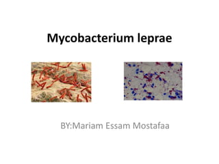
- 1. Mycobacterium leprae BY:Mariam Essam Mostafaa
- 2. Mycobacterium leprae: • Acid-fast staining rod, 1–8 µm long and 0.3 µm in diameter. • Non – motile . • Bacilli are seen in parallel rows and give appearance of cigar bundles. Cell wall consist of four layers: o Intermost layer of peptidoglycan , that give Shape and rigidity to the cell. o Attached to lipoabranomannan , which is Highly immunogenic and use in diagnosis of leprosy. o Adjacent to this is mycolic acid layer. o The outermost layer is made up of a phenolic Glycolipid-1, which is suppresss host celland use In serodiagnosis of leprosy
- 3. Mode of transmission: Transmission by inlalation: Droplet infection Transmission by contact: - Skin to skin contact with infectoius cases. - Contact with soil. Other Routes : -Insect vector e.g. mostique. - Needle sharing.
- 4. Pathogenesis: Leprosy is generally a disease of skin , peripheral nerves and nasal and mucosa. Leprosy is strictly a parasite man and a chronic granulamatous disease. Incubation period is long varies from 3-15 years. The principle target cell of lepra bacilli are schwann cell and resulting nerve damage causes manifestation of leprosy , which include anesthesia and muscle paralysis .
- 5. Virulence factors:- Iron utilization :some iron utilization genes have been conserved to help the pathogen acquire nutrients for growth. Waxy exterior: Mycobacterium Genus are defined by their waxy exterior coat. In Mycobacterium leprae, the exterior allows for intake into the macrophage and into some dendritic cells, in which it can survive. Macrophage invasion: Mycobacterium leprae survives and replicates in macrophages, dividing to approximately 100 organisms per cell. Schwann cell invasion: The major target of Mycobacterium leprae is the Schwann cell, Mycobacterium leprae gets into the lymphatic system and the blood vessels, Once in the area, Mycobacterium leprae binds to the Schwann cell via laminin- binding protein , The bacteria are thought to then enter through the vascular epithelium into the cell. The infection remains localized to the peripheral nervous system by rolling and binding to exposed Schwann cells. Drug resistance : Mycobacterium leprae has many mechanisms of drug resistance to allow it to continue to survive despite antimicrobial presence.
- 6. Clinical significance: Skin: Varible lesions: Macules,Papules,nodules. Single / multiple. Hypopigmented , sometimes reddish. Sensory loss typically(anesthesia / hypothesia) Nerves: Thickened. Loss of sensation. Muscle weakness. Nerve Enlargement Ear nodules Hypopegmented patch
- 7. Laboratory identification: Hansen’s disease (Leprosy) is diagnosed based on clinical presentation and the diagnosis is confirmed by skin or nerve biopsy and acid fast staining. Depending on the form of leprosy suspected by the treating physician, the following specimens may be collected: Skin smears from the earlobes, elbows, and knees. Skin biopsy from edges of active patches. Nerve biopsy from thickened nerves. Acid fast staining The Ziehl-Neelson method using 5% sulphuric acid as decolorizing agent is used. The presence of acid-fast bacilli confirms the diagnosis of Hansen’s disease. This acid-fast-stained photomicrograph of a tissue sample extracted from a patient with leprosy shows a chronic inflammatory lesion known as a granuloma, within which numerous red-colored M. leprae bacteria are visible.
- 8. Lepromin test: Used to determine type of leprosy. Injection of standardized extract of in activated bacilli intradermally in forearm. Positive reaction: 10mm or more induration after48 hrs / or 5mm ormore nodules after 21 days. Treatment: The currently recommended treatment regimen consists of three drugs: dapsone, rifampicin and clofazimine. The combination is referred to as multi-drug therapy (MDT). The duration of treatment is six months for PB and 12 months for MB cases. Prevention: To boost the prevention of leprosy, with the consent of the index case, WHO recommends tracing household contacts along with neighbourhood and social contacts of each patient, accompanied by the administration of a single dose of rifampicin as preventive chemotherapy.