Trichuris spp. in Animals, with Specific Reference to Neo-Tropical Rodents


1
Department of Basic Veterinary Sciences (DBVS), School of Veterinary Medicine (SVM), Faculty of Medical Sciences (FMS), University of the West Indies (UWI), Mt. Hope Campus, Trinidad and Tobago
2
Department of Food Production (DFP), Faculty of Food and Agriculture (FFA), University of the West Indies (UWI), St. Augustine Campus, Trinidad and Tobago
Vet. Sci. 2021, 8(2), 15; https://doi.org/10.3390/vetsci8020015
Submission received: 10 December 2020
/
Revised: 7 January 2021
/
Accepted: 13 January 2021
/
Published: 21 January 2021
Abstract
:Trichuriasis is the clinical disease of animals infected with the parasite of the genus Trichuris. This review attempts to present information on Trichuris spp. infestation in neo-tropical rodents that are utilized for meat consumption by humans. Neo-tropical rodents utilized for meat production can be divided into two categories: those that have been domesticated, which include the guinea pig (Cavia porcellus), and those that are on the verge of domestication, such as the capybara (Hydrochoerus hydrochaeris), lappe (Cuniculus paca/Agouti paca), and agouti (Dasyprocta leporina). This document reviews the literature on the species of Trichuris that affects the rodents mentioned above, as well as the clinical signs observed. The literature obtained spans over sixty years, from 1951 to 2020. Trichuris spp. was found in these neo-tropical rodents mentioned. However, there is a dearth of information on the species of Trichuris that parasitize these animals. The capybara was the only rodent where some molecular techniques were used to identify a new species named T. cutillasae. In most cases, Trichuris spp. was found in combination with other endoparasites, and was found at a low prevalence in the lappe and guinea pig. The presence of Trichuris spp. ranged from 4.62–53.85% in the agouti, 4.21–10.00% in the lappe, 50% in the capybaras, and 1–31% in guinea pigs. Further work must be done towards molecular identification of various Trichuris spp. present in these rodents, as well as the clinical effect of infection on the performance of agouti, lappe, capybara, and guinea pigs.
1. Introduction
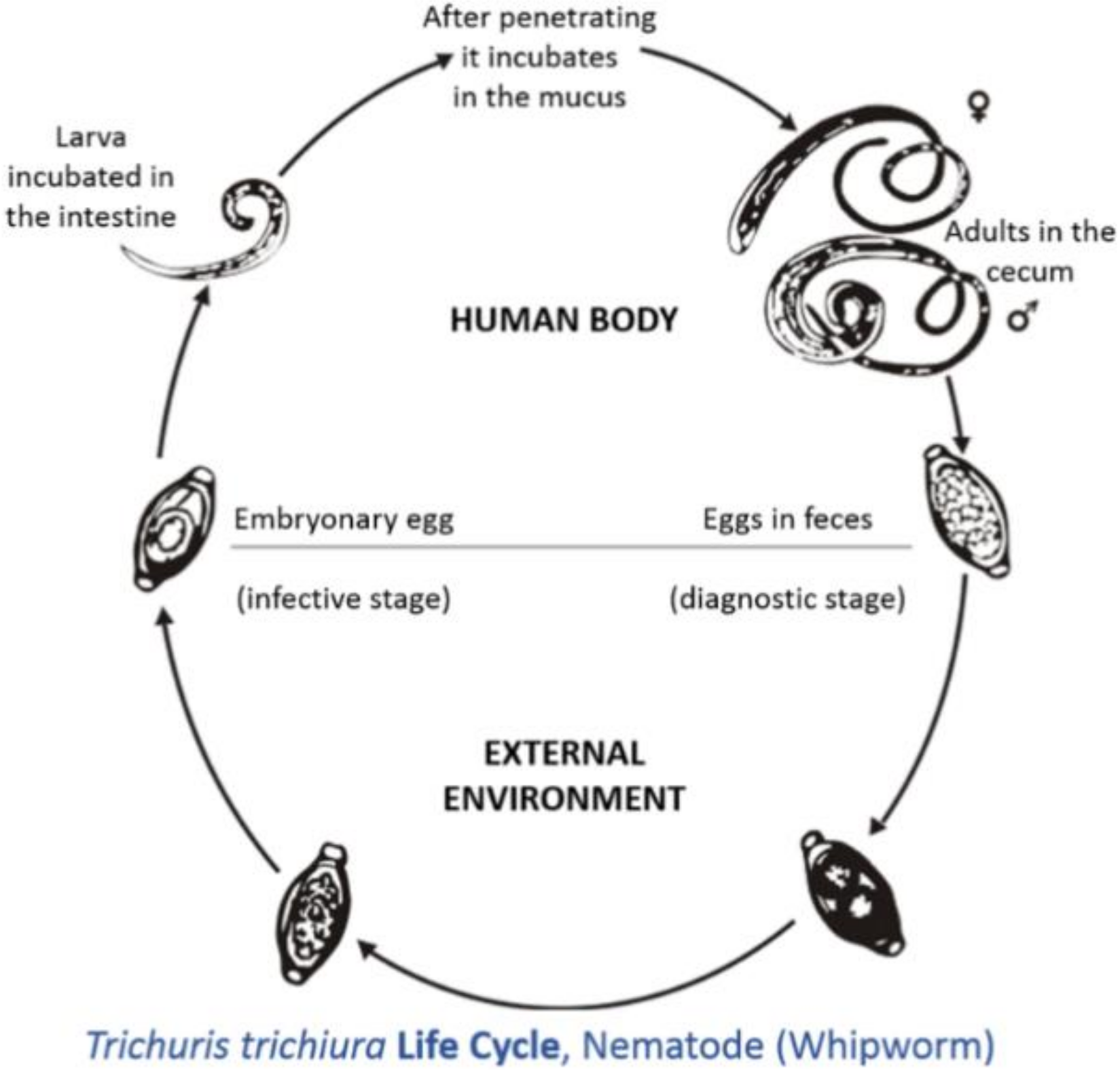
The neo-tropics is a geographical region located in the western hemisphere between the Tropic of Cancer and the Tropic of Capricorn. Geographical territories present within this zone include the southern parts of North America, all of Central America, the northern parts of South America, and all of the Caribbean [1]. Animals that are present in this region can be categorized into three groups: imported domesticated animals [2], domesticated animals originating from the neo-tropics [3], and non-domesticated neo-tropical animals [4]. For the purpose of this review, neo-tropical rodents that are included belong to the domesticated and non-domesticated groups. Domesticated neo-tropical rodents, such as the guinea pig, are utilized in South America for their meat and are reared in captivity to provide meat protein for rural villages. The guinea pig is able to utilize household waste and provide income and food for these communities [5,6]. Neo-tropical rodents on the verge of domestication are the agouti, lappe, and capybara. These animals have been reared in captivity in South America and the Caribbean for their meat [1]. These animals have been able to breed in captivity: the agouti produces four offspring per year [7], the lappe produces two offspring per year [8], and the capybara can produce eight offspring per year [9,10]. These animals are ideal in that they can utilize local feed resources and are adapted to local conditions of high heat and humidity. The meats produced by these rodents are highly nutritious, with high protein values and low fat and cholesterol concentration [11,12,13,14]. Trichuris spp., also known as whipworms, have parasitized many domesticated species, causing enteritis, diarrhea, and weight loss [15]. Trichuris spp. adults live in the caecum and colon; this predilection site has occurred due to evolution. The life cycle is direct; eggs with characteristic bi-polar plugs are passed in the feces and take two to three weeks to become infective (Figure 1) [16]. Animals become infected by the ingestion of infective eggs [16]. However, there has been limited information on the effects of Trichuris spp. on neotropical rodents (domestic and semi-domestic). Thus, the objective of this review is to summarize the species of Trichuris that parasitizes these rodents, the effect of this parasite on these animals, and the zoonotic potential of this pathogen.
2. Methodology
For the purpose of this review, reports and articles were searched for in scholarly publication databases (Google Scholar, PubMed, and UWI linc). Search terms used were specific species names (e.g., rodents, guinea pig (Cavia porcellus), agouti (Dasyprocta leporina), capybrara (Hydrochoerus hydrochaeris), and lappe (Cuniculus paca/ Agouti paca)) combined with the term “Trichuris” or “trichuriasis”. Searches were conducted for articles from 1990 to November 2020. Approximately 220 articles were identified for the review, but only 101 were appropriate to be included in the final manuscript. All sources were assessed by the author for relevance, credibility, and scientific inclusion, to ensure the thoroughness and accuracy of review.
3. Trichuris spp. of Veterinary and Public Health Importance
3.1. Trichuriasis of Man
Trichuriasis is one of the major infectious diseases of children in developing countries [18]. Trichuris trichiura is a major, soil-transmitted helminth targeted by the World Health Organization in their mass drug administration program for pre-school and primary school children in endemic developing countries [18]. There have been several cases of trichuriasis reported in humans. In some cases, it has been due to three Trichuris spp.: T. trichiura, T. vulpis, and T. suis. Humans have been infected with T. vulpis, and the diagnosis was made based on the morphology of the eggs and vulva from an adult female [19]. Molecular techniques were used on Trichuris spp. egg present in feces to identify T. suis and T. trichiura in human populations from Thailand [20]. T. suis has been experimentally given to humans, and the author stated that feces were negative for Trichuris eggs 40 days post-infection [21]. Experimentally treated patients showed no symptoms of gastrointestinal distress [21]. In contrast to the previous studies, Kradin et al. [22] showed that iatrogenic infection with T. suis resulted in a persistent active infection in man. Pathological findings from colonic biopsies showed several round helminths beneath the ileocecal mucosa epithelium [22].
Trichuris trichiura has human and non-human primates as its natural hosts [23]. Mixed infections with various Trichuris spp. in humans have been documented. There have been cases of mixed infections with T. vulpis and T. trichiura [24,25]. The identification of the species of Trichuris spp. was based on the morphology of eggs [24] and polymerase chain reactions of the helminth eggs [25]. Trichuris trichiura and T. vulpis was also found in the stool samples of dogs that roamed around the community. This shows that dogs are key to the transmission of Trichuris spp. to humans, but further work needs to be done to validate this finding [25].
Infections with T. vulpis have been reported in children and adults [19,26,27]. However, all cases of trichuriasis in humans caused by T. vulpis have had some association with dogs, and the diagnosis was made based on morphology of eggs present in the feces. Clinical signs reported in humans are abdominal discomfort, epigastric pain, nausea, vomiting, diarrhea, and poor appetite [24]. Patients with T. vulpis [24,26,27] and T. trichiura [19] have been treated with mebendazole and albendazole with improvements of clinical signs [19,24,26,27]. However, in vivo studies on albendazole and mebendazole have shown little efficacy against T. trichiura [28]. At 14 days post-treatment, there was no difference in the disease prevalence seen between treatments of patients with 400 grams of albendazole [28]. Therefore, alternative anthelmintic treatment against T. trichiura should be investigated. Ivermectin has been used to treat Trichuris spp.; however, it is very ineffective, as these parasites have become resistant to this drug. However, due to the increased prevalence of anthelmintic resistance, the drugs used to treat trichuriasis should be done with caution.
3.2. Morphological and Molecular Identifications of Trichuris spp.
3.2.1. Morphological Identification of Trichuris spp. in Pigs, Dogs, Cats, Humans, and Non-Human Primates
Morphological analysis of Trichuris spp. has been used for identification within various host species. Trichuris trichiura infection has been investigated in humans, non-human primates, and pigs, but based on morphological analysis, the T. trichiura found in humans and non-human primates were indistinguishable [29]. In pigs, T. suis was differentiated from T. trichiura, based on the lack of peri-cloacal papillae in adult specimens. In female specimens, there were no morphological differentiation between T. suis and T. trichiura [29]. Ruminants evaluated in India using morphological analysis identified T. ovis as the major parasite [30].
Further research was done in domestic cats in St. Kitts. Based on the size of the Trichuris spp. identified, authors believed that it was T. campanula, but based on the vulva structure the authors confirmed it was T. serrata. In conclusion, the authors, identified the parasite as T. serrata, but recommended that molecular studies must be done in order to reliably identify this parasite [31]. In dogs, male and female adult T. vulpis could be identified based on nine parameters (including body length, length of cuticular processes, and width of body at tail part) [32]. Male T. vulpis can be distinguished from other species by spicule sheath ornamentation (the dimensions of the spicule) [32].
Recently, the morphometric approach analyzing the adult worms and eggs of Trichuris spp. of non-human primates were analyzed [33,34]. Morphometric data on the adult worms showed that features present in the females made them indistinguishable for species characteristics, but adult male worms may be used to differentiate Trichuris populations [33]. Geometric morphometric analysis is a new diagnostic tool that can be used to differential Trichuris spp. present in non-human primates. However, further data must be collected to determine the sensitivity and specificity of this diagnostic tool [34]. Combination of various techniques, such as the use of molecular and morphological analysis, should be performed for confirmation of various Trichuris spp. [33].
3.2.2. Molecular Identification of Trichuris spp. in Domestic and Non-Domestic Ruminants
Molecular techniques have been used to identify various Trichuris spp. in their animals or human hosts. Such techniques have been applied to Trichuris spp. found in ruminants (both domesticated and non-domesticated). Four Trichuris spp.—T. discolor, T. ovis, T. globulosa and T. skrjabini—have been identified as inhabiting the caecum and colon of ruminants [35,36,37,38,39,40,41,42,43,44,45]. One of the major discoveries was the identification of T. globulosa and T. ovis as the same species by isoenzymes [35], using second, internally transcribed spacer ribosomal DNA (ITS2 rDNA) [38] and ITS1-5.8S-1TS2 [37]. Further molecular analysis was done comparing T. ovis and T. discolor, where the entire mitochondrial DNA (mtDNA) was analyzed [42], and with the use of internally transcribed spacers 1, 2, and 16S, partial DNA sequencing (ITS1, 2, 16rDNA) was completed [44]. Based on mtDNA and rDNA, T. ovis and T. discolor can be classified as two different species.
Trichuris skrjabini, found in small ruminants (sheep and goats), was characterized using isoenzymes [36], ITS1-5.8S-1TS2 [37], and cytochrome oxidase subunit 1 and mitochondrial 16S rDNA [39]. Authors have stated that T. skrjabini is an independent species but has close relations to other Trichuris spp. that parasitize small ruminants. Trichuris discolor has been identified in domestic ruminants with the use of molecular techniques; however, it was recently identified in wild ruminants, such as the roe deer (Capreolus capreolus), sika deer, (Cervus nippon), red deer (Cervus elephus), fallow deer (Dama dama), and mouflons (Ovis orientalis musimon) [43,44,45]. In wild ruminants, T. discolor was identified with use of ITS1-5.8S-1TS2 [43,44,45], but in cattle different populations of T. discolor in Iran, Spain, and Japan were investigated using 16S partial gene mtDNA, as well as ITS1 and 2 [43]. Callejon et al. [43] noted that there were specific populations of T. discolor groups based on geographical location. The author noted that one reason may be due to two cryptic species of T. discolor from Japan and Iran, as well as another from Spain.
3.2.3. Molecular Identification of Trichuris spp. in Cats, Dogs, Pigs, Humans, and Non-Human Primates
Trichuris spp. has also been identified molecularly in pets, such as dogs and cats. In cats it is associated with typhlitis, which also occurs in other animals [46]. Identification of T. serrata (cats) and Trichuris vulpis (dogs) was accomplished through the use of 18S rDNA (cats) and enzyme-linked immunosorbent assay (ELISA) and ITS1-5.8S-1TS2 (dogs) [47,48,49]. Comparative genetic studies were done of the T. vulpis found in dogs and T. suis found in pigs (wild and domesticated). There was a difference seen in amplified ITS1-5.8S-1TS2 rDNA between the T. vulpis found in dogs and T. suis found in pigs. Interestingly, T. suis collected from wild pigs (Sus scrofa scrofa) and domestic pigs (Sus scrofa domestica) showed no sequential genetic differences [49].
Several non-morphological processes were used to identify T. suis found in pigs using isoenzymes [50], ITS 1 and ITS2 regions of rDNA [51], large mitochondrial subunits and ITS2 [52], and nuclear ribosomes (18S, ITS2) [18]. Due to the zoonotic potential of T. suis and its morphological similarity to T. trichiura previous molecular studies have been done in both human and non-human primates [53,54,55]. Trichuris spp. was taken from pigs (wild and domestic) and non-human primates (Colobus guereza kikuyensis and Nomascus gabriellae) and analyzed by amplification of rDNA (ITS1-5.8S-1TS2). The authors confirmed that the T. suis found in pigs was genetically different from T. trichiura in Colobus guereza kikuyensis and Nomascus gabriellae [53]. Nissen et al. [54] conducted a similar study to Cutillas et al. [53], but T. suis and T. trichiura were identified in pigs and humans in Uganda. The gastrointestinal tract of pigs only contained T. suis, while in humans T. trichiura, T. suis, and a heterozygous type was identified [54]. This showed that the use of ITS 2 and β-tubulin allowed the identity of several species of Trichuris in humans to be highlighted.
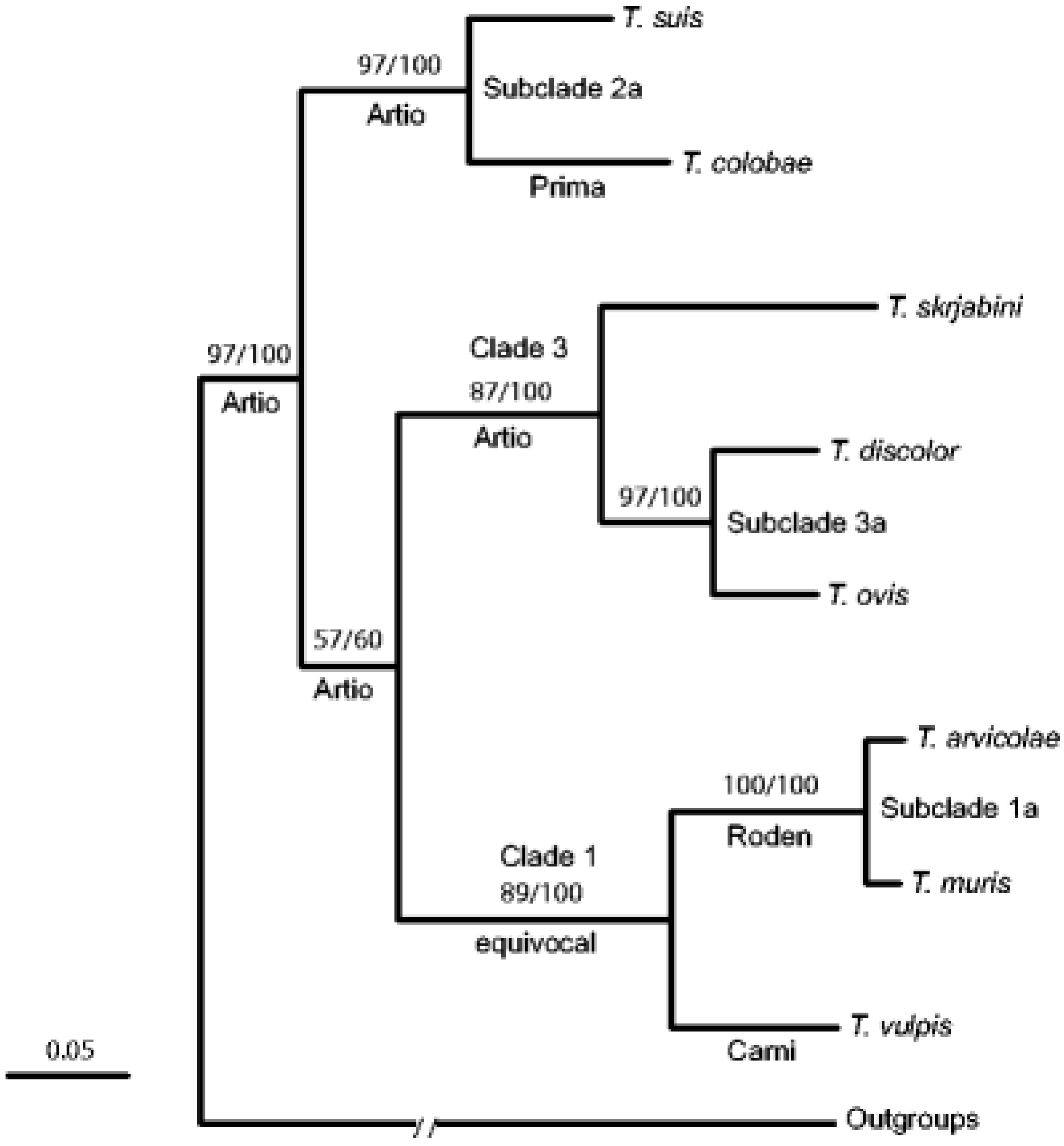
The research done by Cutillas et al. [53] and Nissen et al. [54] highlights the fact that humans and non-human primates may be infected with several species of Trichuris that are generally classified as T. trichiura. This was seen with Trichuris spp. samples taken from the wild Japanese macaques (Macaca fuscata), where the Trichuris spp. identified had genetic (18S rDNA) dissimilarity compared to those found in humans [56]. This new hypothesis sparked scientists to investigate this phenomenon at a molecular level (Figure 2). Ravasi et al. [57] investigated the genotype of human and non-human primates in Central Africa. Sequencing of the rDNA (ITS1-5.8S-1TS2) revealed two Trichuris genotypes that infect both humans and non-primates [57]. Ghai et al. [58] found similar results to Ravasi et al. [57], but three Trichuris genotypes were identified as circulating within human and non-human primates. Humans were infected with two genotypes: one genotype that was only common to human samples (Group 1), and another genotype that infected humans as well as non-human primates (black-and-white colobus (Colobus guereza), blue monkeys (Cercopithecus mitis), grey-cheeked mangabeys (Lophocebus albigena), l’hoest monkeys (Cercopithecus lhoesti), olive baboons (Papio anubis), red colobus (Procolobus rufomitratus), red-tailed guenons (Cercopithecus ascanius), and the chimpanzee (Pan troglodytes)) (Group 3). The intermediary group (Group 2) had a Trichuris genotype that affected non-human primates (black-and-white colobus (Colobus guereza and the red colobus (Procolobus rufomitratus) [58]. Furthermore, this new species of Trichuris was found in the Francois’ leaf monkey (Presbytis francoisi) and the Colobus guereza kikuyensis using mtDNA, rDNA, and morphometry [59,60].
3.2.4. Molecular Identification Trichuris spp. in Rodents
Trichuris spp. has been found in domestic livestock and pets, but there are also species that are specific to rodents. The initial molecular research that was done on the Trichuris spp. present in rodents focused on European rodents [61]. Trichuris muris was identified in Murid rodents in Europe with the use of rDNA (ITS1-5.8S-ITS2). It was found that two lineages had occurred, due to geographical distribution. One was found in northern Spain to Denmark, and the other in the Southern Europe (Croatia, Romania, and Turkey) [61]. In recent years, several new species of Trichuris arvicolae have been found in Arvicolinae rodents using multi-local enzyme electrophoresis [62] and rDNA (ITS1-5.8S-ITS2) [63]. Further investigations were done in the phylogeographic analysis of T. arvicolae in Europe, using the mtDNA cytochrome subunit 1 gene (cox1) and rDNA (ITS1-5.8S-ITS2). Nuclear genetics (ITS1-5.8S-ITS2) suggest that T. arvicolae show two geographic and genetic lineages (Neoarctic and Palaearctic). Mitochondrial results gave further details into the Palaearctic region, giving three geographic and genetic lineages (Northern Europe, Southern and Eastern Europe, and Italy and France) [64].
Scientists also investigated Trichuris present in Sigmodontinae rodents in South America (Argentina). New species, such as Trichuris novonae, were identified based on morphological analysis [65]. Another species that was identified morphologically was T. pardinasi [64]. Based on molecular characteristics, using ITS2 (rDNA), a new species named Trichuris bainae was identified [66]. Molecular analysis using cox1 and mitochondrial cytochrome b (cob) on the Trichuris spp. found in Sigmodontinae rodents found three clades corresponding to three different species, which were T. pardinasi, T. bainae, and T. navonae) [67]. Further to this, T. massoiai was identified in Holochilus chacarius (Cricetidae: Sigmodontinae) using morphological mitochondrial (cox1 and cob) and nuclear (ITS2) markers [68].
Callejon et al. [41,69] investigated nuclear (18S, triose phosphate isomerase) and mitochondrial (cox1, cob1) genes from Trichuris spp. from nine various host species (Colobus guereza kikuyuensis, Papio hamadryas, Homo sapiens, Sus scrofa domesticus, Capra hircus, Canis lupus familiaris, Bos taurus, Mus domesticus, and Myodes glareolus) from Spain. The data show that Trichuris spp. could be divided in three clades: Clade 1 = T. arvicolae, T. muris, and T.vulpis; Clade 2 = T. suis, T. colobae, T. trichiura, and T. spp. ex Papio hamadryas; Clade 3 = T. discolor, T. ovis, and T. skrjabini [69].
3.3. Immunomodulatory Effect of Trichuris spp.
Trichuris spp. has been used in the treatment of gastrointestinal autoimmune diseases, such as inflammatory bowel disease, Crohn’s disease, and ulcerative colitis [70,71,72]. Trichuris suis (pig whipworm) had been experimentally given to humans with no overt sign of gastrointestinal illness. The eggs produced from the feces remained constant, and only a low percentage of these eggs embryonated in vitro [21]. Some authors also noted that treatment of patients with inflammatory bowel disease, ulcerative colitis, and Crohn’s disease with Trichuris suis showed improvement in gastrointestinal signs, and in the management of disease the subjects were given ova every three weeks [70,71,72]. Surprisingly, Kradin et al. [22] noted that a patient that underwent treatment for Crohn’s disease using T. suis had adult worms beneath the ileocecal mucosal epithelium. This case does raise concerns about persistent infection from T. suis in man [22].
Further work was done on the use of excretory secretory products of T. suis in rats [73]. The investigation of the use of excretory products of T. suis in swine epithelium cells was used as a model to be used in humans. It was noted that the excretory secretory products (ESPs) elicited the production of interleukin (IL)-6 and IL-10, which have been identified as anti-inflammatory cytokines that inhibit Th-1 responses. This proved that ESPs from T. suis have immunomodulatory effects and can be used as candidates in the treatment of inflammatory bowel disease [73]. The use of ESPs from T. suis may be safer than the actual treatment with ova.
Subsequent research was done on the immunomodulatory and immunogenic effects of the proteins and ESPs of Trichuris trichiura and Trichuris muris [74,75,76]. Proteins were analyzed from adult worm extract and fragments of T. trichiura. These extracts and fragments were placed in cell cultures of human peripheral blood monocytes, and elicited the production of IL-10, IL-12, and TNF-α. Some fractions showed the inhibition of IL-5 production. The downregulation of IL-5 is a feature of a Th-2 response [74]. Santos et al. [74] concluded that protein fractions of T. trichiura can be used in the treatment and prevention of allergic and autoimmune diseases. Immunogenic research was also conducted on the ESPs of T. muris, and specific immunogenic proteins were identified. The structure of one such protein was Tm16, which was characterized and could be used in the production of a vaccine [75]. Shears et al. [76] noted that ESPs from T. muris elicited production of IL-9 and IL-13 when inoculated into rats. Eleven immunogenic proteins from the ESP of T. muris were also identified, and these could be used in the production of a vaccine [76]. Recent studies show that there is tremendous potential for Trichuris in human autoimmune disease, as well as vaccine development in rural countries where trichuriasis infections are prevalent.
4. Domesticated Neo-Tropical Rodent
4.1. Guinea Pig (Cavia Porcellus)
The guinea pig is a domesticated rodent that is utilized for its meat in rural communities in South America and Africa. In rural communities, it provides food security and economic opportunity. These animals can be reared on local feed by-products and can produce four to nine offspring per female per year [5,6]. Several gastrointestinal parasites have been reported to inhabit these animals, with few reports on the clinical effect on these animals [3].
Several authors that have done work investigating the gastrointestinal parasites present in captive reared guinea pigs have failed to find Trichuris present [77,78,79,80,81,82]. Endoparasites of wild and captive reared guinea pigs were investigated in many countries, including the Democratic Republic of Congo [77], Cameroon [78], Iran [80], and Brazil [81,82]. In Peru, 3.5% of wild guinea pigs were infected with Trichuris gracilis [83], but in captivity infection rates of 5% [84] and 31% [85] were recorded. However, in captive reared guinea pigs present in Cameroon, 0.3% [86] and 3.3% [87] were positive for Trichuris spp. In Benin, guinea pigs reared in captivity using traditional and modern housing arrangements had an infection rate for Trichuris spp. of 11.18% [88] (Table 1). Infection with elevated levels of Trichuris in domesticated animals can lead to diarrhea, weight loss, enteritis, and colitis.
The authors who failed to identify Trichuris in guinea pigs used the fecal floatation technique [77,78,79,80,81,82] (using sodium chloride and zinc sulphate solution), gross identification of adult worms, as well as a combination of both methods mentioned above [80]. Trichuris gracilis in some studies was identified by the gross identification of adult worms in wild animals and captive animals [83,84,85], but in Cameroon and Benin Trichuris spp. was identified using fecal floatation and fecal sedimentation [86,87,88,94]. The weights, clinical conditions, or pathological findings of guinea pigs infected with Trichuris gracilis was not described by investigators. In most cases of trichuriasis in the guinea pig, there were parasites (Paraspidodera uncinata, Capillaria spp., or Trichostrongylus colubriformis) that co-infected the hosts’ gastrointestinal tract. However, Garcia et al. [95] reported that 55% of the animals were infected only with Trichuris spp.
Trichuris spp. was identified in several countries, including those found in the African and South American continent. Guinea pigs that were wild and captive reared were both found to have Trichuris spp. However, the species of Trichuris was not identified in most cases, due to a lack of molecular techniques in the detection of this parasite. The genus Trichuris has zoonotic significance, since human and non-human primates are infected with Trichuris trichiura [54]. Future work should focus on molecular technique in the identification of parasites in the guinea pig and their potential immunoregulatory effects in experimental studies.
4.2. Semi-Domesticated Neo-Tropical Rodents
4.2.1. Agouti (Dasyprocta leporina)
The agouti is a robust rodent, with adults weighing 2–4 kg [8]. These animals are omnivorous [96], practice cecotrophy, and possess a large cecum [97]. Some authors have even classified these animals as opportunistic omnivores [98]. These animals have been successfully fed in captivity [89], and several endoparasites have been identified in this animal [4]. Parasites that have been found in the agouti seem to have no effect on the animals clinically or sub-clinically [4]. Animals that have parasites living within their digestive tract appear to be well-fleshed, with the absence of any gastrointestinal disturbances.
Early parasitic investigation done on the agouti using the morphological data of adult specimens in the digestive tract identified Trichuris gracilis var. trinitatae from wild agoutis in Trinidad [99]. Further to this initial work, Suepaul et al. [100] found T. gracilis var. trinitatae in hunted agouti in Trinidad, and Goncalves et al. [90] identified T. gracilis var. trinitatae in wild agouti in Brazil. In recent times, the identification of different Trichuris spp. using the morphological analysis of adult worms and eggs have proven to be inadequate [90]. In wild animals Trichuris spp. was identified in conjunction with other gastrointestinal helminths. However, authors have failed to record data on the health of the animals or the pathology of the gastrointestinal tract.
Trichuris spp. was also identified using fecal floatation techniques in wild and captive reared agoutis. Species identification was impossible with the use of fecal floatation. In Brazil, Trichuris spp. was identified in wild agoutis, with eggs having their characteristic bi-polar plugs [101]. Further research done in Trinidad identified Trichuris spp. in farmed agoutis [102]. Trichuris spp. was identified along with Strongyloides spp., Eimeria spp., and Paraspidodera uncinata [103]. Infected animals had an average fecal egg count of 2.2 × 10², and animals were in good body condition, with no gross pathological lesions and blood values within normal reference ranges [103,104].
The prevalence of Trichuris spp. in captive reared animals in Trinidad was 4.62% [102]. Suepaul et al. [100] obtained a higher prevalence of 53.95% in free range agoutis (Table 1). These studies are the only record in the literature that report the prevalence of Trichuris in the agouti. This shows the limited research done on trichuriasis in this neo-tropical rodent. In the agouti, there has been an absence of molecular identification of endoparasites, and in particular to Trichuris. The agouti has been grown in captivity, with close contact with humans and domesticated animals. Thus, species identification of the presence of this parasite is paramount. Trichuris appears to be ubiquitous in captive and wild environments of the agouti, and proper analysis of the effect of this parasite must be documented.
4.2.2. Lappe (Agouti paca/Cuniculus paca)
The lappe is a robust neotropical rodent with adults weighing 4–8 kg [8]. These rodents practice cecotrophy and consume locally available fruits and crops [98]. The majority of parasitic investigations done on the lappe have focused on echinococcosis. This is because the lappe is the intermediate host for Echinococcus spp. [103,104,105,106,107], with the dog as the final host. Humans can become dead-end intermediate hosts following ingestion of eggs shed by dogs, but cannot become infected by meat or organ consumption.
In the lappe, adult Trichuris worms have been found in the cecum of the gastrointestinal tract [91], and eggs have been found in the feces using fecal floatation [92,108]. However, in the studies conducted, scientists failed to identify the species of Trichuris and its effect on the lappe. It is impossible to distinguish different species of Trichuris through the use of morphological identification of the adult worms or the eggs that are produced.
Trichuris spp. was found in both captive [105,106] and wild lappe populations [91]. Trichuris was found in conjunction with several endoparasites in the digestive tract, with no cases being reported of lappe infected with only Trichuris spp. The research work reported on the lappe was done in the neotropics in Mexico, Costa Rica, and Brazil. The prevalence of this disease varied between locations, with 10% of lappe in the study in Mexico being infected [92], while in Costa Rica, 2.13% (Table 1) of the sampled animals had Trichuris spp. [108]. In Brazil, animals had an average fecal egg load of 4.15 eggs per gram (EPG) [108].
The differences in prevalence of Trichuris seen between Costa Rica and Mexico can be due to firstly, the method of identification, with Ramirez-Herrera et al. [92] utilizing fecal floatation and Matamoros et al. [105] utilized gross identification of adult parasites. Secondly, these can be variations in the number of infected animals within the respective countries that contribute to the contamination of the environment with infective eggs. Thirdly, the environment in which the sampled animals inhabit may be different, with the lappes studied in Costa Rica being raised in the wild and lappes sampled in Mexico being maintained in a captive environment. Further research must be done to obtain the prevalence of trichuriasis in the lappes of various countries within the neo-tropical region. The species identification must also be performed in these investigations, as well as a comparison of prevalence between captive and wild populations, using molecular techniques for identification.
4.2.3. Capybara (Hydrochoerus hydrochaeris)
The capybara is the largest rodent in the world. Lall et al. [96] summarized these animals as semi-aquatic herbivorous rodents that practice cecotrophy. These animals are hindgut fermenters that possess a mucus trap separation mechanism [109]. Various parasites have been found in the capybara, but the majority of research views these animals as reservoirs for specific pathogens that have zoonotic potential or can cause disease in domestic livestock species.
The major parasites investigated have included helminths and protozoa. Helminths like Fasciola hepatica has been found in both capybara and cattle, but cause major disease problems in cattle [110,111,112]. Protozoan parasites of zoonotic importance reported in capybara include Cryptosporidium parvum [113]. Protozoan parasites of found in the capybara that can negatively affect livestock include Eimeria spp., Eimeria ichiloensis, Eimeria boliviensis, and Eimeria trinidadensis [114,115,116,117,118,119]. However, few reports have been made on the identification of Trichuris or the clinical effect of this pathogen.
Brazilian capybaras reared in captivity had Trichuris spp. in 50% of samples [117] (Table 1). Trichuris spp. was found in conjunction with several other parasites in the gastrointestinal tract. The effects of these endoparasites on the capybara have not been documented. Surprisingly, the capybara is the only rodent where molecular techniques have been used in the identification of Trichuris spp. Eberhardt et al. [93] identified a new species of Trichuris from capybara using molecular characteristics and phylogenetic relationships. The new species was identified as Trichuris cutillasae, and this was found in the cecum of capybara in Argentina. This new species has veterinary importance, and emphasizes the fact that further work has to be done on the genetic identification of Trichuris spp. in the capybara at different geographical locations in the neo-tropics. This new species must be investigated to provide clarity on the effect of this parasites on the health and performance of the capybara. Information on the parasitic load of Trichuris cutillasae in the capybara that will precipitate disease need to be investigated.
5. Conclusions
This review revealed that Trichuris has been found in the guinea pig (C. porcellus), agouti (D. leporina), lappe (A. paca/C. paca), and capybara (H. hydrochaeris). However, there is a dearth of information on the species of Trichuris that parasitize these animals. The capybara was the only rodent where some molecular techniques were used to identify a new species of Trichuris, named T. cutillasae. In most cases, Trichuris was found in combination with other endoparasites, and had a prevalence ranging from 4.62–53.85% in the agouti, 4.21–10.00% in the lappe, 50% in the capybara, and 1–31% in the guinea pig.
6. Recommendations
Further work must be done on the molecular identification of various Trichuris spp. present in neo-tropical rodents, as well as the effect of Trichuris on the performance of agouti, lappe, capybara, and guinea pigs.
Funding
This research was funded by the University of the West Indies, Campus Research and Publication (CRP) unit.
Institutional Review Board Statement
Ethical review was not applicable because this was a review and did not involve experimentation on humans or animals.
Informed Consent Statement
Informed consent was not applicable to this study.
Data Availability Statement
All data used are presented in the document.
Acknowledgments
The Staff at the Alma Jordan Library, University of the West Indies, St. Augustine Campus. Special thanks must be given to Sheeba Sreenivasan for her assistance in locating journal articles that were not easily accessible.
Conflicts of Interest
There are no conflicts of interest among the authors.
References
- Brown-Uddenberg, R.C.; Garcia, G.W.; Baptiste, Q.S.; Counand, T.; Adogwa, A.O.; Sampson, T. The Agouti (Dasyprocta leporina, D. aguti) Booklet and Producers’ Manual; GWG Publications: Champs Fleurs, Trinidad and Tobago, 2014; Available online: http://ostasp.brinkster.net/ (accessed on 17 June 2020).
- Jones, K.R.; Garcia, G.W. Gastrointestinal parasites of domesticated animals introduced into the Neo-tropics (New World Tropics). Concepts Dairy Vet. Sci. 2018, 1, 56–78. [Google Scholar]
- Jones, K.R.; Garcia, G.W. Endoparasites of domesticated animals that originated in the neo-tropics (new world tropics). Vet. Sci. 2019, 6, 24. [Google Scholar] [CrossRef] [PubMed] [Green Version]
- Jones, K.R.; Lall, K.R.; Garcia, G.W. Endoparasites of selective native non-domesticated mammals in the neo-tropics (new world tropics). Vet. Sci. 2019, 6, 87. [Google Scholar] [CrossRef] [PubMed] [Green Version]
- Paterson, R.T.; Joaquin, N.; Chamon, K.; Palomino, E. The productivity of small animal species in small-scale mixed farming systems in subtropical Bolivia. Trop. Anim. Health Prod. 2001, 33, 1–14. [Google Scholar] [CrossRef] [PubMed]
- Manjeli, Y.; Tchoumboue, J.; Njwe, R.M.; Teguia, A. Guinea-pig productivity under traditional management. Trop. Anim. Health Prod. 1998, 30, 115–122. [Google Scholar] [CrossRef]
- Jones, K.R.; Garcia, G.W. Anthelmintic usage on the reproductive parameter in captive reared agoutis (Dasyprocta leporina) in Trindad and Tobago, West Indies. Trop. Agric. 2020, 97, in Press. [Google Scholar]
- Govoni, G.; Fielding, D. Paca (Agouti paca) and Agouti (Dasyprocta spp.)-Minilivestock Production in the Amazonas State of Venezuela: 1. Biology. Tropicultura 2001, 19, 56–60. [Google Scholar]
- Chapman, C.A. Biology of Capybaras. J. Mammal. 1991, 72, 206–208. [Google Scholar] [CrossRef]
- Alvarez, M.R.; Kravetz, F.O. Reproductive performance of capybaras (Hyrochoerus hydrochaeris) in captivity under different management systems in Argentina. Anim. Res. 2006, 55, 153–164. [Google Scholar] [CrossRef] [Green Version]
- Ali, A.J.; Jones, K.R. Nutritive value and physical properties of Neo-tropical rodent meat-with emphasis on the Capybara (Hydrochoerus hydrochaeris). Animals 2020, 10, 2134. [Google Scholar] [CrossRef]
- Dalle Zotte, A. Perception of rabbit meat quality and major factors influencing the rabbit carcass and meat quality. Livest. Prod. Sci. 2002, 75, 11–32. [Google Scholar] [CrossRef]
- Pla, M.; Pascual, M.; Arino, B. Protein, Fat and Moisture Content of Retail Cuts of Rabbit Meat Evaluated with the NIRS Methodology. World Rabbit. Sci. 2004, 12, 149–158. [Google Scholar] [CrossRef] [Green Version]
- Nogueira-Filho, S.L.G.; Nogueira, S.S.C. Capybara meat: An extraordinary resource for food security in South America. Meat Sci. 2018, 145, 329–333. [Google Scholar] [CrossRef]
- Soulsby, E.J. Helminths, Arthropods and Protozoa of Domesticated Animals, 6th ed.; Baillière Tindall & Cassell; The American Society of Tropical Medicine and Hygiene: Arlington, VA, USA, 1968. [Google Scholar]
- Zajac, A.M.; Conboy, G.A. Veterinary Clinical Parasitology; John Wiley & Sons: Hoboken, NJ, USA, 2012. [Google Scholar]
- Life Cycle of Trichuris Found inside the Body. Available online: https://en.wikipedia.org/wiki/Trichuris_trichiura#/media/File:Trichuris_trichiura_Life_Cycle.tif (accessed on 7 January 2021).
- Albonico, M.; Allen, H.; Chitsulo, L.; Engels, D.; Gabrielli, A.F.; Savioli, L. Controlling soil-transmitted helminthiasis in pre-school-age children through preventive chemotherapy. PLoS Negl. Trop. Dis. 2008, 2, e126. [Google Scholar] [CrossRef] [PubMed] [Green Version]
- Hall, J.E.; Sonnenberg, B. An apparent case of human infection with whip-worm of dogs, Trichuris vulpis (Froelich, 1789). J. Parasitol. 1956, 42, 197–199. [Google Scholar] [CrossRef]
- Phosuk, I.; Sanpool, O.; Thanchomnang, T.; Sadaow, L.; Rodpai, R.; Anamnart, W.; Janwan, P.; Wijit, A.; Laymanivong, S.; Aung, W.P.P.; et al. Molecular identification of Trichuris suis and Trichuris trichiura eggs in human populations from Thailand, Lao PDR, and Myanmar. Am. J. Trop. Med. Hyg. 2018, 98, 39–44. [Google Scholar] [CrossRef] [Green Version]
- Beer, R.J. Experimental infection of man with pig whipworm. Br. Med. J. 1971, 2, 44. [Google Scholar] [CrossRef] [Green Version]
- Kradin, R.L.; Badizadegan, K.; Auluck, P.; Korzenik, J.; Lauwers, G.Y. Iatrogenic Trichuris suis infection in a patient with Crohn disease. Arch. Pathol. Lab. Med. 2006, 130, 718–720. [Google Scholar]
- Sunkara, T.; Sharma, S.R.; Ofosu, A. Trichuris trichiura—An Unwelcome Surprise during Colonoscopy. Am. J. Trop. Med. Hyg. 2018, 99, 555. [Google Scholar] [CrossRef]
- Vásquez, O.T.; Martínez, I.B.; Romero, R.C.; Valencia, S.R.; Tay, J.Z. Mixed infection by Trichuris trichiura and Trichuris vulpis. Rev. Gastroenterol. Peru Organo Soc. Gastroenterol. Peru 1997, 17, 255–258. [Google Scholar]
- Areekul, P.; Putaporntip, C.; Pattanawong, U.; Sitthicharoenchai, P.; Jongwutiwes, S. Trichuris vulpis and T. trichiura infections among schoolchildren of a rural community in north-western Thailand: The possible role of dogs in disease transmission. Asian Biomed. 2010, 4, 49–60. [Google Scholar] [CrossRef] [Green Version]
- Dunn, J.J.; Columbus, S.T.; Aldeen, W.E.; Davis, M.; Carroll, K.C. Trichuris vulpis recovered from a patient with chronic diarrhea and five dogs. J. Clin. Microbiol. 2002, 40, 2703–2704. [Google Scholar] [CrossRef] [PubMed] [Green Version]
- Márquez-Navarro, A.; García-Bracamontes, G.; Álvarez-Fernández, B.E.; Ávila-Caballero, L.P.; Santos-Aranda, I.; Díaz-Chiguer, D.L.; Sánchez-Manzano, R.M.; Rodríguez-Bataz, E.; Nogueda-Torres, B. Trichuris vulpis (Froelich, 1789) infection in a child: A case report. Korean J. Parasitol. 2012, 50, 69–71. [Google Scholar] [CrossRef] [PubMed]
- Olsen, A.; Namwanje, H.; Nejsum, P.; Roepstorff, A.; Thamsborg, S.M. Albendazole and mebendazole have low efficacy against Trichuris trichiura in school-age children in Kabale District, Uganda. Trans. R. Soc. Trop. Med. Hyg. 2009, 103, 443–446. [Google Scholar] [CrossRef] [PubMed] [Green Version]
- Ooi, H.K.; Tenora, F.; Itoh, K.; Kamiya, M. Comparative study of Trichuris trichiura from non-human primates and from man, and their difference with T. suis. J. Vet. Med. Sci. 1993, 55, 363–366. [Google Scholar] [CrossRef] [Green Version]
- Kuchai, J.A.; Ahmad, F.; Chishti, M.Z.; Dar, J.A.; Tak, H. On morphology and morphometry of Trichuris ovis Abildgaard, 1795 recovered from ruminants of Ladakh, India. J. Buffalo Sci. 2013, 2, 49–52. [Google Scholar] [CrossRef]
- Ketzis, J.K. Trichuris spp. infecting domestic cats on St. Kitts: Identification based on size or vulvar structure? SpringerPlus 2015, 4, 115. [Google Scholar] [CrossRef] [Green Version]
- Yevstafieva, V.A.; Kravchenko, S.O.; Gutyj, B.V.; Melnychuk, V.V.; Kovalenko, P.N.; Volovyk, L.B. Morphobiological analysis of Trichuris vulpis (Nematoda, Trichuridae), obtained from domestic dogs. Regul. Mech. Biosyst. 2019, 10, 165–171. [Google Scholar] [CrossRef]
- García-Sánchez, A.M.; Rivero, J.; Callejón, R.; Zurita, A.; Reguera-Gomez, M.; Valero, M.A.; Cutillas, C. Differentiation of Trichuris species using a morphometric approach. Int. J. Parasitol. Parasites Wildl. 2019, 9, 218–223. [Google Scholar] [CrossRef]
- García-Sánchez, A.M.; Reguera-Gomez, M.; Valero, M.A.; Cutillas, C. Differentiation of Trichuris species eggs from non-human primates by geometric morphometric analysis. Int. J. Parasitol. Parasites Wildl. 2020, 12, 214–219. [Google Scholar] [CrossRef]
- Cutillas, C.; German, P.; Arias, P.; Guevara, D. Trichuris ovis and Trichuris globulosa: Morphological, biometrical, and genetic studies. Exp. Parasitol. 1995, 81, 621–625. [Google Scholar] [CrossRef] [PubMed]
- Cutillas, C.; German, P.; Arias, P.; Guevara, D. Characterization of Trichuris skrjabini by isoenzyme gel electrophoresis: Comparative study with Trichuris ovis. Acta Trop. 1996, 62, 63–69. [Google Scholar] [CrossRef]
- Cutillas, C.; Oliveros, R.; De Rojas, M.; Guevara, D.C. Determination of Trichuris skrjabini by sequencing of the ITS1–5.8 S–ITS2 segment of the ribosomal DNA: Comparative molecular study of different species of trichurids. J. Parasitol. 2004, 90, 648–652. [Google Scholar] [CrossRef]
- Oliveros, R.; Cutillas, C.; De Rojas, M.; Arias, P. Characterization of four species of Trichuris (Nematoda: Enoplida) by their second internal transcribed spacer ribosomal DNA sequence. Parasitol. Res. 2000, 86, 1008–1013. [Google Scholar] [CrossRef]
- Callejón, R.; De Rojas, M.; Ariza, C.; Ubeda, J.M.; Guevara, D.C.; Cutillas, C. Cytochrome oxidase subunit 1 and mitochondrial 16S rDNA sequences of Trichuris skrjabini (Tricocephalida: Trichuridae). Parasitol. Res. 2009, 104, 715–716. [Google Scholar] [CrossRef] [PubMed]
- Callejón, R.; Halajian, A.; De Rojas, M.; Marrugal, A.; Guevara, D.; Cutillas, C. 16S partial gene mitochondrial DNA and internal transcribed spacers ribosomal DNA as differential markers of Trichuris discolor populations. Vet. Parasitol. 2012, 186, 350–363. [Google Scholar] [CrossRef] [PubMed]
- Callejón, R.; Nadler, S.; De Rojas, M.; Zurita, A.; Petrášová, J.; Cutillas, C. Molecular characterization and phylogeny of whipworm nematodes inferred from DNA sequences of cox1 mtDNA and 18S rDNA. Parasitol. Res. 2013, 112, 3933–3949. [Google Scholar] [CrossRef] [PubMed]
- Liu, G.H.; Wang, Y.; Xu, M.J.; Zhou, D.H.; Ye, Y.G.; Li, J.Y.; Song, H.Q.; Lin, R.Q.; Zhu, X.Q. Characterization of the complete mitochondrial genomes of two whipworms Trichuris ovis and Trichuris discolor (Nematoda: Trichuridae). Infect. Genet. Evol. 2012, 12, 1635–1641. [Google Scholar] [CrossRef]
- Salaba, O.; Rylková, K.; Vadlejch, J.; Petrtýl, M.; Scháňková, Š.; Brožová, A.; Jankovská, I.; Jebavý, L.; Langrová, I. The first determination of Trichuris sp. from roe deer by amplification and sequenation of the ITS1-5.8 S-ITS2 segment of ribosomal DNA. Parasitol. Res. 2013, 112, 955–960. [Google Scholar] [CrossRef]
- Vejl, P.; Nechybová, S.; Peřinková, P.; Melounová, M.; Sedláková, V.; Vašek, J.; Čílová, D.; Rylková, K.; Jankovská, I.; Vadlejch, J.; et al. Reliable molecular differentiation of Trichuris ovis and Trichuris discolor from sheep (Ovis orientalis aries) and roe deer (Capreolus capreolus) and morphological characterisation of their females: Morphology does not work sufficiently. Parasitol. Res. 2017, 116, 2199–2210. [Google Scholar] [CrossRef]
- Nechybová, S.; Vejl, P.; Hart, V.; Melounová, M.; Čílová, D.; Vašek, J.; Jankovská, I.; Vadlejch, J.; Langrová, I. Long-term occurrence of Trichuris species in wild ruminants in the Czech Republic. Parasitol. Res. 2018, 117, 1699–1708. [Google Scholar] [CrossRef] [PubMed]
- Wulcan, J.M.; Ketzis, J.K.; Dennis, M.M. Typhlitis Associated with Natural Trichuris sp. Infection in Cats. Vet. Pathol. 2020, 57, 266–271. [Google Scholar] [CrossRef] [PubMed]
- Ketzis, J.K.; Verma, A.; Burgess, G. Molecular characterization of Trichuris serrata. Parasitol. Res. 2015, 114, 1993–1995. [Google Scholar] [CrossRef] [PubMed]
- Cutillas, C.; de Rojas, M.; Ariza, C.; Ubeda, J.M.; Guevara, D. Molecular identification of Trichuris vulpis and Trichuris suis isolated from different hosts. Parasitol. Res. 2007, 100, 383–389. [Google Scholar] [CrossRef] [PubMed]
- Elsemore, D.A.; Geng, J.; Flynn, L.; Cruthers, L.; Lucio-Forster, A.; Bowman, D.D. Enzyme-linked immunosorbent assay for coproantigen detection of Trichuris vulpis in dogs. J. Vet. Diagn. Investig. 2014, 26, 404–411. [Google Scholar] [CrossRef] [Green Version]
- Oliveros, R.; Cutillas, C.; Arias, P.; Guevara, D. Morphologic, biometric, and isoenzyme characterization of Trichuris suis. Parasitol. Res. 1998, 84, 513–515. [Google Scholar] [CrossRef]
- Liu, G.H.; Zhou, W.; Nisbet, A.J.; Xu, M.J.; Zhou, D.H.; Zhao, G.H.; Wang, S.K.; Song, H.Q.; Lin, R.Q.; Zhu, X.Q. Characterization of Trichuris trichiura from humans and T. suis from pigs in China using internal transcribed spacers of nuclear ribosomal DNA. J. Helminthol. 2014, 88, 64–68. [Google Scholar] [CrossRef]
- Muramatsu, R.; Sato, R.; Onuma, N.; Sasai, K.; Shibahara, T.; Matsubayashi, M. Molecular Identification of Trichuris suis Worms and Eggs in Pig Feces, Infected Intestines, and Farm Environments in Japan. Jpn. Agric. Res. Q. JARQ 2020, 54, 271–275. [Google Scholar] [CrossRef]
- Cutillas, C.; Callejon, R.; De Rojas, M.; Tewes, B.; Ubeda, J.M.; Ariza, C.; Guevara, D.C. Trichuris suis and Trichuris trichiura are different nematode species. Acta Trop. 2009, 111, 299–307. [Google Scholar] [CrossRef]
- Nissen, S.; Al-Juburry, A.; Hansen, V.A.; Olsen, A.; Christensen, H.; Thamsborg, S.M.; Nejsum, P. Genetic analysis of Trichuris suis and Trichuris trichiura recovered from humans and pigs in a sympatric setting in Uganda. Vet. Parasitol. 2012, 188, 68–77. [Google Scholar] [CrossRef]
- Liu, G.H.; Gasser, R.B.; Su, A.; Nejsum, P.; Peng, L.; Lin, R.Q.; Li, M.W.; Xu, M.J.; Zhu, X.Q. Clear genetic distinctiveness between human-and pig-derived Trichuris based on analyses of mitochondrial datasets. PLoS Negl. Trop. Dis. 2012, 6, e1539. [Google Scholar] [CrossRef] [PubMed] [Green Version]
- Arizono, N.; Yamada, M.; Tegoshi, T.; Onishi, K. Molecular identification of Oesophagostomum and Trichuris eggs isolated from wild Japanese macaques. Korean J. Parasitol. 2012, 50, 253–257. [Google Scholar] [CrossRef] [PubMed]
- Ravasi, D.F.; O’Riain, M.J.; Davids, F.; Illing, N. Phylogenetic evidence that two distinct Trichuris genotypes infect both humans and non-human primates. PLoS ONE 2012, 7, e44187. [Google Scholar] [CrossRef] [PubMed] [Green Version]
- Ghai, R.R.; Simons, N.D.; Chapman, C.A.; Omeja, P.A.; Davies, T.J.; Ting, N.; Goldberg, T.L. Hidden population structure and cross-species transmission of whipworms (Trichuris sp.) in humans and non-human primates in Uganda. PLoS Negl. Trop. Dis. 2014, 8, e3256. [Google Scholar] [CrossRef] [PubMed] [Green Version]
- Liu, G.H.; Gasser, R.B.; Nejsum, P.; Wang, Y.; Chen, Q.; Song, H.Q.; Zhu, X.Q. Mitochondrial and nuclear ribosomal DNA evidence supports the existence of a new Trichuris species in the endangered françois’ leaf-monkey. PLoS ONE 2013, 8, e 66249. [Google Scholar] [CrossRef] [Green Version]
- Cutillas, C.; de Rojas, M.; Zurita, A.; Oliveros, R.; Callejón, R. Trichuris colobae n. sp. (Nematoda: Trichuridae), a new species of Trichuris from Colobus guereza kikuyensis. Parasitol. Res. 2014, 113, 2725–2732. [Google Scholar] [CrossRef]
- Callejón, R.; de Rojas, M.; Nieberding, C.; Foronda, P.; Feliú, C.; Guevara, D.; Cutillas, C. Molecular evolution of Trichuris muris isolated from different Muridae hosts in Europe. Parasitol. Res. 2010, 107, 631–641. [Google Scholar] [CrossRef]
- Feliu, C.; Spakulová, M.; Casanova, J.C.; Renaud, F.; Morand, S.; Hugot, J.P.; Santalla, F.; Durand, P. Genetic and morphological heterogeneity in small rodent whipworms in southwestern Europe: Characterization of Trichuris muris and description of Trichuris arvicolae n. sp. (Nematoda: Trichuridae). J. Parasitol. 2000, 86, 442–449. [Google Scholar] [CrossRef]
- Cutillas, C.; Oliveros, R.; De Rojas, M.; Guevara, D.C. Determination of Trichuris muris from murid hosts and T. arvicolae (Nematoda) from arvicolid rodents by amplification and sequentiation of the ITS1–5.8 S-ITS2 segment of the ribosomal DNA. Parasitol. Res. 2002, 88, 574–582. [Google Scholar] [CrossRef]
- Callejon, R.; de Rojas, M.; Feliu, C.; Balao, F.; Marrugal, A.; Henttonen, H.; Guevara, D.; Cutillas, C. Phylogeography of Trichuris populations isolated from different Cricetidae rodents. Parasitology 2012, 139, 1795–1813. [Google Scholar] [CrossRef] [Green Version]
- Robles, M.D.R. New species of Trichuris (Nematoda: Trichuridae) from Akodon montensis Thomas, 1913, of the Paranaense forest in Argentina. J. Parasitol. 2011, 97, 319–327. [Google Scholar] [CrossRef] [PubMed]
- Robles, M.D.R.; Cutillas, C.; Panei, C.J.; Callejón, R. Morphological and molecular characterization of a new Trichuris species (Nematoda-Trichuridae), and phylogenetic relationships of Trichuris species of cricetid rodents from Argentina. PLoS ONE 2014, 9, e112069. [Google Scholar] [CrossRef] [PubMed] [Green Version]
- Callejón, R.; Robles, M.D.R.; Panei, C.J.; Cutillas, C. Molecular diversification of Trichuris spp. from Sigmodontinae (Cricetidae) rodents from Argentina based on mitochondrial DNA sequences. Parasitol. Res. 2016, 115, 2933–2945. [Google Scholar] [CrossRef] [PubMed]
- Robles, M.D.R.; Cutillas, C.; Callejón, R. Morphological-molecular characterization and phylogenetic relationships of a new Trichuris species (Nematoda: Trichuridae) parasitic on Holochilus chacarius (Cricetidae: Sigmodontinae) from the Chaco ecoregion (Argentina). Infect. Genet. Evol. 2018, 58, 66–76. [Google Scholar] [CrossRef] [PubMed]
- Callejón, R.; Cutillas, C.; Nadler, S.A. Nuclear and mitochondrial genes for inferring Trichuris phylogeny. Parasitol. Res. 2015, 114, 4591–4599. [Google Scholar] [CrossRef] [PubMed]
- Summers, R.W.; Elliott, D.E.; Qadir, K.; Urban, J.F., Jr.; Thompson, R.; Weinstock, J.V. Trichuris suis seems to be safe and possibly effective in the treatment of inflammatory bowel disease. Am. J. Gastroenterol. 2003, 98, 2034–2041. [Google Scholar] [CrossRef]
- Summers, R.W.; Elliott, D.E.; Urban, J.F.; Thompson, R.; Weinstock, J.V. Trichuris suis therapy in Crohn’s disease. Gut 2005, 54, 87–90. [Google Scholar] [CrossRef] [Green Version]
- Summers, R.W.; Elliott, D.E.; Urban, J.F., Jr.; Thompson, R.A.; Weinstock, J.V. Trichuris suis therapy for active ulcerative colitis: A randomized controlled trial. Gastroenterology 2005, 128, 825–832. [Google Scholar] [CrossRef] [Green Version]
- Parthasarathy, G.; Mansfield, L.S. Trichuris suis excretory secretory products (ESP) elicit interleukin-6 (IL-6) and IL-10 secretion from intestinal epithelial cells (IPEC-1). Vet. Parasitol. 2005, 131, 317–324. [Google Scholar] [CrossRef]
- Santos, L.N.; Gallo, M.B.; Silva, E.S.; Figueiredo, C.A.V.; Cooper, P.J.; Barreto, M.L.; Loureiro, S.; Pontes-de-Carvalho, L.C.; Alcantara-Neves, N.M. A proteomic approach to identify proteins from Trichuris trichiura extract with immunomodulatory effects. Parasite Immunol. 2013, 35, 188–193. [Google Scholar] [CrossRef]
- Liu, Z.; Kelleher, A.; Tabb, S.; Wei, J.; Pollet, J.; Hotez, P.J.; Bottazzi, M.E.; Zhan, B.; Asojo, O.A. Identification, Characterization, and Structure of Tm16 from Trichuris muris. J. Parasitol. Res. 2017, 2017, 4342789. [Google Scholar] [CrossRef] [PubMed] [Green Version]
- Shears, R.K.; Bancroft, A.J.; Sharpe, C.; Grencis, R.K.; Thornton, D. Vaccination against whipworm: Identification of potential immunogenic proteins in Trichuris muris excretory/secretory material. Sci. Rep. 2018, 8, 1–10. [Google Scholar] [CrossRef] [PubMed] [Green Version]
- Umba, J.M.; Kashala, J.C.K.; Ilaka, A.N.; Ngulu, A.N.; Tshikung, K.M.D.; Atangana, A.; Khasa, D. Mortality of young guinea pigs (Cavia porcellus) crossed and its main causes around Kinshasa, DR Congo. Livest. Res. Rural Dev. 2016, 7, 1–8. [Google Scholar]
- Kouman, M.K.; Meutchieye, F.; Nguafack, T.T.; Miegoue, E.; Tchoumboue, J.; Theodoropoulos, G. Parasitic fauna of domestic cavies in the western highland of Cameroon (Central Africa). BMC Vet. Res. 2015, 11, 288. [Google Scholar]
- d’ Ovidio, D.; Noviello, E.; Ianniello, D.; Cringoli, G.; Rinaldi, L. Survey of endoparasites in pet guinea pigs in Italy. Parasitol. Res. 2015, 114, 1213–1216. [Google Scholar] [CrossRef] [Green Version]
- Matamedi, G.; Moharami, M.; Paykari, H.; Eslampanah, M.; Omraninava, A. A survey of the gastrointestinal parasites of rabbits and guinea pigs in a laboratory animal house. Arch. Razi Inst. 2014, 69, 77–81. [Google Scholar]
- Gressler, L.T.; Silva, A.S.D.; Silva, M.K.D.; Tonin, A.A.; Monteiro, S.G. Gastrointestinal parasites of cavy (Cavia aperea aperea) in southern Brazil. Res. Vet. Sci. 2010, 89, 206–208. [Google Scholar] [CrossRef]
- Pinto, R.M.; Gomes, D.C.; Muniz-Pereira, L.C.; Noronha, D. Helminths of guinea pigs, Cavia porcellus (Linnaeus), in Brazil. Rev. Bras. Zool. 2002, 19, 261–269. [Google Scholar] [CrossRef] [Green Version]
- Dittmar, K. Arthropod and helminths parasites of the wild guinea pig, Cavia aperea, from the Andes and the Cordillera in Peru, South America. J. Parasitol. 2002, 88, 409–411. [Google Scholar] [CrossRef]
- Vargas, M.R.; Chavez, A.V.; Pinedo, R.S.; Morales, S.C.; Suarez, F.A. Gastrointestinal parasitism in two seasons in guinea pigs (Cavia porcellus) of Oxapampa, Pasco. Rev. Int. Vet. Peru 2014, 25, 276–283. [Google Scholar]
- Garcia, C.J.; Chavez, A.V.; Pinedo, R.S.; Suarez, F.A. Gastrointestinal helminthiasis in guinea pig (Cavia porcellus) family-commercial breeding farms in Ancash, Peru. Rev. Int. Vet. Peru 2013, 24, 473–479. [Google Scholar]
- Meutchieye, F.; Kouman, M.K.; Miegoue, E.; Nguafack, T.T.; Tchoumboue, J.; Teguia, A.; Theodoropoulos, G. A survey of potentially zoonotic gastrointestinal parasites in domestic cavies in Cameroon (Central Africa). BMC Vet. Res. 2017, 13, 196. [Google Scholar] [CrossRef] [PubMed] [Green Version]
- Payne, V.K.; Germaine, N.; Ngwa, F.; Yamssi, C.; Megwi, L.; Florence, F.A.; Mbida, M.; Felix, B.B.C. Prevalence and intensity of infection of gastrointestinal parasites in cavies from Menoua division-west region of Cameroon. J. Agri. Ecol. Res. 2016, 5, 1–12. [Google Scholar] [CrossRef]
- Faihun, A.M.L.; Zoffoun, G.A.; Adenile, A.D.; Anago, D.E.; Hounzangbe-Adote, M.S. Gastrointestinal parasites of guinea pigs (Cavia porcellus) reared in different breeding systems in Benin. Livest. Res. Rural Dev. 2019, 31, 11. [Google Scholar]
- Suepaul, R.; Charles, C.; Dziva, F. Aerobic microflora and endoparasites of freshly shot wild agouti (Dasyprocta leporina) in Trinidad, West Indies. J. Zoo Wild. Med. 2016, 47, 1044–1048. [Google Scholar] [CrossRef] [PubMed]
- Jones, K.R.; Garcia, G.W. A survey of the gastrointestinal parasites present in the Agouti (Dasyprocta leporina) reared intensively in Trinidad. Livest. Res. Rural Dev. 2017, 29, 1–7. [Google Scholar]
- Matamoro, Y.; Velazquez, J.; Pashov, B. Parasitos intestinalis del tepezcuinte, Agouti paca (Rodentia: Dasyproctidae) en Costa Rica. Rev. Biol. Trop. 1991, 39, 173–176. [Google Scholar]
- Ramirez-Hererra, O.; Rodriguez-Vivas, R.I.; Montes-Perez, R.; Torres-Acosta, J.F. Seguimiento annual de la parasitosis gastrointestinal del tepezcuintle, Agouti paca (Rodentia: Agoutidae) en cautiverio en la tropic mexicano. Rev. Biol. Trop. 2001, 49, 1171–1176. [Google Scholar]
- Eberhardt, A.T.; Robles, M.D.S.; Monje, L.D.; Beldomenico, P.M.; Callejon, R. A new Trichuris species (Nematoda: Trichuridae) from capybaras: Morphological-molecular characterization and polygenetic relationships. Acta Trop. 2019, 190, 244–252. [Google Scholar] [CrossRef]
- Jones, K.R.; Lall, K.R.; Garcia, G.W. Omnivorous Behaviour of the Agouti (Dasyprocta leporina): A Neotropical Rodent with the Potential for Domestication. Scientifica 2019, 2019, 3759783. [Google Scholar] [CrossRef] [Green Version]
- Garcia, G.W.; Baptiste, Q.S.; Adogwa, A.O.; Kakuni, M.; Arishima, K.; Makita, T. The digestive system of the agouti (Dasyprocta leporina)-gross anatomy and histology. Jpn. J. Zoo Wild. Med. 2000, 5, 55–66. [Google Scholar] [CrossRef]
- Lall, K.R.; Jones, K.R.; Garcia, G.W. Nutrition of six selected neo-tropical mammals in Trinidad and Tobago with the potential for domestication. Vet. Sci. 2018, 5, 52. [Google Scholar] [CrossRef] [PubMed] [Green Version]
- Brown-Uddenberg, R.C. Conceptualisation of an Intensive Production Model for the Agouti (Dasyprocta leporina) a Neotropical Rodent in Trinidad, West Indies. Ph.D. Thesis, University of the West Indies, Mona, Jamaica, 2001. [Google Scholar]
- Cameron, T.W.M.; Reesal, M.R. Studies on the endoparasitic fauna of Trinidad mammals. Can. J. Zool. 1951, 29, 276–289. [Google Scholar] [CrossRef]
- Goncalves, A.Q.; Bola, M.N.; Coura, J.R.; Pinto, R.M. New records of helminths if hystricomorphic rodent from the middle and high Rio Negro microregion, State of Amazonas, Brazil. Rev. Bras. Zool. 2006, 23, 716–726. [Google Scholar] [CrossRef] [Green Version]
- Noronha, D.; Vincente, J.J.; Pinto, R.M. A survey of new host records of nematodes form mammals deposited in the Helminthological Collection of the Oswaldo Cruz Institute (CHIOC). Rev. Bras. Zool. 2002, 19, 945–949. [Google Scholar] [CrossRef] [Green Version]
- Jones, K.R.; Garcia, G.W. Observations on the endoparasitic load in captive reared agoutis (Dasyprocta leporina) without anthelminthic exposure in Trinidad, Republic of Trinidad and Tobago. Livest. Res. Rural Dev. 2018, 30, 1–8. Available online: http://www.lrrd.org/lrrd30/10/kegan30181.html (accessed on 2 August 2020).
- Jones, K.R.; Lall, K.R.; Garcia, G.W. Haematological and Serum biochemical values of the agouti (Dasyprocta leporina) reared intensively in Trinidad, Republic of Trinidad and Tobago. Livest. Res. Rural Dev. 2019, 31, 1–8. [Google Scholar]
- Morales, G.A.; Guzman, V.H.; Wells, E.A.; Angel, D. Polycystic echinococcus in Columbia: The larval cestodes in infected rodents. J. Wild. Dis. 1979, 15, 421–428. [Google Scholar] [CrossRef] [Green Version]
- Gardner, S.L.; Rausch, R.L.; Camacho, O.C.J. Echinococcus vogeli Rausch and Bernstein, 1972, from the paca, Cuniculus paca L. (Rodentia: Dasyproctidae), in the Departmento de Santa Cruz, Bolivia. J. Parasitol. 1988, 74, 399–402. [Google Scholar] [CrossRef]
- Meneghelli, U.G.; Martinelli, A.L.C.; Velludo, M.A.S.L. Cisto de Echinococcus vogeli em figado de paca (Cuniculus paca) origniaria do estado do Acre, Brazil. Rev. Soc. Bras. Med. Trop. 1990, 23, 153–155. [Google Scholar] [CrossRef]
- Tantalean, M.V.; Angulo, J.V.; Martinez, R.R.; Diaz, S.M. First record of the Echinococcus vogeli (Cesotda, Taeniidae) metacestod in finding in Iquitos, Peru. Peruv. J. Parasitol. 2012, 20, 74–76. [Google Scholar]
- Almeida, F.; Caldas, R.; Corre, C.; Rodriguez- Silva, R.; Siqueira, N.; Machado-Silva, J.R. Co-infection of the cestode Echinoccocus vogeli and the nematode Calodium hepaticum in the hystricomorphic rodent Agouti paca from a forest reserve in Acre, Brazil. J. Helminthol. 2013, 87, 489–493. [Google Scholar] [CrossRef] [PubMed]
- Ribiero, V.M.F.; de Souza, S.F.; Pinto, N.N.M.; Alves, A.L.F.; de Araujo Santos, F.G. Monitoring of the intestinal tract parasites load and of the sanitary management at the pacific breeding farm. Braz. Anim. Sci. 2015, 16, 608–614. [Google Scholar]
- Kiani, A.; Clauss, M.; Ortmann, S.; Vendl, C.; Congdon, E.R.; Herrera, E.A.; Kreuzer, M.; Schwarm, A. Digestive physiology of captive capybara (Hydrochoerus hydrochaeris). Zoo Biol. 2019, 38, 167–179. [Google Scholar] [CrossRef]
- Santarem, V.A.; Tostes, R.A.; Alberti, H.; Sanches, O.C. Fasciola hepatica in capybara. Acta Trop. 2006, 98, 311–313. [Google Scholar] [CrossRef]
- Bellato, V.; de Souza, A.P.; Sartor, A.P.; Veiga, L.P.H.N.; Centenaro, F. Fasciola hepatica occurrence in capybaras (Hydrochaeris hydrochaeris) and bovines (Bos taurus) in Timbo, SC, Brazil. Rev. Cienc. Agrovet. 2009, 8, 66–70. [Google Scholar]
- Dracz, R.M.; Ribeiro, V.M.A.; Pereira, C.A.J.; Lima, W.D.S. Occurrence of Fasciola hepatica (Linnaeus, 1758) in capybara (Hydrochoerus hydrochaeris) (Linnaeus, 1766) in Minas Gerais, Brazil. Braz. J. Vet. Parasitol. 2016, 25, 364–367. [Google Scholar] [CrossRef] [Green Version]
- Meireles, M.V.; Soares, R.M.; Bonello, F.; Gennari, S.M. Natural infection with zoonotic subtype of Cryptosporidium parvum in Capybara (Hydrochoerus hydrochaeris) from Brazil. Vet. Parasitol. 2007, 147, 166–170. [Google Scholar] [CrossRef]
- Casas, M.C.; Duszynski, D.W.; Zalles, L.M. Three new eimerians in capybara (Hydrochaeris hydrochaeris) populations form eastern Bolivia and southern Venezuela. J. Parasitol. 1995, 81, 247–251. [Google Scholar] [CrossRef] [Green Version]
- Silva, M.K.D.; da Silva, A.S.; Oliveira, C.B.; Soares, J.F.; Monteiro, S.G. Occorrencia de Eimeria ichiloensis em capivara (Hydrochaeris hydrochaeris) de criatorio. Arq. Cienc. Vet. Zool. 2007, 10, 129–131. [Google Scholar]
- Gurgel, A.C.F.; Sartori, A.D.S.; de Araujo, F.A.P. Eimeriosis in Capybara (Hydrochaeris hydrochaeris) in the state of Rio Grande do Sul, Brazil. Parasitol. Lat. 2007, 62, 76–78. [Google Scholar]
- Albuquerque, G.R.; Berto, B.P.; Catenacci, L.S.; Nogueira, S.S.C.; Nogueira-Filho, S.L.G.; Lopes, C.W.G. Eimerid coccidian from capybaras (Hydrochoerus hydrochaeris) in southern Bahia, Brazil. Presq. Vet. Bras. 2008, 26, 323–328. [Google Scholar] [CrossRef]
- Rodriguez-Duran, A.; Palma, L.C.B.; Florez, R.P. Main gastrointestinal protozoa in wild capybara (Hydrochoerus hydrochaeris) in a village in the municipality of Arauca, Columbia. Zootec. Trop. 2015, 33, 261–268. [Google Scholar]
- Sinkoc, A.L.; Brum, J.G.W.; Muller, G. Gastrointestinal Helminths of Capybara (Hydrochoerus hydrochaeris, Linnaeus, 1766) in Cattle Breeding Farms in the Area of the Ecological Reserve of Taim, Rio Grande. Braz. Arch. Biol. Technol. 2009, 52, 327–333. [Google Scholar] [CrossRef] [Green Version]
Figure 1.
Life cycle of Tichuris trichiura (taken from [17]).
Figure 1.
Life cycle of Tichuris trichiura (taken from [17]).

Figure 2.
Phylogenic tree of Trichuris spp. (taken from Cutillas et al. [53]).
Figure 2.
Phylogenic tree of Trichuris spp. (taken from Cutillas et al. [53]).


Table 1.
Prevalence of Trichuris spp. in different locations in neo-tropical rodents.
| Species | Geographical Location | Environment | Sample Size (n) | Prevalence (%) | Reference |
|---|---|---|---|---|---|
| Cavia porcellus | Benin | Captive reared | 18 | 2/18 (11.11) | [88] |
| Cavia porcellus | Peru | Captive reared | 400 | 20/400 (5.00) | [84] |
| Cavia porcellus | Peru | Captive reared | 100 | 31/100 (31.00) | [85] |
| Cavia porcellus | Cameroon | Captive reared | 397 | 4/397 (1.00) | [86] |
| Cavia porcellus | Cameroon | Captive reared | 300 | 10 (3.30) | [87] |
| Cavia aperera | Peru | Free range | 143 | 5/143 (3.50) | [83] |
| Dasyprocta leporina | Trinidad | Free range | 13 | 11/13 (53.85) | [89] |
| Dasyprocta leporina | Trinidad | Captive reared | 65 | 3/65 (4.62) | [90] |
| Agouti paca | Costa Rica | Captive reared | 140 | 3/140 (2.41) | [91] |
| Agouti paca | Mexico | Captive reared | 10 | 1/10 (10.00) | [92] |
| Hyrdochoerus hydrochaeris | Brazil | Captive reared | 24 | 12/24 (50.00) | [93] |
Publisher’s Note: MDPI stays neutral with regard to jurisdictional claims in published maps and institutional affiliations. |
© 2021 by the author. Licensee MDPI, Basel, Switzerland. This article is an open access article distributed under the terms and conditions of the Creative Commons Attribution (CC BY) license (http://creativecommons.org/licenses/by/4.0/).
Share and Cite
MDPI and ACS Style
Jones, K.R. Trichuris spp. in Animals, with Specific Reference to Neo-Tropical Rodents. Vet. Sci. 2021, 8, 15. https://doi.org/10.3390/vetsci8020015
AMA Style
Jones KR. Trichuris spp. in Animals, with Specific Reference to Neo-Tropical Rodents. Veterinary Sciences. 2021; 8(2):15. https://doi.org/10.3390/vetsci8020015
Chicago/Turabian StyleJones, Kegan Romelle. 2021. "Trichuris spp. in Animals, with Specific Reference to Neo-Tropical Rodents" Veterinary Sciences 8, no. 2: 15. https://doi.org/10.3390/vetsci8020015
Note that from the first issue of 2016, this journal uses article numbers instead of page numbers. See further details here.