Review

Metal Nanoparticles to Combat Candida albicans Infections: An Update

by
Paulo Henrique Fonseca do Carmo
1,*,
Maíra Terra Garcia
1,
Lívia Mara Alves Figueiredo-Godoi
1,
Anna Carolina Pinheiro Lage
2,
Newton Soares da Silva
3 and
Juliana Campos Junqueira
1
1
Department of Biosciences and Oral Diagnosis, Institute of Science and Technology, São Paulo State University (Unesp), São José dos Campos 12245-000, SP, Brazil
2
Instituto René Rachou, Fiocruz Minas Gerais, Belo Horizonte 30190-002, MG, Brazil
3
Department of Environmental Engineering, Institute of Science and Technology, São Paulo State University (Unesp), São José dos Campos 12245-000, SP, Brazil
*
Author to whom correspondence should be addressed.
Microorganisms 2023, 11(1), 138; https://doi.org/10.3390/microorganisms11010138
Submission received: 7 December 2022 / Revised: 29 December 2022 / Accepted: 3 January 2023 / Published: 5 January 2023
(This article belongs to the Special Issue Candida Species Virulence Factors and Their Pathogenicity)

Abstract

:
Candidiasis is an opportunistic mycosis with high annual incidence worldwide. In these infections, Candida albicans is the chief pathogen owing to its multiple virulence factors. C. albicans infections are usually treated with azoles, polyenes and echinocandins. However, these antifungals may have limitations regarding toxicity, relapse of infections, high cost, and emergence of antifungal resistance. Thus, the development of nanocarrier systems, such as metal nanoparticles, has been widely investigated. Metal nanoparticles are particulate dispersions or solid particles 10–100 nm in size, with unique physical and chemical properties that make them useful in biomedical applications. In this review, we focus on the activity of silver, gold, and iron nanoparticles against C. albicans. We discuss the use of metal nanoparticles as delivery vehicles for antifungal drugs or natural compounds to increase their biocompatibility and effectiveness. Promisingly, most of these nanoparticles exhibit potential antifungal activity through multi-target mechanisms in C. albicans cells and biofilms, which can minimize the emergence of antifungal resistance. The cytotoxicity of metal nanoparticles is a concern, and adjustments in synthesis approaches or coating techniques have been addressed to overcome these limitations, with great emphasis on green synthesis.

1. Introduction

Candidiasis is an opportunistic mycosis caused by Candida spp. and affects the oral cavity, skin, reproductive tract, and gastrointestinal tract [1,2,3,4]. Candida infections can also occur systemically and lead to life-threatening candidaemia [5,6]. The annual incidence of Candida infections is approximately four million cases worldwide [7] with a mortality rate about 40% [8,9], resulting in a significant economic burden owing to high drug costs and long hospital stay [10].
Among the aetiologic agents of candidiasis, C. albicans is the chief pathogen due to its high prevalence in infections and phenotypic plasticity [7,11]. The colonization and infection by C. albicans are influenced by fungal virulence factors that include the ability to grow at 37 °C, morphological transition, secretion of hydrolytic enzymes, haemolytic activity, tissue adhesion and invasion, evasion from the host immune system, filamentation ability, and biofilm formation [12,13] (Figure 1).
C. albicans forms biofilms under different environmental conditions, and on biotic and abiotic surfaces that include host tissues, medical devices, dentures, and catheters [14,15]. Biofilms are communities of yeast, pseudohyphae, and hyphae that are encapsulated in a self-secreted polymeric extracellular matrix [16]. C. albicans biofilms are complex structures that protect cells from the surrounding environment, providing a high level of resistance to conventional antifungal agents and host immune system components. These biofilms contribute to recurrent candidiasis and represents a challenge for antifungal therapy [17,18].
Usually, C. albicans infections are treated using antifungal drugs, including azoles, polyenes and echinocandins. However, these antifungals may be limited in terms of toxicity, limited spectrum, relapse of infections, route of administration, high cost, availability, and the emergence of antifungal resistant strains of C. albicans [19,20]. To overcome these limitations, several studies have sought to identify new therapeutic strategies for candidiasis with a focus on nanotechnology [21,22].
In this context, metal nanoparticles have been widely studied because of their intrinsic antifungal activity and potential to deliver antifungal drugs, with a focus on increasing the biocompatibility and effectiveness of these compounds [23,24,25]. In this review, we discuss different types of metal nanoparticles, mechanisms of action, methods of synthesis, cytotoxicity for host cells, and their use as drug delivery systems to treat candidiasis.

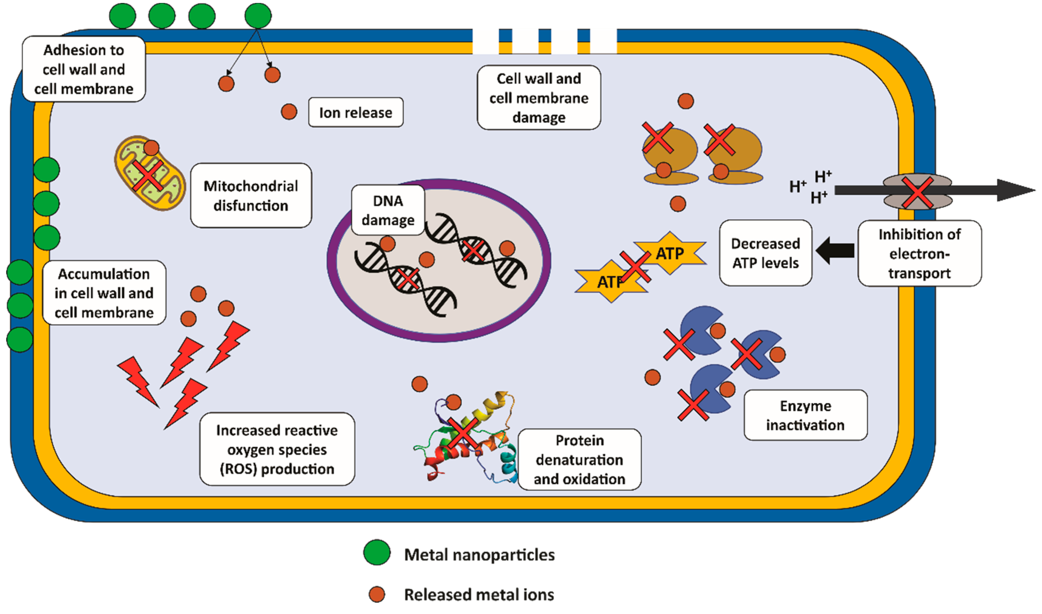
2. Metal Nanoparticles

The development of nanodrugs and nanocarriers has provided new perspectives in the biomedical field. Metal nanoparticles have received much attention because of their potential activity against fungal species of interest in the medical, pharmaceutical, and agricultural fields [26,27,28,29]. A wide range of nanoparticles with inorganic cores has been proposed, including noble metals, magnetic materials, and metal oxide nanoparticles [30].
Nanoparticles are particulate dispersions or solid particles 10–100 nm in size. They are defined as objects with three external nanoscale dimensions [31]. Metal nanoparticles have unique physical and chemical properties. Adjusting their synthesis process can modify their size, deformability, charge, hydrophobicity, and porosity, which can affect particle stability, biocompatibility, drug charge, circulation time, biodistribution, cellular localization, and internalization [23].
Metal nanoparticles exhibit antifungal activity through mechanisms involving the release of ions, oxidative and nitrosative stress, membrane and cell wall damage, enzymatic activity inhibition, gene expression regulation, decreasing ATP levels, DNA, protein, and mitochondrial dysfunction (Figure 2) [27,32,33,34].
Based on these properties, metal nanoparticles have broad applications in the drug delivery of antimicrobial agents [29,35,36,37]. The use of metal nanoparticles in combination with amphotericin B (AMB), caspofungin (CAS) and fluconazole (FCZ) may enhance its effectiveness against C. albicans [14,38,39]. Metal nanoparticles can play an important role in combating C. albicans strains resistant to antifungal drugs and biofilms formed on mucosa and medical devices [14,15]. Here, recent studies focused on the use of metal nanoparticles to combat C. albicans infections are highlighted, with a special focus on silver (Ag), gold (Au), and iron (Fe) nanoparticles.

2.1. Silver Nanoparticles

Silver nanoparticles (AgNPs) have been widely studied for their potential applications in biomedical areas such as anticancer drug nanocarriers, vaccines, and drug delivery systems [37,40]. Recent improvements in their properties of biocompatibility and stability through surface modifications make them candidates for carrying a large number of compounds with antifungal activity [35].

2.1.1. AgNPs as Systems to Carrier Antifungal Drugs

AgNPs have been used as carrier systems for conventional antifungals such as voriconazole [41], nystatin [42], and AMB [43]. The use of AgNPs to carrier antifungals makes it possible to increase the efficacy of therapy with reductions in drug concentration, toxicity and cost [35].
Recently, Jia and Sun [44] evaluated FCZ alone and in combination with AgNPs on two FCZ-resistant C. albicans strains. AgNPs had a size ranging from 8 to 12 nm. The minimum inhibitory concentration (MIC) of FCZ decreased when it was used in combination with AgNPs against planktonic cells, indicating a synergistic effect between AgNPs and FCZ. Combination therapy also exhibited synergism against biofilms in the early stages of formation 6 and 12 h, but not in mature biofilms. In addition, exposure to AgNPs reduced ergosterol levels, impaired membrane structure by downregulating the ERG1, ERG11, and ERG25 genes [45], and increasing ROS production, resulting in oxidative degradation and cellular damage [46,47]. Interestingly, these authors also verified that AgNPs treatment reduced the membrane content of Cdr1p and Cdr2p, and thus efflux pump activity. FCZ-resistant C. albicans strains usually exhibit overexpression or increased efflux pump activity, which reduces their susceptibility to antifungal agents [48,49]. In this case, exposure to AgNPs restored the FCZ susceptibility phenotype of the resistant strains. Finally, the combination treatment of AgNPs and FCZ improved the survival rate of infected mice, and markedly reduced the fungal burden in the liver and kidneys. Therefore, combination therapy was a useful strategy for the treatment of FCZ-resistant strains in vitro and in vivo.
Falcão et al. [14] developed poly(methacrylic acid)-silver nanoparticles (PMAA-AgNPs) capped with FCZ and evaluated their in vitro effects on C. albicans and mammalian cells. PMAA-AgNPs had a spherical shape, mean diameter of 15 nm, and a Zeta potential of −41.83 mV. Zeta potential is a significant physiochemical attribute of nanosystems that can predict their interactions with different surfaces, playing a crucial role in drug delivery systems [21]. PMAA-AgNPs and FCZ reportedly exhibited synergistic interactions against C. albicans strains resistant to FCZ, leading to a reduction in MIC values and inhibition of germ tube formation. PMAA-AgNPs were toxic in a dose-dependent manner to J774.A1 macrophages and fibroblasts, but the concentrations required to decrease cell viability were higher than those that inhibited C. albicans growth [14]. This combination therapy provided a convincing way to reduce the amount of antifungal drugs compared to antifungal monotherapy, and most importantly, overcome antifungal drug resistance in C. albicans [25,50].
While AgNPs exhibit promising results as drug delivery systems for antifungal agents, their potential cytotoxicity is a concern [51]. Therefore, development of AgNPs with antifungal activity should consider the toxicity levels and eco-friendly characteristics [52].

2.1.2. Green Synthesis of AgNPs

Green synthesis is a promising approach in many aspects of modern nanobiotechnology. Green synthesis uses compounds of natural origin, such as proteins, alkaloids, flavonoids, and polyphenols, such as metal ion reducing and stabilizing agents to produce nanoparticles. Green synthesis is an eco-friendly and inexpensive method that can be easily incorporated into the industrial scale production of nanoparticles [53]. In addition to natural compounds, microbial cells and cell-free extracts of microorganisms can also be a source of reducing and stabilizing agents for green synthesis [52,54]. Metal nanoparticles synthetized using green methods that are coated with organic compounds can have increased biological activity and reduced toxicity [55].
Ahamad et al. [56] produced biogenic silver nanoparticles from Anabaena variabilis (AV-AgNPs), a prokaryotic photosynthetic filamentous cyanobacterium. The resulting AV-AgNPs were spherical and 11–15 nm in size. Antifungal activity against C. albicans planktonic cells and synergism with FCZ have been demonstrated. In a subsequent study, Ahamad and Bano [57] verified the capacity of AV-AgNPs to inhibit biofilms and suppress the filamentation of C. albicans. C. albicans cells exposed to AV-AgNPs were wrinkled and deformed, with nuclear abnormalities and membrane damage. These observations reinforce the ability of AV-AgNPs to damage C. albicans in both planktonic and biofilm stages.
Similarly, Abdallah and Ali [27] used the leaf extract of Erodium glaucophyllum to biosynthesize AgNPs (EG-AgNPs). The authors characterized the EG-AgNPs and assessed their antifungal effect against C. albicans. The EG-AgNPs exhibited a diameter of 50 nm, Zeta potential of –10 mV, and higher anti-Candida activity than AMB. The MIC value of EG-AgNPs and AMB was 50 and 100 μg/mL, respectively. In addition, EG-AgNPs completely eliminated the fungal burden within 10 h post-treatment, with complete elimination achieved within 20 h of AMB treatment. The dimorphic transition of C. albicans was inhibited by 56.36% at 25 μg/mL EG-AgNPs and the viability of biofilms was reduced by 52% at 50 μg/mL. EG-AgNPs applied at 50 μg/mL also decreased the enzymatic activity of both proteinases and phospholipases. Transmission electron microscopy (TEM) images revealed that EG-AgNPs treatment (50 μg/mL) led to modifications in the morphology of fungal cells, including cytoplasmic disintegration, vacuolation, perinuclear, and granular nuclear alteration. In addition, the C. albicans cell wall was swollen and the outer layer appeared to be separated from the cell [12,58]. EG-AgNPs cytotoxicity was not evident for the human gingival fibroblast (HGF-1) when used at concentrations up to 150 μg/mL. The efficiency of topical treatment with 50 μg/mL of EG-AgNPs was examined in an in vivo mouse model of oral candidiasis. Treatment with EG-AgNPs resulted in a reduction in the number of fungal cells and lesions in the tongue dorsum, suggesting the potential use of EG-AgNPs in the treatment of oral candidiasis.
Alqarni [59] used Gum acacia, which is a material enriched in polysaccharide from acacia plants. Gum acacia was used as a dispersal and reduction agent to synthetize rutin-AgNPs. Rutin, also known as vitamin P or rutoside, exhibits several pharmacological effects, including anti-Candida activity [60]. Rutin-AgNPs had an average size of 59.67 nm, and Zeta potential of –11.2 mV. Rutin-AgNPs were incorporated into base gels to produce rutin-loaded AgNPs gel to evaluate their activity in skin candidiasis. Both formulations exhibited antifungal activity against C. albicans. However, this activity was enhanced in the rutin-loaded AgNPs gel. This gel increased drug release, probably due to the capsizing effects of rutin on the formed AgNPs. Altogether, the effectiveness of this formulation offers new perspectives for the topical delivery of antifungal agents for the treatment of Candida wound infections.
Miškovská et al. [61] synthesized AgNPs using Vitis vinifera cane extract (VV-AgNPs). These nanoparticles had various shapes, with an average size ranging from 34.43 to 101.63 nm, and a Zeta potential ranging from –30.04 to –21.24 mV. VV-AgNPs were active against planktonic cells and biofilms of C. albicans at a concentration of 20 µg/mL. Scanning electron microscopy (SEM) images demonstrated that C. albicans cells treated with VV-AgNPs (5 µg/mL) visibly adhered to the surface, while other cells were clearly disrupted, reinforcing the antifungal effect of VV-AgNPs against C. albicans.

2.2. Gold Nanoparticles

Gold nanoparticles (AuNPs) have also been extensively studied because of their good physicochemical properties, chemical resistivity, ease of synthesis, and minimal size [62,63,64]. The Food and Drug Administration (FDA) approval for the use of AuNPs in various biomedical applications has boosted research and development focused on these nanoparticles [65]. The unique properties of AuNPs include adjustable size, shape, surface properties, biocompatibility, low cytotoxicity, and high stability [24]. Furthermore, they can be easily combined or recovered with several biomolecules, including peptides, enzymes, and DNA, enabling their use in inducing drug stability and reducing adverse effects [66,67]. While AuNPs exhibit a variety of biomedical effects, cytotoxicity remains a concern [68]. Strategies that include changing the charge, size, and dispersion of AuNPs can be easily employed to minimize toxicity, and/or enhance antifungal activity [65,69].

2.2.1. AuNPs as Carrier Systems for Antifungal Drugs

AuNPs have been typically investigated for the delivery of conventional antifungals. For example, Salehi et al. [38] formulated the antifungal caspofungin by incorporating it into AuNPs (CAS-AuNPs). CAS, a member of the echinocandin class, inhibits the biosynthesis of the fungal cell wall component β-1,3-D-glucan [70,71,72]. In the study, resulting CAS-AuNPs had a spherical shape with an average size of 20 nm and the Zeta potential of –38.2 mV. CAS-AuNPs exhibited anti-Candida activity at lower concentrations than CAS alone. SEM images demonstrated membrane and cell wall damage in yeast cells after exposure to CAS-AuNPs, indicating the improved antifungal activity associated with reduced antifungal concentrations and decreased toxicity [38].
Similarly, Hamad et al. [73] described the formation of functionalized AuNPs with poly(ethylene) glycol (PEG) and FCZ. The resulting PEG-FCZ-AuNPs were loaded into a poloxamer hydrogel (PEG-FCZ-AuNP hydrogel). PEG-FCZ-AuNP formed nanorods, with a Zeta potential of +1.6 mV, and an average length of 80 nm. Hydrogel loading did not affect the size and Zeta potential of the PEG-FCZ-AuNP. The MIC value of PEG-FCZ-AuNPs was 0.12 nM, which corresponded to a nine-fold reduction compared to FCZ alone. TEM images revealed that the nanoformulation enhanced the delivery of FCZ to the cell wall and accelerated its cellular uptake. The exposure of C. albicans to the PEG-FCZ-AuNP hydrogel resulted in a 1.3 log reduction in the viability of fungal cells. The cytotoxicity of PEG-FCZ-AuNP was evaluated against CCD-1064Sk human dermal fibroblasts. Exposure of these cells to PEG-FCZ-AuNPs for 24 h maintained cell viability by more than 75% at the MIC value against C. albicans, suggesting that it is a promising approach for the effective treatment of mucosal candidiasis.

2.2.2. Chitosan-AuNPs as Anti-C. albicans Nanosystems

The association of AuNPs with organic polymers such as chitosan has also been studied. Chitosan is a natural cationic polysaccharide used as a biopolymer for the synthesis of colloidal nanoparticles. Chitosan act as a reducing and stabilizing agent [74], and exhibits intrinsic in vitro anti-Candida activity owing to its polycationic nature. Interaction with the negatively charged cell membrane of microorganisms via electrostatic interactions leads to extensive cell surface modifications [75,76]. Furthermore, chitosan exhibits anti-inflammatory activity, good biocompatibility, mucoadhesive and haemostatic properties, and the capacity to modulate fibroblast growth factors. These attributes have led to the use of chitosan as a wound dressing material [77].
Hashem et al. [69] synthetized spherical chitosan-based AuNPs (Chi-AuNPs) with an average particle size ranging from 20 to 120 nm and a Zeta potential of −52.39 mV. Evaluation of the antifungal effect of Chi-AuNPs was assessed for C. albicans using two methods. In the first method, Chi-AuNPs produced a 25 mm inhibition zone when applied at 2000 µg/mL, with a MIC value of 62.5 µg/mL. In the microdilution toxicity assays, Chi-AuNPs were non-toxic to the BJ-1 normal human skin cell line, with a cytotoxic concentration for 50% of the cells of 111.10 µg/mL. The findings highlight the feasibility of Chi-AuNPs as a promising agent against C. albicans infections [69].
Yadav et al. [21] designed tyrosol-functionalized chitosan gold nanoparticles (Chi-TY-AuNPs). The intent was to use the Chi-TY-AuNPs as an alternative treatment strategy for Candida infections. Tyrosol is a molecule produced by C. albicans; it is involved in quorum sensing, which stimulates hyphal formation. In contrast, exogenous tyrosol administration can inhibit planktonic cells and biofilms of C. albicans and enhance their susceptibility to antifungal agents [78]. However, the exogenous mechanism underlying the antagonistic action of tyrosol against C. albicans has not been fully elucidated. Yadav et al. [21] reported that Chi-TY-AuNPs had a Zeta potential of +45.5 mV and were spherical, with an average diameter in the range of 10−15 nm. These nanoparticles showed activity against C. albicans at 200 μg/mL, and the minimum fungicidal concentration was 800 μg/mL. Chi-TY-AuNPs were also able to inhibit biofilm development and eradicate mature biofilms of C. albicans at 200 and 400 μg/mL, respectively. In addition, Chi-TY-AuNPs inhibited germ tube development and transition from yeast to hyphae at a subinhibitory concentration of 200 μg/mL. The ability of Chi-TY-AuNPs to inhibit C. albicans was associated with increased ROS production. Yadav et al. [21] also evaluated the cytotoxicity of Chi-TY-AuNPs to NIH-3T3 fibroblast cells, and reported the absence of reduced cell viability, even when these nanoparticles were tested in higher concentrations. The finding highlighted the good biocompatibility of Chi-TY-AuNPs.

2.2.3. Green Synthesis of AuNPs

As with AgNPs, green methods have been used to synthetize AuNPs. Kareem, Samaka and Abdulridha [28] produced gold nanoparticles using olive leaf extract (OL-AuNPs). The OL-AuNPs displayed a particle size of 29.16 nm. These nanoparticles inhibited C. albicans growth at 40.77 ng/mL. The antifungal effects on the cutaneous candidiasis infections were evaluated. OL-AuNPs showed good pharmacological efficacy in the treatment of murine cutaneous candidiasis, with superior efficacy compared to the antifungal nystatin. The candidiasis lesions of the group treated with 326.12 ng/mL OL-AuNPs were entirely cured three days post-treatment, confirming the anti-Candida effects of OL-AuNPs in vitro and in vivo.
In a thorough study, Cruz et al. [18] evaluated the ability of fifteen ethnobotanical crude extracts from the leaves, roots and/or bark of Hydrocotyle vulgaris, Eluesine indica, Mikania micrantha, Dillenia philippinensis, Ceiba pentandra, Cymbopogon winterianus, Senna alata, Urena lobata, Premna odorata, Stachytarpeta jamaicensis, Diplazium esculentum, and Phyllanthus urinaria as well as their gold nanoparticles (CE-AuNPs) on biofilms and quorum sensing-related gene expression. The CE-AuNPs exhibited a variety of shapes and sizes depending on the crude extract used. None of the extracts or their CE-AuNPs exhibited antifungal activity against planktonic C. albicans. However, 14 CE-AuNPs decreased C. albicans biofilm formation at concentrations of 67.00 to 80.00 µg/mL. The expressions of BCR1 and HSP90 were downregulated after exposure to 13 crude extracts, and to 14 CE-AuNPs. BCR1 and HSP90 genes act as transcription factors involved in biofilm formation. BCR1 downregulation affected the development of biofilms as well as the production of polymeric extracellular matrix [79,80]. Apparently, the extracts and CE-AuNPs may acted as quorum sensing inhibitor molecules that blocked the BCR1 and HSP90 pathways [81,82]. It is possible that the difference in CE-AuNPs activities was influenced by the composition of the extracts, and by the final nanoparticle size.
While not as well studied as AgNPs, available data indicate that AuNPs have effective antifungal activity, and so may be a key strategy for drug development against C. albicans infections.

2.3. Iron Nanoparticles

Iron oxide nanoparticles (IONP) are recognized by their high chemical and thermal stabilities, low cost, very high area-to-volume and area-to-weight ratios, and superparamagnetic behaviour. These attributes are valuable for biomedical applications [83,84]. Accordingly, IONPs have been explored as drug delivery systems to increase drug effectiveness and reduce therapeutic drug concentrations [36]. Ferumoxytol is an intravenous IONP formulation that is intravenously administrated to treat anaemia in patients with chronic kidney disease. Ferumoxytol is approved by the FDA, Health Canada, and other agencies to treat anaemia [85,86], and has also been proposed as a candidate for drug repurposing therapy owing to its excellent inherent properties and good safety and clearance profiles [85]. For example, topical ferumoxytol was able to disrupt oral biofilms by generating ROS and killing bacteria by membrane disruption, demonstrating the potential to prevent dental caries [87].
The antimicrobial activity of IONPs has also been proven against fungal species, such as C. albicans [33,88,89]. While there are concerns related to biocompatibility and toxicity, these characteristics can be modified by altering their size, shape, stability, and coating [90].
IONPs can be functionalized with different organic and inorganic compounds to modify their general properties, particularly their antimicrobial properties [91]. For example, Arias et al. [92] synthetized iron nanoparticles functionalized with chitosan as nanocarriers for the antifungal miconazole (IONP-Chi-MCZ). The resulting IONP-Chi-MCZ had a diameter < 50 nm and promoted an 8-fold reduction in C. albicans planktonic cells compared to miconazole. In contrast, IONP and quitosan alone did not inhibit C. albicans growth at 140 μg/mL, and the combination of IONP, quitosan, and miconazole was characterized as synergistic against C. albicans. IONP-Chi-MCZ also reduced the metabolic activity and viability of C. albicans biofilms. However, treatment with IONP-Chi-MCZ did not affect the protein, carbohydrate, or DNA content of the polymeric extracellular matrix.
Similarly, Balabathula et al. [29] designed IONP functionalized with bovine serum albumin and targeted amphotericin B (AMB-IONP). The synthetized AMB-IONP were spherical, monodisperse, had ≤36 nm in size, and a Zeta potential of −20 mV. AMB-IONPs exhibited improved efficacy (16–25-fold) compared to AMB against C. albicans. In addition, TEM and confocal laser scanning microscopy revealed intracellular trafficking of AMB-IONPs inside fungal cells, confirming successful drug delivery. TEM also revealed that the AMB-IONPs were localized inside the cytoplasm or near the cell wall and cell membrane, indicating that the uptake pathway mechanism of AMB-IONPs in C. albicans was controlled by receptor-mediated endocytosis. AMB-IONP treatment also led to the disorganization of the cytoplasm with deformed nuclei and other intracellular components, along with a significant increase in the number of vacuoles [29].

2.4. Other Metal Nanoparticles

In addition to the traditional silver, gold, and iron nanoparticles, several other metal nanoparticles have been investigated for the in vitro and in vivo treatment of C. albicans infections. Nanomaterials such as zinc oxide (ZnO), titanium dioxide (TiO2), zirconium dioxide (ZrO2), and copper (cupric oxide [CuO] and cuprous oxide [Cu2O]) have been used successfully to inhibit and kill C. albicans cells [93,94,95].
Padmavathi et al. [13] synthetized oxide copper nanoparticles (CuO-NP and Cu2O-NP) having an average size of 10.7 nm and 36 nm, respectively, and a Zeta potential of −38.35 mV, and +7.9 mV, respectively. CuO-NPs and Cu2O-NPs completely inhibited C. albicans growth at 150 and 200 µg/mL, respectively. Furthermore, CuO-NPs and Cu2O-NPs showed significant anti-biofilm activity against C. albicans at 1 µg/mL, which was comparable to that of AMB [96]. Macromorphologically, Cu2O-NP treatment decreased spore and hyphal formation on solid agar media. Microstructural analysis demonstrated that exposure of C. albicans to Cu2O-NPs led to cell shrinkage, possibly due to the treatment-induced increase in ROS production. This enhanced activity was attributed to the positive surface charge of Cu2O-NPs, since C. albicans cells are electronegative; their cell walls effectively bind positively charged ions, facilitating electrostatic interactions [97,98]. The CuO-NPs were shown to be internalized more than were Cu2O-NPs, probably because of their smaller size. Both CuO-NPs and Cu2O-NPs reduced ergosterol production in C. albicans, and downregulated the genes involved in morphogenesis and virulence.
Aati, Shrestha and Fawzy [99] synthesized zirconium dioxide nanoparticles (ZrO2NPs) and studied their antimicrobial and cytotoxic effects in a modified three-dimensional printed resin, which is routinely used in dentistry. The diameters of ZrO2NPs ranged from 20 to 40 nm before and after functionalization. The metabolic activity of C. albicans was remarkably reduced by the addition of ZrO2NPs, even after aging for three months. SEM confirmed the reduction in C. albicans biofilm viability when the resin was modified with ZrO2NPs. This antifungal activity may be related to the ability of ZrO2NPs to produce ROS and interfere with cell functions, leading to deformation of the fungal hyphae [100,101]. The authors measured cytotoxicity to human oral fibroblast. The higher concentration of ZrO2NPs used slightly reduced the number of viable cells, whereas low concentrations did not affect cell viability. Importantly, cell viability was re-established after aging for 3 months. Resin modified with ZrO2NPs exhibited a critical ability to inhibit biofilm formation without inducing any adverse cellular effects.
Seeking to refine the biomaterials used in dentistry, AlQahtani et al. [102] designed titanium dioxide nanoparticles and added them to polymethylmethacrylate (PMMA-TiO2NPs). Polymethylmethacrylate (PMMA) is commonly used as a denture base material owing to its low cost, reduced weight, colour-matching ability, and ease of finishing and polishing [95,103]. TiO2NPs exhibited a spherical shape with an average diameter of 26 nm. PMMA-TiO2NPs reduced C. albicans adhesion, probably because of the antifungal properties of TiO2NPs [104,105].
Taken together, these studies indicate that the successful use of different types of metallic nanoparticles in biomedical applications demands modulation of their properties to maximize the antifungal effect and decrease its toxicity [106].

2.5. Bimetallic Nanoparticles

Based on the antifungal activity exhibited by AgNPs, AuNPs, INOPs and other metal nanoparticles, several studies have addressed the use of bimetallic nanoparticles. In addition to antifungal drugs or natural biomolecules, metallic nanoparticles may be functionalized with different inorganic compounds, resulting in core–shell nanoparticles and bimetallic nanoparticles. In this context, a variety of metals, such as zinc (Zn), nickel (Ni), manganese (Mn), dysprosium (Dy), and copper (Cu) have been associated with AgNPs and IONPs to investigate their effects on C. albicans.
Padilla-Cruz et al. [25] synthesized AgNPs, iron (Fe), and Ag–Fe bimetallic nanoparticles (Ag-FeNPs) using Gardenia jasminoides extract as the reducing agent. The synthesized nanoparticles displayed homogenously distributed spherical core–shell structures with an average diameter of 13 nm and were magnetic. The activities of AgNPs, FeNPs, and Ag-FeNPs were evaluated. The MIC of AgNPs was 125 ppm and FeNPs exhibited no activity against C. albicans at the maximum concentration tested (250 ppm). Interestingly, Ag-FeNPs showed fungicidal activity against C. albicans at 62.5 ppm and were biocompatible. The findings indicate the potential of Ag-FeNPs as a nanomaterial for biomedical applications.
Other studies have evaluated the antifungal and cytotoxic activities of different silver chromite (Ag-CrNPs) nanocomposites [107]. Nanocomposites are hybrid materials in which at least one component has nanometric dimensions [108]. The synthetized Ag-CrNPs were spherical, with an average particle size of 93.14 nm. In addition, they showed significant antifungal activity against C. albicans, and concentrations < 31.25 μg/mL were not cytotoxicity against Vero Cells.
Kamli et al. [22] synthetized silver and nickel bimetallic nanoparticles (Ag-NiNPs) using an extract of Salvia officinalis leaves. Ag-NiNPs were generated as semi-spherical agglomerated clusters that harboured smaller and larger nanoparticles ranging in average size from 31.84 to 47.85 nm. Importantly, Ag-NiNPs showed potent antifungal activity against C. albicans strains resistant and susceptible to FCZ with effective concentrations ranging from 0.19 to 1.56 µg/mL. These nanoparticles also decreased hyphae production and biofilm formation. In addition to antifungal effects at low concentrations, Ag-NiNPs acted synergistically with FCZ against C. albicans, re-establishing the susceptibility of the fungus to FCZ. Furthermore, Ag-NiNPs modulated multi-drug resistance efflux transporters reducing ATP-dependent efflux pumps (ABC superfamily) in C. albicans at 1.56 µg/mL. Exposure to Ag-NiNPs also resulted in disruption of the Candida cell membrane accompanied by the generation of ROS, as confirmed by SEM. Cells treated with Ag-NiNPs exhibited a variety of sizes and shapes, surface depression, and leakage of intracellular material, reflecting the antifungal effect of Ag-NiNPs.
Based on the promising optoelectronic, electrochemical, catalytic and antibacterial properties of tin (Sn) and tin dioxide (SnO2) nanomaterials [109,110], and the well-established antimicrobial activity of AgNPs, Pandey et al. [111] evaluated the antifungal activity of bimetallic (Ag/Sn-SnO2) composite nanoparticles (Ag/Sn-SnO2NPs). Ag/Sn-SnO2NPs formed with a rod or needle shape, with ranging in size from 1 to 18 nm. The Zeta potential varied from −18.4 to −17.3 mV. Ag/Sn-SnO2NPs prepared easily and inexpensively inhibited C. albicans. In silico molecular docking evaluations of adhesion and invasion revealed that Ag/Sn-SnO2NPs exhibited interaction with several target amino acid residues of lanosterol 14-α-demethylase, an enzyme involved in the biosynthesis of ergosterol [112]. This binding directly inhibited the growth of C. albicans strains. Ag/Sn-SnO2NPs also interacted with Als3, Hsp90, and Cyp51, which are related to the growth of C. albicans, biofilm formation and tissue invasion [113], suggesting that the Ag/Sn–SnO2NPs exhibited potential inhibitory properties by targeting several proteins in different molecular pathways.
Sayed, Abdelsalam and El-Bassuony [114] developed several types of iron bimetallic nanoparticles to obtain spinel nanoferrites with diameters ranging from 22 to 97 nm. Among them, nickel-zinc-iron nanoparticles (Ni-Zn-IONPs) and manganese-zinc-dysprosium-iron nanoparticles (Mn-Zn-Dy-IONPs) exhibited activity against C. albicans at concentrations of 200 [34], and 8 mg/mL [115], respectively. Ni-Zn-IONPs had nanomagnetic surfaces containing nanospheres and nanosheets with an average particle size of 30 nm. Exposure of C. albicans to Ni-Zn-IONPs prevented fungal growth and increased leakage of intracellular proteins. Furthermore, Ni-Zn-IONPs increased ROS production, resulting in DNA and mitochondrial damage [34]. Mn-Zn-Dy-IONPs adopted a cubic shape with an average particle size < 20 nm. SEM demonstrated that exposure of C. albicans to Mn-Zn-Dy-IONPs resulted in deformed and distorted cells, indicating the loss of cell membrane integrity, which resulted in cell death [116]. Al-Jameel et al. [115] hypothesized that Mn-Zn-Dy-IONPs could bind to the cell surface and target ergosterol.
Ansari et al. [117] synthetized cube-shaped nickel-copper-zinc-iron nanoparticles (Ni-Cu-Zn-IONP) with an average size between 10 and 19 nm. Ni-Cu-Zn-IONP significantly reduced the number of planktonic cells of C. albicans at 2 mg/mL and inhibited biofilm formation at 1 mg/mL. Interestingly, SEM demonstrated inhibition of hyphae in treated biofilms with severely damaged treated yeast cells, including deformation, distortion and separation of the cell wall and membrane, indicative of a significant loss of membrane integrity. TEM revealed complete lysis of yeast cell and confirmed in SEM data. The inhibition of C. albicans was probably due to the adsorption and penetration of Ni-Cu-Zn-IONPs by yeast cells, which may have led to the disorganization of the cell membrane and wall, followed by the leakage of various cytoplasmic contents [116,118,119].
These collective findings confirmed that bimetallic nanoparticles can have antifungal activity against C. albicans strains via multiple mechanisms. These antifungal effects can be attributed to the unique physicochemical properties of each type of metal nanoparticles that, when combined, can interact with fungal cells through different pathways [120].

2.6. Emerging Concerns and Future Directions for Metal Nanoparticles

The antifungal activity against C. albicans and physical-chemical characteristics of metallic nanoparticles discussed in this review are summarized in the Table 1. The physical–chemical properties of nanoparticles have been considered key factors for their biological applications [121]. It is known that variations in physical and chemical parameters have a significant effect on cellular uptake and antimicrobial efficacy [121,122]. Metal nanoparticles are actively incorporated into the microbial cell through different endocytic pathways, and their size, shape and surface chemistry can influence the internalization process [121,123]. Typically, smaller nanoparticles have higher antimicrobial activity, while larger NPs require specific functionalization to increase their delivery into cells [62,124]. While there is no consensus about the optimum size that maximizes the cellular uptake, it has been suggested that small sized silver (5, 9, 10, 12, and 13.5 nm), gold (8.4 nm), zinc-oxide (12 nm), and titanium-oxide (12 and 17 nm) nanoparticles have high antimicrobial activities against bacterial cells [125]. However, little is known about the optimum size and other physical-chemical properties of metal nanoparticles that can maximize the cellular uptake by Candida albicans, and additional studies are required in this field.
Another issue that requires more investigation is the action of metal nanoparticles on polymicrobial biofilms. In these biofilms, C. albicans and bacterial species can establish synergistic interactions that provide protection to one or both species against antimicrobial agents and make polymicrobial biofilms more resistant than those formed by a single species. Competitive interactions can also be established between fungus and bacteria through the production of quorum sensing molecules [126]. Some microorganisms can secrete small molecules specialized in metal uptake named metallophores, which represent an important mechanism in the competitive interactions since iron, zinc and other metal ions are essential for the microbial survival. The most well-characterized metallophore family is the siderophore with high affinity for iron. Siderophores molecules are synthesized within the microbial cytoplasm and exported to the extracellular environment to scavenge the iron, then the siderophore-iron complex is transported into the periplasm by specific transporters [127,128]. Interestingly, it was reported that iron-based nanoparticles (Fe3O4 NPs) with antimicrobial activity were able to penetrate in Gram-negative bacteria through the siderophore transporters on the outer membrane [129]. While it is unclear whether C. albicans synthetizes its own siderophores, it possesses siderophore transporters and can take iron by siderophores produced by other microorganisms (xenosiderophores) [130,131]. These data instigate future studies to understand the role of metallophores in the antifungal activity of metal nanoparticles on Candida cells and polymicrobial biofilms.
Attention must also be given to possible adverse effects of metal nanoparticles. While most studies indicate that the toxicity of metal nanoparticles for mammalian cells is low, translation to clinical applications will require additional data concerning their possible accumulation in body tissues and adverse effects [132]. The absorption of metal ions by the mucosal surface can be associated with localized and generalized argyria, ocular irritation, contact dermatitis, and genotoxic, hepatic, renal, neurological, and haematological effects [133]. Thus, regulatory clarity concerning the use of metal nanoparticles is urgently needed. It is essential to encourage the development of clinical trials that use metal nanoparticles with proven in vitro and in vivo efficacy. These studies are crucial for the evaluation, knowledge, and follow-up of their effects on the body and environment.

3. Conclusions

Several in vitro and in vivo studies have shown the efficacy of metal nanoparticles in combating C. albicans at both planktonic and biofilm stages. Promisingly, most of the synthesized silver, gold, iron, and other metal nanoparticles inhibit the growth and lessen virulence of C. albicans, improving the development of antifungal resistance. Some studies have also demonstrated the in vivo efficacy of metal nanoparticles on oral and cutaneous candidiasis in rodent models, reinforcing their potential use as a candidate for the topical treatment of superficial candidiasis.
For clinical applications, improvements and adjustments in the antifungal activity and cytotoxicity properties of metal nanoparticles are still needed. To overcome these limitations, most studies have focused on modifications of the synthesis parameters and development of coating methods. Green synthesis and bimetallic coating are being intensively studied. The development of metal nanosystems could be a key strategy to circumvent problems related to antifungal resistance and recurrent candidiasis.

Author Contributions

Conceptualization: P.H.F.d.C., M.T.G., L.M.A.F.-G., A.C.P.L., N.S.d.S. and J.C.J.; Figure design: P.H.F.d.C.; Writing—original draft preparation: P.H.F.d.C.; Writing—review & editing: P.H.F.d.C., M.T.G., L.M.A.F.-G., A.C.P.L., N.S.d.S. and J.C.J. All authors have read and agreed to the published version of the manuscript.

Funding

This study was supported by the U.S. Office of Naval Research Global (ONRglobal N62909-20-1-2034) and the Brazilian National Council for Scientific and Technological Development (CNPq 306330/2018-0).

Institutional Review Board Statement

Not applicable.

Informed Consent Statement

Not applicable.

Data Availability Statement

Not applicable.

Conflicts of Interest

The authors declare no conflict of interest.

References

  1. Taverne-Ghadwal, L.; Kuhns, M.; Buhl, T.; Schulze, M.H.; Mbaitolum, W.J.; Kersch, L.; Weig, M.; Bader, O.; Groß, U. Epidemiology and Prevalence of Oral Candidiasis in HIV Patients from Chad in the Post-HAART Era. Front. Microbiol. 2022, 13, 844069. [Google Scholar] [CrossRef] [PubMed]
  2. Ghaddar, N.; El Roz, A.; Ghssein, G.; Ibrahim, J.-N. Emergence of Vulvovaginal Candidiasis among Lebanese Pregnant Women: Prevalence, Risk Factors, and Species Distribution. Infect. Dis. Obstet. Gynecol. 2019, 2019, 5016810. [Google Scholar] [CrossRef] [PubMed] [Green Version]
  3. Arendorf, T.M.; Walker, D.M. The Prevalence and Intra-Oral Distribution of Candida Albicans in Man. Arch. Oral Biol. 1980, 25, 1–10. [Google Scholar] [CrossRef] [PubMed]
  4. Darwazeh, A.M.G.; Darwazeh, T.A. What Makes Oral Candidiasis Recurrent Infection? A Clinical View. J. Mycol. 2014, 2014, 758394. [Google Scholar] [CrossRef]
  5. Joshi, K.M.; Shelar, A.; Kasabe, U.; Nikam, L.K.; Pawar, R.A.; Sangshetti, J.; Kale, B.B.; Singh, A.V.; Patil, R.; Chaskar, M.G. Biofilm Inhibition in Candida albicans with Biogenic Hierarchical Zinc-Oxide Nanoparticles. Biomater. Adv. 2022, 134, 112592. [Google Scholar] [CrossRef] [PubMed]
  6. Bhattacharya, S.; Sae-Tia, S.; Fries, B.C. Candidiasis and Mechanisms of Antifungal Resistance. Antibiotics 2020, 9, 312. [Google Scholar] [CrossRef]
  7. Bongomin, F.; Gago, S.; Oladele, R.O.; Denning, D.W. Global and Multi-National Prevalence of Fungal Diseases-Estimate Precision. J. Fungi 2017, 3, 57. [Google Scholar] [CrossRef]
  8. Xiao, Z.; Wang, Q.; Zhu, F.; An, Y. Epidemiology, Species Distribution, Antifungal Susceptibility and Mortality Risk Factors of Candidemia among Critically Ill Patients: A Retrospective Study from 2011 to 2017 in a Teaching Hospital in China. Antimicrob. Resist. Infect. Control 2019, 8, 89. [Google Scholar] [CrossRef]
  9. Koehler, P.; Stecher, M.; Cornely, O.A.; Koehler, D.; Vehreschild, M.J.G.T.; Bohlius, J.; Wisplinghoff, H.; Vehreschild, J.J. Morbidity and Mortality of Candidaemia in Europe: An Epidemiologic Meta-Analysis. Clin. Microbiol. Infect. 2019, 25, 1200–1212. [Google Scholar] [CrossRef]
  10. Benedict, K.; Jackson, B.R.; Chiller, T.; Beer, K.D. Estimation of Direct Healthcare Costs of Fungal Diseases in the United States. Clin. Infect. Dis. Off. Publ. Infect. Dis. Soc. Am. 2019, 68, 1791–1797. [Google Scholar] [CrossRef]
  11. Mba, I.E.; Nweze, E.I.; Eze, E.A.; Anyaegbunam, Z.K.G. Genome Plasticity in Candida Albicans: A Cutting-Edge Strategy for Evolution, Adaptation, and Survival. Infect. Genet. Evol. 2022, 99, 105256. [Google Scholar] [CrossRef]
  12. Garcia-Rubio, R.; de Oliveira, H.C.; Rivera, J.; Trevijano-Contador, N. The Fungal Cell Wall: Candida, Cryptococcus, and Aspergillus Species. Front. Microbiol. 2020, 10, 2993. [Google Scholar] [CrossRef]
  13. Padmavathi, A.R.; Murphy P., S.; Das, A.; Priya, A.; Sushmitha, T.J.; Pandian, S.K.; Toleti, S.R. Impediment to Growth and Yeast-to-Hyphae Transition in Candida albicans by Copper Oxide Nanoparticles. Biofouling 2020, 36, 56–72. [Google Scholar] [CrossRef]
  14. Falcão, C.M.C.; Andrade, A.; Holanda, V.N.; de Figueiredo, R.C.B.Q.; Ximenes, E.A.; Gomes, A.S.L. Activity of Poly(Methacrylic Acid)-Silver Nanoparticles on Fluconazole-Resistant Candida albicans Strains: Synergistic and Cytotoxic Effects. J. Appl. Microbiol. 2022, 132, 4300–4309. [Google Scholar] [CrossRef]
  15. Marassi, V.; Di Cristo, L.; Smith, S.G.J.; Ortelli, S.; Blosi, M.; Costa, A.L.; Reschiglian, P.; Volkov, Y.; Prina-Mello, A. Silver Nanoparticles as a Medical Device in Healthcare Settings: A Five-Step Approach for Candidate Screening of Coating Agents. R. Soc. Open Sci. 2018, 5, 171113. [Google Scholar] [CrossRef] [Green Version]
  16. Sardi, J.C.O.; Scorzoni, L.; Bernardi, T.; Fusco-Almeida, A.M.; Mendes Giannini, M.J.S. Candida Species: Current Epidemiology, Pathogenicity, Biofilm Formation, Natural Antifungal Products and New Therapeutic Options. J. Med. Microbiol. 2013, 62, 10–24. [Google Scholar] [CrossRef]
  17. Ceylan, O.; Tamfu, A.N.; Doğaç, Y.İ.; Teke, M. Antibiofilm and Anti-Quorum Sensing Activities of Polyethylene Imine Coated Magnetite and Nickel Ferrite Nanoparticles. 3 Biotech 2020, 10, 513. [Google Scholar] [CrossRef]
  18. Judan Cruz, K.G.; Alfonso, E.D.; Fernando, S.I.D.; Watanabe, K. Candida albicans Biofilm Inhibition by Ethnobotanicals and Ethnobotanically-Synthesized Gold Nanoparticles. Front. Microbiol. 2021, 12, 665113. [Google Scholar] [CrossRef]
  19. Peron, I.H.; Reichert-Lima, F.; Busso-Lopes, A.F.; Nagasako, C.K.; Lyra, L.; Moretti, M.L.; Schreiber, A.Z. Resistance Surveillance in Candida albicans: A Five-Year Antifungal Susceptibility Evaluation in a Brazilian University Hospital. PLoS ONE 2016, 11, e0158126. [Google Scholar] [CrossRef] [Green Version]
  20. Nguyen, D.H.; Vo, T.N.N.; Nguyen, N.T.; Ching, Y.C.; Hoang Thi, T.T. Comparison of Biogenic Silver Nanoparticles Formed by Momordica charantia and Psidium guajava Leaf Extract and Antifungal Evaluation. PLoS ONE 2020, 15, e0239360. [Google Scholar] [CrossRef]
  21. Yadav, T.C.; Gupta, P.; Saini, S.; Mohiyuddin, S.; Pruthi, V.; Prasad, R. Plausible Mechanistic Insights in Biofilm Eradication Potential against Candida spp. Using In Situ-Synthesized Tyrosol-Functionalized Chitosan Gold Nanoparticles as a Versatile Antifouling Coating on Implant Surfaces. ACS Omega 2022, 7, 8350–8363. [Google Scholar] [CrossRef] [PubMed]
  22. Kamli, M.R.; Alzahrani, E.A.; Albukhari, S.M.; Ahmad, A.; Sabir, J.S.M.; Malik, M.A. Combination Effect of Novel Bimetallic Ag-Ni Nanoparticles with Fluconazole against Candida albicans. J. Fungi 2022, 8, 733. [Google Scholar] [CrossRef] [PubMed]
  23. Kumar, M.; Singh Dosanjh, H.; Sonika; Singh, J.; Monir, K.; Singh, H. Review on Magnetic Nanoferrites and Their Composites as Alternatives in Waste Water Treatment: Synthesis, Modifications and Applications. Environ. Sci. Water Res. Technol. 2020, 6, 491–514. [Google Scholar] [CrossRef]
  24. Bansal, S.; Kumar, V.; Karimi, J.; Singh, A.; Kumar, S. Role of Gold Nanoparticles in Advanced Biomedical Applications. Nanoscale Adv. 2020, 2, 3764–3787. [Google Scholar] [CrossRef] [PubMed]
  25. Padilla-Cruz, A.L.; Garza-Cervantes, J.A.; Vasto-Anzaldo, X.G.; García-Rivas, G.; León-Buitimea, A.; Morones-Ramírez, J.R. Synthesis and Design of Ag–Fe Bimetallic Nanoparticles as Antimicrobial Synergistic Combination Therapies against Clinically Relevant Pathogens. Sci. Rep. 2021, 11, 5351. [Google Scholar] [CrossRef]
  26. Cruz-Luna, A.R.; Cruz-Martínez, H.; Vásquez-López, A.; Medina, D.I. Metal Nanoparticles as Novel Antifungal Agents for Sustainable Agriculture: Current Advances and Future Directions. J. Fungi 2021, 7, 1033. [Google Scholar] [CrossRef]
  27. Abdallah, B.M.; Ali, E.M. Therapeutic Effect of Green Synthesized Silver Nanoparticles Using Erodium glaucophyllum Extract against Oral Candidiasis: In Vitro and In Vivo Study. Molecules 2022, 27, 4221. [Google Scholar] [CrossRef]
  28. Kareem, H.A.; Samaka, H.M.; Abdulridha, W.M. Evaluation of the Effect of the Gold Nanoparticles Prepared by Green chemistry on the treatment of Cutaneous Candidiasis. Curr. Med. Mycol. 2021, 7, 1–5. [Google Scholar]
  29. Balabathula, P.; Whaley, S.G.; Janagam, D.R.; Mittal, N.K.; Mandal, B.; Thoma, L.A.; Rogers, P.D.; Wood, G.C. Lyophilized Iron Oxide Nanoparticles Encapsulated in Amphotericin B: A Novel Targeted Nano Drug Delivery System for the Treatment of Systemic Fungal Infections. Pharmaceutics 2020, 12, 247. [Google Scholar] [CrossRef] [Green Version]
  30. Rodrigues, G.R.; López-Abarrategui, C.; de la Serna Gómez, I.; Dias, S.C.; Otero-González, A.J.; Franco, O.L. Antimicrobial Magnetic Nanoparticles Based-Therapies for Controlling Infectious Diseases. Int. J. Pharm. 2019, 555, 356–367. [Google Scholar] [CrossRef]
  31. Jeevanandam, J.; Barhoum, A.; Chan, Y.S.; Dufresne, A.; Danquah, M.K. Review on Nanoparticles and Nanostructured Materials: History, Sources, Toxicity and Regulations. Beilstein J. Nanotechnol. 2018, 9, 1050–1074. [Google Scholar] [CrossRef]
  32. Lee, K.X.; Shameli, K.; Yew, Y.P.; Teow, S.-Y.; Jahangirian, H.; Rafiee-Moghaddam, R.; Webster, T.J. Recent Developments in the Facile Bio-Synthesis of Gold Nanoparticles (AuNPs) and Their Biomedical Applications. Int. J. Nanomed. 2020, 15, 275–300. [Google Scholar] [CrossRef]
  33. Gholami, A.; Mohammadi, F.; Ghasemi, Y.; Omidifar, N.; Ebrahiminezhad, A. Antibacterial Activity of SPIONs versus Ferrous and Ferric Ions under Aerobic and Anaerobic Conditions: A Preliminary Mechanism Study. IET Nanobiotechnol. 2020, 14, 155–160. [Google Scholar] [CrossRef]
  34. Abou Hammad, A.B.; Hemdan, B.A.; El Nahrawy, A.M. Facile Synthesis and Potential Application of Ni0.6Zn0.4Fe2O4 and Ni0.6Zn0.2Ce0.2Fe2O4 Magnetic Nanocubes as a New Strategy in Sewage Treatment. J. Environ. Manag. 2020, 270, 110816. [Google Scholar] [CrossRef]
  35. Brown, P.K.; Qureshi, A.T.; Moll, A.N.; Hayes, D.J.; Monroe, W.T. Silver Nanoscale Antisense Drug Delivery System for Photoactivated Gene Silencing. ACS Nano 2013, 7, 2948–2959. [Google Scholar] [CrossRef]
  36. Yun, Y.H.; Lee, B.K.; Park, K. Controlled Drug Delivery: Historical Perspective for the next Generation. J. Control. Release 2015, 219, 2–7. [Google Scholar] [CrossRef] [Green Version]
  37. Rai, M.; Ingle, A.P.; Gupta, I.; Brandelli, A. Bioactivity of Noble Metal Nanoparticles Decorated with Biopolymers and Their Application in Drug Delivery. Int. J. Pharm. 2015, 496, 159–172. [Google Scholar] [CrossRef]
  38. Salehi, Z.; Fattahi, A.; Lotfali, E.; Kazemi, A.; Shakeri-Zadeh, A.; Ahmad Nasrollahi, S. Susceptibility Pattern of Caspofungin-Coated Gold Nanoparticles Against Clinically Important Candida Species. Adv. Pharm. Bull. 2021, 11, 693–699. [Google Scholar] [CrossRef]
  39. Alshahrani, S.M.; Khafagy, E.-S.; Riadi, Y.; Al Saqr, A.; Alfadhel, M.M.; Hegazy, W.A.H. Amphotericin B-PEG Conjugates of ZnO Nanoparticles: Enhancement Antifungal Activity with Minimal Toxicity. Pharmaceutics 2022, 14, 1646. [Google Scholar] [CrossRef]
  40. Thayath, J.; Pavithran, K.; Nair, S.V.; Koyakutty, M. Cancer Nanomedicine Developed from Total Human Serum: A Novel Approach for Making Personalized Nanomedicine. Nanomedicine 2021, 16, 997–1015. [Google Scholar] [CrossRef]
  41. Sun, L.; Liao, K.; Li, Y.; Zhao, L.; Liang, S.; Guo, D.; Hu, J.; Wang, D. Synergy Between Polyvinylpyrrolidone-Coated Silver Nanoparticles and Azole Antifungal Against Drug-Resistant Candida albicans. J. Nanosci. Nanotechnol. 2016, 16, 2325–2335. [Google Scholar] [CrossRef] [PubMed]
  42. Monteiro, D.R.; Silva, S.; Negri, M.; Gorup, L.F.; de Camargo, E.R.; Oliveira, R.; Barbosa, D.B.; Henriques, M. Antifungal Activity of Silver Nanoparticles in Combination with Nystatin and Chlorhexidine Digluconate against Candida albicans and Candida glabrata Biofilms. Mycoses 2013, 56, 672–680. [Google Scholar] [CrossRef] [PubMed]
  43. Leonhard, V.; Alasino, R.V.; Munoz, A.; Beltramo, D.M. Silver Nanoparticles with High Loading Capacity of Amphotericin B: Characterization, Bactericidal and Antifungal Effects. Curr. Drug Deliv. 2018, 15, 850–859. [Google Scholar] [CrossRef] [PubMed]
  44. Jia, D.; Sun, W. Silver Nanoparticles Offer a Synergistic Effect with Fluconazole against Fluconazole-Resistant Candida albicans by Abrogating Drug Efflux Pumps and Increasing Endogenous ROS. Infect. Genet. Evol. J. Mol. Epidemiol. Evol. Genet. Infect. Dis. 2021, 93, 104937. [Google Scholar] [CrossRef]
  45. Li Quentin, Q.; Tsai, H.; Mandal, A.; Walker, A.B.; Noble, A.J.; Fukuda, Y.; Bennett, E.J. Sterol Uptake and Sterol Biosynthesis Act Coordinately to Mediate Antifungal Resistance in Candida glabrata under Azole and Hypoxic Stress. Mol. Med. Rep. 2018, 17, 6585–6597. [Google Scholar]
  46. Kim, S.; Woo, E.-R.; Lee, D.G. Synergistic Antifungal Activity of Isoquercitrin: Apoptosis and Membrane Permeabilization Related to Reactive Oxygen Species in Candida albicans. IUBMB Life 2019, 71, 283–292. [Google Scholar] [CrossRef] [Green Version]
  47. Ferreira, G.F.; Baltazar, L.d.M.; Santos, J.R.A.; Monteiro, A.S.; Fraga, L.A.d.O.; Resende-Stoianoff, M.A.; Santos, D.A. The Role of Oxidative and Nitrosative Bursts Caused by Azoles and Amphotericin B against the Fungal Pathogen Cryptococcus gattii. J. Antimicrob. Chemother. 2013, 68, 1801–1811. [Google Scholar] [CrossRef] [Green Version]
  48. Kakar, M.U.; Khan, K.; Akram, M.; Sami, R.; Khojah, E.; Iqbal, I.; Helal, M.; Hakeem, A.; Deng, Y.; Dai, R. Synthesis of Bimetallic Nanoparticles Loaded on to PNIPAM Hybrid Microgel and Their Catalytic Activity. Sci. Rep. 2021, 11, 14759. [Google Scholar] [CrossRef]
  49. Maheronnaghsh, M.; Teimoori, A.; Dehghan, P.; Fatahinia, M. The Evaluation of the Overexpression of the ERG-11, MDR-1, CDR-1, and CDR-2 Genes in Fluconazole-Resistant Candida albicans Isolated from Ahvazian Cancer Patients with Oral Candidiasis. J. Clin. Lab. Anal. 2022, 36, e24208. [Google Scholar] [CrossRef]
  50. Hussain, M.A.; Ahmed, D.; Anwar, A.; Perveen, S.; Ahmed, S.; Anis, I.; Shah, M.R.; Khan, N.A. Combination Therapy of Clinically Approved Antifungal Drugs Is Enhanced by Conjugation with Silver Nanoparticles. Int. Microbiol. Off. J. Span. Soc. Microbiol. 2019, 22, 239–246. [Google Scholar] [CrossRef]
  51. Punjabi, K.; Mehta, S.; Chavan, R.; Chitalia, V.; Deogharkar, D.; Deshpande, S. Efficiency of Biosynthesized Silver and Zinc Nanoparticles Against Multi-Drug Resistant Pathogens. Front. Microbiol. 2018, 9, 2207. [Google Scholar] [CrossRef] [Green Version]
  52. Różalska, B.; Sadowska, B.; Budzyńska, A.; Bernat, P.; Różalska, S. Biogenic Nanosilver Synthesized in Metarhizium robertsii Waste Mycelium Extract—As a Modulator of Candida albicans Morphogenesis, Membrane Lipidome and Biofilm. PLoS ONE 2018, 13, e0194254. [Google Scholar] [CrossRef]
  53. Dauthal, P.; Mukhopadhyay, M. Noble Metal Nanoparticles: Plant-Mediated Synthesis, Mechanistic Aspects of Synthesis, and Applications. Ind. Eng. Chem. Res. 2016, 55, 9557–9577. [Google Scholar] [CrossRef]
  54. Dikshit, P.K.; Kumar, J.; Das, A.K.; Sadhu, S.; Sharma, S.; Singh, S.; Gupta, P.K.; Kim, B.S. Green Synthesis of Metallic Nanoparticles: Applications and Limitations. Catalysts 2021, 11, 902. [Google Scholar] [CrossRef]
  55. Ferreira, M.D.; Neta, L.C.d.S.; Brandão, G.C.; dos Santos, W.N.L. Evaluation of the Antimicrobial Activity of Silver Nanoparticles Biosynthesized from the Aqueous Extract of Schinus terebinthifolius Raddi Leaves. Biotechnol. Appl. Biochem. 2022. Online ahead of print. [Google Scholar] [CrossRef]
  56. Ahamad, I.; Aziz, N.; Zaki, A.; Fatma, T. Synthesis and Characterization of Silver Nanoparticles Using Anabaena variabilis as a Potential Antimicrobial Agent. J. Appl. Phycol. 2021, 33, 829–841. [Google Scholar] [CrossRef]
  57. Ahamad, I.; Bano, F.; Anwer, R.; Srivastava, P.; Kumar, R.; Fatma, T. Antibiofilm Activities of Biogenic Silver Nanoparticles Against Candida albicans. Front. Microbiol. 2021, 12, 741493. [Google Scholar] [CrossRef]
  58. Rella, A.; Farnoud, A.M.; Del Poeta, M. Plasma Membrane Lipids and Their Role in Fungal Virulence. Prog. Lipid Res. 2016, 61, 63–72. [Google Scholar] [CrossRef] [Green Version]
  59. Alqarni, M.H.; Foudah, A.I.; Alam, A.; Salkini, M.A.; Muharram, M.M.; Labrou, N.E.; Kumar, P. Development of Gum-Acacia-Stabilized Silver Nanoparticles Gel of Rutin against Candida albicans. Gels 2022, 8, 472. [Google Scholar] [CrossRef]
  60. Ganeshpurkar, A.; Saluja, A.K. The Pharmacological Potential of Rutin. Saudi Pharm. J. 2017, 25, 149–164. [Google Scholar] [CrossRef] [Green Version]
  61. Miškovská, A.; Rabochová, M.; Michailidu, J.; Masák, J.; Čejková, A.; Lorinčík, J.; Maťátková, O. Antibiofilm Activity of Silver Nanoparticles Biosynthesized Using Viticultural Waste. PLoS ONE 2022, 17, e0272844. [Google Scholar] [CrossRef] [PubMed]
  62. Wani, I.A.; Ahmad, T.; Manzoor, N. Size and Shape Dependant Antifungal Activity of Gold Nanoparticles: A Case Study of Candida. Colloids Surf. B Biointerfaces 2013, 101, 162–170. [Google Scholar] [CrossRef] [PubMed]
  63. Yu, Q.; Li, J.; Zhang, Y.; Wang, Y.; Liu, L.; Li, M. Inhibition of Gold Nanoparticles (AuNPs) on Pathogenic Biofilm Formation and Invasion to Host Cells. Sci. Rep. 2016, 6, 26667. [Google Scholar] [CrossRef] [PubMed]
  64. Chamundeeswari, M.; Sobhana, S.S.L.; Jacob, J.P.; Kumar, M.G.; Devi, M.P.; Sastry, T.P.; Mandal, A.B. Preparation, Characterization and Evaluation of a Biopolymeric Gold Nanocomposite with Antimicrobial Activity. Biotechnol. Appl. Biochem. 2010, 55, 29–35. [Google Scholar] [CrossRef] [PubMed]
  65. Sani, A.; Cao, C.; Cui, D. Toxicity of Gold Nanoparticles (AuNPs): A Review. Biochem. Biophys. Rep. 2021, 26, 100991. [Google Scholar] [CrossRef]
  66. Vigderman, L.; Zubarev, E.R. Therapeutic Platforms Based on Gold Nanoparticles and Their Covalent Conjugates with Drug Molecules. Adv. Drug Deliv. Rev. 2013, 65, 663–676. [Google Scholar] [CrossRef]
  67. Graczyk, A.; Pawlowska, R.; Jedrzejczyk, D.; Chworos, A. Gold Nanoparticles in Conjunction with Nucleic Acids as a Modern Molecular System for Cellular Delivery. Molecules 2020, 25, 204. [Google Scholar] [CrossRef] [Green Version]
  68. Zhang, X.; Wu, H.; Wu, D.; Wang, Y.-Y.; Chang, J.-H.; Zhai, Z.-B.; Meng, A.; Liu, P.-X.; Zhang, L.-A.; Fan, F.-Y. Toxicologic Effects of Gold Nanoparticles in Vivo by Different Administration Routes. Int. J. Nanomed. 2010, 5, 771–781. [Google Scholar] [CrossRef] [Green Version]
  69. Hashem, A.H.; Shehabeldine, A.M.; Ali, O.M.; Salem, S.S. Synthesis of Chitosan-Based Gold Nanoparticles: Antimicrobial and Wound-Healing Activities. Polymers 2022, 14, 2293. [Google Scholar] [CrossRef]
  70. Bachmann, S.P.; VandeWalle, K.; Ramage, G.; Patterson, T.F.; Wickes, B.L.; Graybill, J.R.; López-Ribot, J.L. In Vitro Activity of Caspofungin against Candida albicans Biofilms. Antimicrob. Agents Chemother. 2002, 46, 3591–3596. [Google Scholar] [CrossRef] [Green Version]
  71. Betts, R.F.; Nucci, M.; Talwar, D.; Gareca, M.; Queiroz-Telles, F.; Bedimo, R.J.; Herbrecht, R.; Ruiz-Palacios, G.; Young, J.-A.H.; Baddley, J.W.; et al. A Multicenter, Double-Blind Trial of a High-Dose Caspofungin Treatment Regimen versus a Standard Caspofungin Treatment Regimen for Adult Patients with Invasive Candidiasis. Clin. Infect. Dis. 2009, 48, 1676–1684. [Google Scholar] [CrossRef]
  72. Clancy, C.J.; Huang, H.; Cheng, S.; Derendorf, H.; Nguyen, M.H. Characterizing the Effects of Caspofungin on Candida albicans, Candida parapsilosis, and Candida glabrata Isolates by Simultaneous Time-Kill and Postantifungal-Effect Experiments. Antimicrob. Agents Chemother. 2006, 50, 2569–2572. [Google Scholar] [CrossRef]
  73. Hamad, K.M.; Mahmoud, N.N.; Al-Dabash, S.; Al-Samad, L.A.; Abdallah, M.; Al-Bakri, A.G. Fluconazole Conjugated-Gold Nanorods as an Antifungal Nanomedicine with Low Cytotoxicity against Human Dermal Fibroblasts. RSC Adv. 2020, 10, 25889–25897. [Google Scholar] [CrossRef]
  74. Cheung, R.C.F.; Ng, T.B.; Wong, J.H.; Chan, W.Y. Chitosan: An Update on Potential Biomedical and Pharmaceutical Applications. Mar. Drugs 2015, 13, 5156–5186. [Google Scholar] [CrossRef]
  75. Shih, P.-Y.; Liao, Y.-T.; Tseng, Y.-K.; Deng, F.-S.; Lin, C.-H. A Potential Antifungal Effect of Chitosan Against Candida albicans Is Mediated via the Inhibition of SAGA Complex Component Expression and the Subsequent Alteration of Cell Surface Integrity. Front. Microbiol. 2019, 10, 602. [Google Scholar] [CrossRef] [Green Version]
  76. Soliman, A.M.; Fahmy, S.R.; Mohamed, W.A. Therapeutic Efficacy of Chitosan against Invasive Candidiasis in Mice. J. Basic Appl. Zool. 2015, 72, 163–172. [Google Scholar] [CrossRef] [Green Version]
  77. Croisier, F.; Jérôme, C. Chitosan-Based Biomaterials for Tissue Engineering. Eur. Polym. J. 2013, 49, 780–792. [Google Scholar] [CrossRef] [Green Version]
  78. Cordeiro, R.d.A.; Teixeira, C.E.C.; Brilhante, R.S.N.; Castelo-Branco, D.S.C.M.; Alencar, L.P.; de Oliveira, J.S.; Monteiro, A.J.; Bandeira, T.J.P.G.; Sidrim, J.J.C.; Moreira, J.L.B.; et al. Exogenous Tyrosol Inhibits Planktonic Cells and Biofilms of Candida Species and Enhances Their Susceptibility to Antifungals. FEMS Yeast Res. 2015, 15, fov012. [Google Scholar] [CrossRef] [Green Version]
  79. de Barros, P.P.; Rossoni, R.D.; Garcia, M.T.; Kaminski, V.d.L.; Loures, F.V.; Fuchs, B.B.; Mylonakis, E.; Junqueira, J.C. The Anti-Biofilm Efficacy of Caffeic Acid Phenethyl Ester (CAPE) In Vitro and a Murine Model of Oral Candidiasis. Front. Cell. Infect. Microbiol. 2021, 11, 700305. [Google Scholar] [CrossRef]
  80. Nobile, C.J.; Andes, D.R.; Nett, J.E.; Smith, F.J.; Yue, F.; Phan, Q.-T.; Edwards, J.E.; Filler, S.G.; Mitchell, A.P. Critical Role of Bcr1-Dependent Adhesins in C. albicans Biofilm Formation in Vitro and in Vivo. PLoS Pathog. 2006, 2, e63. [Google Scholar] [CrossRef]
  81. Zhong, S.; He, S. Quorum Sensing Inhibition or Quenching in Acinetobacter Baumannii: The Novel Therapeutic Strategies for New Drug Development. Front. Microbiol. 2021, 12, 558003. [Google Scholar] [CrossRef] [PubMed]
  82. Mallick, E.M.; Bennett, R.J. Sensing of the Microbial Neighborhood by Candida albicans. PLoS Pathog. 2013, 9, e1003661. [Google Scholar] [CrossRef] [PubMed]
  83. Rodrigues, R.B.; Gioppo, N.M.; Busato, P.M.R.; Mendonça, M.J.; Camilotti, V. In Vitro Evaluation of the Antibacterial Behavior of a Self-Etch Adhesive Associated with Chlorhexidine. Rev. Odontol. UNESP 2019, 48, e20170094. [Google Scholar] [CrossRef]
  84. Riboni, N.; Spadini, C.; Cabassi, C.S.; Bianchi, F.; Grolli, S.; Conti, V.; Ramoni, R.; Casoli, F.; Nasi, L.; de Julián Fernández, C.; et al. OBP-Functionalized/Hybrid Superparamagnetic Nanoparticles for Candida albicans Treatment. RSC Adv. 2021, 11, 11256–11265. [Google Scholar] [CrossRef] [PubMed]
  85. Huang, Y.; Hsu, J.C.; Koo, H.; Cormode, D.P. Repurposing Ferumoxytol: Diagnostic and Therapeutic Applications of an FDA-Approved Nanoparticle. Theranostics 2022, 12, 796–816. [Google Scholar] [CrossRef]
  86. Lu, M.; Cohen, M.H.; Rieves, D.; Pazdur, R. FDA Report: Ferumoxytol for Intravenous Iron Therapy in Adult Patients with Chronic Kidney Disease. Am. J. Hematol. 2010, 85, 315–319. [Google Scholar] [CrossRef]
  87. Liu, Y.; Naha, P.C.; Hwang, G.; Kim, D.; Huang, Y.; Simon-Soro, A.; Jung, H.-I.; Ren, Z.; Li, Y.; Gubara, S.; et al. Topical Ferumoxytol Nanoparticles Disrupt Biofilms and Prevent Tooth Decay in Vivo via Intrinsic Catalytic Activity. Nat. Commun. 2018, 9, 2920. [Google Scholar] [CrossRef] [Green Version]
  88. Golipour, F.; Habibipour, R.; Moradihaghgou, L. Investigating Effects of Superparamagnetic Iron Oxide Nanoparticles on Candida albicans Biofilm Formation. Med. Lab. J. 2019, 13, 44–50. [Google Scholar] [CrossRef] [Green Version]
  89. Seddighi, N.S.; Salari, S.; Izadi, A.R. Evaluation of Antifungal Effect of Iron-oxide Nanoparticles against Different Candida Species. IET Nanobiotechnol. 2017, 11, 883–888. [Google Scholar] [CrossRef]
  90. Raghunath, A.; Perumal, E. Metal Oxide Nanoparticles as Antimicrobial Agents: A Promise for the Future. Int. J. Antimicrob. Agents 2017, 49, 137–152. [Google Scholar] [CrossRef]
  91. Arias, L.S.; Pessan, J.P.; Vieira, A.P.M.; de Lima, T.M.T.; Delbem, A.C.B.; Monteiro, D.R. Iron Oxide Nanoparticles for Biomedical Applications: A Perspective on Synthesis, Drugs, Antimicrobial Activity, and Toxicity. Antibiotics 2018, 7, 46. [Google Scholar] [CrossRef] [Green Version]
  92. Arias, L.S.; Pessan, J.P.; de Souza Neto, F.N.; Lima, B.H.R.; de Camargo, E.R.; Ramage, G.; Delbem, A.C.B.; Monteiro, D.R. Novel Nanocarrier of Miconazole Based on Chitosan-Coated Iron Oxide Nanoparticles as a Nanotherapy to Fight Candida Biofilms. Colloids Surf. B Biointerfaces 2020, 192, 111080. [Google Scholar] [CrossRef]
  93. Mba, I.E.; Nweze, E.I. The Use of Nanoparticles as Alternative Therapeutic Agents against Candida Infections: An up-to-Date Overview and Future Perspectives. World J. Microbiol. Biotechnol. 2020, 36, 163. [Google Scholar] [CrossRef]
  94. Jalal, M.; Ansari, M.A.; Ali, S.G.; Khan, H.M.; Rehman, S. Anticandidal Activity of Bioinspired ZnO NPs: Effect on Growth, Cell Morphology and Key Virulence Attributes of Candida Species. Artif. Cells Nanomed. Biotechnol. 2018, 46, 912–925. [Google Scholar] [CrossRef] [Green Version]
  95. Gad, M.M.; Abualsaud, R. Behavior of PMMA Denture Base Materials Containing Titanium Dioxide Nanoparticles: A Literature Review. Int. J. Biomater. 2019, 2019, 6190610. [Google Scholar] [CrossRef]
  96. Kuhn, D.M.; George, T.; Chandra, J.; Mukherjee, P.K.; Ghannoum, M.A. Antifungal Susceptibility of Candida Biofilms: Unique Efficacy of Amphotericin B Lipid Formulations and Echinocandins. Antimicrob. Agents Chemother. 2002, 46, 1773–1780. [Google Scholar] [CrossRef] [Green Version]
  97. Klis, F.M.; De Groot, P.; Hellingwerf, K. Molecular Organization of the Cell Wall of Candida albicans. Med. Mycol. 2001, 39, 1–8. [Google Scholar] [CrossRef] [Green Version]
  98. Jones, L.; O’Shea, P. The Electrostatic Nature of the Cell Surface of Candida albicans: A Role in Adhesion. Exp. Mycol. 1994, 18, 111–120. [Google Scholar] [CrossRef]
  99. Aati, S.; Shrestha, B.; Fawzy, A. Cytotoxicity and Antimicrobial Efficiency of ZrO2 Nanoparticles Reinforced 3D Printed Resins. Dent. Mater. 2022, 38, 1432–1442. [Google Scholar] [CrossRef]
  100. Veeraapandian, S.; Sawant, S.N.; Doble, M. Antibacterial and Antioxidant Activity of Protein Capped Silver and Gold Nanoparticles Synthesized with Escherichia coli. J. Biomed. Nanotechnol. 2012, 8, 140–148. [Google Scholar] [CrossRef]
  101. Jangra, S.L.; Stalin, K.; Dilbaghi, N.; Kumar, S.; Tawale, J.; Singh, S.P.; Pasricha, R. Antimicrobial Activity of Zirconia (ZrO2) Nanoparticles and Zirconium Complexes. J. Nanosci. Nanotechnol. 2012, 12, 7105–7112. [Google Scholar] [CrossRef] [PubMed]
  102. AlQahtani, G.M.; AlSuhail, H.S.; Alqater, N.K.; AlTaisan, S.A.; Akhtar, S.; Khan, S.Q.; Gad, M.M. Polymethylmethacrylate Denture Base Layering as a New Approach for the Addition of Antifungal Agents. J. Prosthodont. 2022. [Google Scholar] [CrossRef] [PubMed]
  103. Alhareb, A.O.; Akil, H.M.; Ahmad, Z.A. Impact Strength, Fracture Toughness and Hardness Improvement of PMMA Denture Base through Addition of Nitrile Rubber/Ceramic Fillers. Saudi J. Dent. Res. 2017, 8, 26–34. [Google Scholar] [CrossRef]
  104. Ahmad, N.S.; Abdullah, N.; Yasin, F.M. Antifungal Activity of Titanium Dioxide Nanoparticles against Candida albicans. Bioresources 2019, 14, 8866–8878. [Google Scholar]
  105. Mohammadi, S.; Mohammadi, P.; Hosseinkhani, S.; Shipour, R. Antifungal Activity of TiO2 Nanoparticles and EDTA on Candida albicans Biofilms. Infect. Epidemiol. Med. 2013, 1, 33–38. [Google Scholar]
  106. Fatrekar, A.P.; Morajkar, R.; Krishnan, S.; Dusane, A.; Madhyastha, H.; Vernekar, A.A. Delineating the Role of Tailored Gold Nanostructures at the Biointerface. ACS Appl. Bio Mater. 2021, 4, 8172–8191. [Google Scholar] [CrossRef]
  107. Sayed, M.A.; El-Rahman, T.M.A.A.; Abdelsalam, H.K.; Ali, A.M.; Hamdy, M.M.; Badr, Y.A.; Rahman, N.H.A.E.; El-Latif, S.M.A.; Mostafa, S.H.; Mohamed, S.S.; et al. Attractive Study of the Antimicrobial, Antiviral, and Cytotoxic Activity of Novel Synthesized Silver Chromite Nanocomposites. BMC Chem. 2022, 16, 39. [Google Scholar] [CrossRef]
  108. Du, T.; Lu, J.; Liu, L.; Dong, N.; Fang, L.; Xiao, S.; Han, H. Antiviral Activity of Graphene Oxide–Silver Nanocomposites by Preventing Viral Entry and Activation of the Antiviral Innate Immune Response. ACS Appl. Bio Mater. 2018, 1, 1286–1293. [Google Scholar] [CrossRef]
  109. Das, S.; Jayaraman, V. SnO2: A Comprehensive Review on Structures and Gas Sensors. Prog. Mater. Sci. 2014, 66, 112–255. [Google Scholar] [CrossRef]
  110. Khan, D.; Rehman, A.; Rafiq, M.Z.; Khan, A.M.; Ali, M. Improving the Optical Properties of SnO2 Nanoparticles through Ni Doping by Sol-Gel Technique. Curr. Res. Green Sustain. Chem. 2021, 4, 100079. [Google Scholar] [CrossRef]
  111. Pandey, M.; Wasnik, K.; Gupta, S.; Singh, D.M.; Patra, S.; Gupta, P.; Pareek, D.; Maity, S.; Tilak, R.; Paik, P. Targeted Specific Inhibition of Bacterial and Candida Species by Mesoporous Ag/Sn–SnO2 Composite Nanoparticles: In Silico and in Vitro Investigation. RSC Adv. 2022, 12, 1105–1120. [Google Scholar] [CrossRef]
  112. Keniya, M.V.; Sabherwal, M.; Wilson, R.K.; Woods, M.A.; Sagatova, A.A.; Tyndall, J.D.A.; Monk, B.C. Crystal Structures of Full-Length Lanosterol 14α-Demethylases of Prominent Fungal Pathogens Candida albicans and Candida glabrata Provide Tools for Antifungal Discovery. Antimicrob. Agents Chemother. 2018, 62, e01134-18. [Google Scholar] [CrossRef] [Green Version]
  113. Mayer, F.L.; Wilson, D.; Hube, B. Candida albicans Pathogenicity Mechanisms. Virulence 2013, 4, 119–128. [Google Scholar] [CrossRef] [Green Version]
  114. Sayed, M.A.; Abdelsalam, H.K.; El-Bassuony, A.A.H. Antimicrobial Activity of Novel Spinel Nanoferrites against Pathogenic Fungi and Bacteria. World J. Microbiol. Biotechnol. 2020, 36, 25. [Google Scholar] [CrossRef]
  115. Al-Jameel, S.S.; Rehman, S.; Almessiere, M.A.; Khan, F.A.; Slimani, Y.; Al-Saleh, N.S.; Manikandan, A.; Al-Suhaimi, E.A.; Baykal, A. Anti-Microbial and Anti-Cancer Activities of Mn(0.5)Zn(0.5)Dy(x)Fe(2-x)O(4) (x ≤ 0.1) Nanoparticles. Artif. Cells Nanomed. Biotechnol. 2021, 49, 493–499. [Google Scholar] [CrossRef]
  116. Rehman, S.; Jermy, B.R.; Akhtar, S.; Borgio, J.F.; Abdul Azeez, S.; Ravinayagam, V.; Al Jindan, R.; Alsalem, Z.H.; Buhameid, A.; Gani, A. Isolation and Characterization of a Novel Thermophile; Bacillus haynesii, Applied for the Green Synthesis of ZnO Nanoparticles. Artif. Cells Nanomed. Biotechnol. 2019, 47, 2072–2082. [Google Scholar] [CrossRef] [Green Version]
  117. Ansari, M. Sol–Gel Synthesis of Dy-Substituted Ni0.4Cu0.2Zn0.4(Fe2-XDyx)O4 Nano Spinel Ferrites and Evaluation of Their Antibacterial, Antifungal, Antibiofilm and Anticancer Potentialities for Biomedical Application. Int. J. Nanomed. 2021, 2021, 5633–5650. [Google Scholar] [CrossRef]
  118. Ansari, M.A.; Baykal, A.; Asiri, S.; Rehman, S. Synthesis and Characterization of Antibacterial Activity of Spinel Chromium-Substituted Copper Ferrite Nanoparticles for Biomedical Application. J. Inorg. Organomet. Polym. Mater. 2018, 28, 2316–2327. [Google Scholar] [CrossRef]
  119. Barathiraja, C.; Manikandan, A.; Mohideen, A.M.U.; Jayasree, S.S.; Antony, S.A. Magnetically Recyclable Spinel MnxNi1−xFe2O4 (X= 0.0–0.5) Nano-Photocatalysts: Structural, Morphological and Opto-Magnetic Properties. J. Supercond. Nov. Magn. 2016, 29, 477–486. [Google Scholar] [CrossRef]
  120. Arora, N.; Thangavelu, K.; Karanikolos, G.N. Bimetallic Nanoparticles for Antimicrobial Applications. Front. Chem. 2020, 8, 412. [Google Scholar] [CrossRef]
  121. Mosquera, J.; García, I.; Liz-Marzán, L.M. Cellular Uptake of Nanoparticles versus Small Molecules: A Matter of Size. Acc. Chem. Res. 2018, 51, 2305–2313. [Google Scholar] [CrossRef] [PubMed] [Green Version]
  122. Slavin, Y.N.; Asnis, J.; Häfeli, U.O.; Bach, H. Metal Nanoparticles: Understanding the Mechanisms behind Antibacterial Activity. J. Nanobiotechnol. 2017, 15, 65. [Google Scholar] [CrossRef] [PubMed]
  123. Behzadi, S.; Serpooshan, V.; Tao, W.; Hamaly, M.A.; Alkawareek, M.Y.; Dreaden, E.C.; Brown, D.; Alkilany, A.M.; Farokhzad, O.C.; Mahmoudi, M. Cellular Uptake of Nanoparticles: Journey inside the Cell. Chem. Soc. Rev. 2017, 46, 4218–4244. [Google Scholar] [CrossRef] [PubMed]
  124. Chithrani, B.D.; Ghazani, A.A.; Chan, W.C.W. Determining the Size and Shape Dependence of Gold Nanoparticle Uptake into Mammalian Cells. Nano Lett. 2006, 6, 662–668. [Google Scholar] [CrossRef] [PubMed]
  125. Shaikh, S.; Nazam, N.; Rizvi, S.M.D.; Ahmad, K.; Baig, M.H.; Lee, E.J.; Choi, I. Mechanistic Insights into the Antimicrobial Actions of Metallic Nanoparticles and Their Implications for Multidrug Resistance. Int. J. Mol. Sci. 2019, 20, 2468. [Google Scholar] [CrossRef] [Green Version]
  126. Lohse, M.B.; Gulati, M.; Johnson, A.D.; Nobile, C.J. Development and Regulation of Single- and Multi-Species Candida albicans Biofilms. Nat. Rev. Microbiol. 2018, 16, 19–31. [Google Scholar] [CrossRef] [Green Version]
  127. Ghssein, G.; Matar, S.F. Chelating Mechanisms of Transition Metals by Bacterial Metallophores “Pseudopaline and Staphylopine”: A Quantum Chemical Assessment. Computation 2018, 6, 56. [Google Scholar] [CrossRef] [Green Version]
  128. Carvalho, S.D.; Castillo, J.A. Influence of Light on Plant-Phyllosphere Interaction. Front. Plant Sci. 2018, 9, 1482. [Google Scholar] [CrossRef] [Green Version]
  129. Franco, D.; Calabrese, G.; Guglielmino, S.P.P.; Conoci, S. Metal-Based Nanoparticles: Antibacterial Mechanisms and Biomedical Application. Microorganisms 2022, 10, 1778. [Google Scholar] [CrossRef]
  130. Leon-Sicairos, N.; Reyes-Cortes, R.; Guadrón-Llanos, A.M.; Madueña-Molina, J.; Leon-Sicairos, C.; Canizalez-Román, A. Strategies of Intracellular Pathogens for Obtaining Iron from the Environment. Biomed Res. Int. 2015, 2015, 476534. [Google Scholar] [CrossRef] [Green Version]
  131. Martínez-Pastor, M.T.; Puig, S. Adaptation to Iron Deficiency in Human Pathogenic Fungi. Biochim. Biophys. Acta Mol. Cell Res. 2020, 1867, 118797. [Google Scholar] [CrossRef]
  132. Vlamidis, Y.; Voliani, V. Bringing Again Noble Metal Nanoparticles to the Forefront of Cancer Therapy. Front. Bioeng. Biotechnol. 2018, 6, 143. [Google Scholar] [CrossRef]
  133. Hadrup, N.; Sharma, A.K.; Loeschner, K. Toxicity of Silver Ions, Metallic Silver, and Silver Nanoparticle Materials after in Vivo Dermal and Mucosal Surface Exposure: A Review. Regul. Toxicol. Pharmacol. 2018, 98, 257–267. [Google Scholar] [CrossRef]
Figure 1. Major virulence factors of C. albicans. C. albicans is a fungal pathogen that is able to grow at 37 °C, transition from the yeast stage to hyphae or pseudohyphae (polymorphism), form biofilms on biotic and abiotic surfaces, evade the immune system by filamentation, adhere and invade tissues and surfaces, secrete several hydrolytic enzymes (phospholipase, haemolysin, lipase, protease, and mainly secreted aspartyl protease—Sap), and enzymes of the fungal antioxidant system (catalase, superoxide dismutase, glutathione peroxidase, and glutathione reductase). ROS: reactive oxygen species.
Microorganisms 11 00138 g001
Figure 2. Main mechanisms of action of metallic nanoparticles against C. albicans. Metal nanoparticles exhibit activity against C. albicans by metal ion release, adhesion, accumulation and damage to the cell wall and cell membrane, inhibition of electron-transport with consequent reduction in ATP levels, enzyme inactivation, protein oxidation and denaturation, increased production of reactive oxygen species (ROS), mitochondria dysfunction, and DNA damage.
Microorganisms 11 00138 g002
Table 1. Characteristics and main activities of metal nanoparticles against C. albicans included in the study.
NanoparticleAntifungal CompoundsSize (nm)Zeta Potential (mV)Main Activities Against C. albicansReference
SilverFCZ8 to 12ND
-
Growth inhibition, anti-biofilm in vitro
-
Downregulation of ergosterol biosynthesis genes
-
ROS production
-
Reduction in efflux pump activity
-
Antifungal activity in vivo
[44]
PMAA15−41.83
-
Growth inhibition in vitro
-
Germ tube inhibition
[14]
Anabaena variabilis11 to 15NDGrowth inhibition in vitro [56]
A. variabilis11 to 15NDAnti-biofilm in vitro [57]
Erodium glaucophyllum50–10
-
Growth inhibition, anti-biofilm in vitro
-
Enzymatic activity reduction
-
Filamentation inhibition
-
Antifungal activity in vivo
[27]
Rutin59.67–11.2Growth inhibition in vitro [59]
Vitis vinifera34.43 to 101.63–30.04 to –21.24Growth inhibition, anti-biofilm in vitro [61]
GoldCAS20–38.2Growth inhibition in vitro [38]
PEG and FCZ80+1.6Growth inhibition in vitro [73]
Chitosan20 to 120−52.39Growth inhibition in vitro [69]
Chitosan and tyrosol10 to 15+45.5
-
Growth inhibition, anti-biofilm in vitro
-
Germ tube and morphogenesis inhibition
-
ROS production
[21]
Olive leaf extract 29.16ND
-
Growth inhibition in vitro
-
Antifungal activity in vivo
[28]
Crude extracts NDND
-
Anti-biofilm in vitro
-
Downregulation of biofilm formation genes
[18]
IronChitosan and miconazole<50ND
-
Growth inhibition in vitro
-
Metabolic activity reduced
[92]
Bovine serum albumin and AMB≤36−20 - Growth inhibition in vitro [29]
Other Copper oxide10.7 to 36−38.35 to +7.9
-
Growth inhibition and anti-biofilm in vitro
-
Morphogenesis inhibition
-
ROS production
-
Downregulation of morphogenesis and virulence genes
[13]
Zirconium dioxide 20 to 40 ND
-
Reduction of metabolic activity
-
Anti-biofilm in vitro
-
ROS production
[99]
PMMA and titanium dioxide26NDReduction of adhesion[102]
Bimetallic Silver and iron13ND- Growth inhibition in vitro [25]
Silver and chromium93.14 ND- Growth inhibition in vitro [107]
Silver, nickel and Salvia officinalis 31.84 to 47.85 ND
-
Growth inhibition and anti-biofilm in vitro
-
Morphogenesis inhibition
-
Downregulation of efflux pump genes
-
ROS production
[22]
Tin dioxide1 to 18 −18.4 to −17.3 Growth inhibition and anti-biofilm in vitro [111]
Nickel, zinc, manganese, dysprosium and iron20 to 30ND
-
Growth inhibition in vitro
-
ROS production
[114]
Nickel, copper, zinc and iron10 to 19NDGrowth inhibition and anti-biofilm in vitro [117]
ND: not determined.
Disclaimer/Publisher’s Note: The statements, opinions and data contained in all publications are solely those of the individual author(s) and contributor(s) and not of MDPI and/or the editor(s). MDPI and/or the editor(s) disclaim responsibility for any injury to people or property resulting from any ideas, methods, instructions or products referred to in the content.

