Article

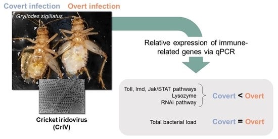
Induction of Multiple Immune Signaling Pathways in Gryllodes sigillatus Crickets during Overt Viral Infections

1
National Center for Agricultural Utilization Research, Crop BioProtection Research Unit, USDA-ARS, 1815 N. University St., Peoria, IL 61604, USA
2
School of Biological Sciences, Illinois State University, Normal, IL 61761, USA
3
Microscopy Services Laboratory, National Animal Disease Center, USDA-ARS, Ames, IA 50010, USA
4
School of Science, Western Sydney University, Hawkesbury Campus, Richmond, NSW 2753, Australia
*
Author to whom correspondence should be addressed.
Viruses 2022, 14(12), 2712; https://doi.org/10.3390/v14122712
Submission received: 9 September 2022 / Revised: 28 November 2022 / Accepted: 30 November 2022 / Published: 3 December 2022
(This article belongs to the Special Issue Insect Virus Discovery)

Abstract

:
Despite decades of focus on crickets (family: Gryllidae) as a popular commodity and model organism, we still know very little about their immune responses to microbial pathogens. Previous studies have measured downstream immune effects (e.g., encapsulation response, circulating hemocytes) following an immune challenge in crickets, but almost none have identified and quantified the expression of immune genes during an active pathogenic infection. Furthermore, the prevalence of covert (i.e., asymptomatic) infections within insect populations is becoming increasingly apparent, yet we do not fully understand the mechanisms that maintain low viral loads. In the present study, we measured the expression of several genes across multiple immune pathways in Gryllodes sigillatus crickets with an overt or covert infection of cricket iridovirus (CrIV). Crickets with overt infections had higher relative expression of key pathway component genes across the Toll, Imd, Jak/STAT, and RNAi pathways. These results suggests that crickets can tolerate low viral infections but can mount a robust immune response during an overt CrIV infection. Moreover, this study provides insight into the immune strategy of crickets following viral infection and will aid future studies looking to quantify immune investment and improve resistance to pathogens.

Graphical Abstract

1. Introduction

Although the practice of mass-produced insects has been long-standing (e.g., silkworm farming, apiculture, biocontrol agents) [1,2], its application has recently expanded to include uses as livestock and pet feed ingredients [3,4], protein for human consumption [5], chitin for numerous industrial applications [6], and remediation of wastes [7]. Due to the increasing popularity of and demand for insect-based products, there are considerable efforts to maximize insect mass-production [8]. For example, within rearing facilities, microbial pathogens (e.g., viruses, bacteria, fungi) can cause significant mortality, in addition to reducing fecundity and body size. Thus, increasing disease prevention and resistance of insects is essential to the success of this burgeoning industry [9,10,11].
Despite the threat that entomopathogenic infections pose to insect mass-production, we know little about disease prevalence in these settings as systematic screening efforts are currently absent. Crickets (family: Gryllidae) are an especially popular farmed insect; however, they are known to be susceptible to multiple microbial pathogens that can cause disease outbreaks [12,13,14]. For example, the Acheta domesticus densovirus (AdDNV), a parvovirus, was implicated to have caused large disease outbreaks in farmed house cricket (Acheta domesticus) colonies globally, resulting in wholesale product losses. As a direct response to these outbreaks, many producers switched to farming alternative species, including Gryllodes sigillatus in North America, due to reports that they are less susceptible to AdDNV [15]. Furthermore, the number of reports of covert (or, silent) viral infections has increased in reared populations as molecular screening of viruses has expanded to asymptomatic populations [13]. Covert infections by a broad range of microorganisms and other infectious agents are widespread and can manifest as latent infections (e.g., remain within the host cell or integrate into the host genome) or persistent infections with low levels of replication [16]. Importantly, covert infections may become activated resulting in detectable pathology, including mortality.
Beyond agricultural and industrial applications, several species of crickets have long been a model organism within several fields of research (e.g., evolutionary ecology, ecological immunology, insect physiology), where immune effectors have been evaluated across numerous contexts [17,18,19,20,21,22,23,24,25,26,27,28,29]. From these studies, we know that crickets mount an immune response to some microbial pathogens by, for example, exhibiting increased lysozyme-like activity of their hemolymph [30,31], producing a melanization and/or encapsulation response [32,33], and/or increasing circulating hemocytes [34] after controlled exposure to live, inactivated, or simulated (e.g., nylon filaments) pathogens. Although these studies provide insight on the functional downstream outcomes of infection, few have identified and quantified immune gene expression in response to pathogens in these insects [35,36], which is foundational to understanding the molecular basis of their defensive repertoire. Additionally, we do not yet know how crickets respond to naturally acquired pathogenic infections, as almost all assessments have been conducted following controlled inoculation.
Insects possess a suite of cellular and humoral immune defenses in response to viral infection [37] and most of what we know about gene expression as the basis of these defenses comes from work in Drosophila melanogaster [38,39], lepidopterans [40], and several mosquito species [41]. Once a pathogen is detected by the insect host, a series of immune signaling pathways are activated intracellularly to respond to infection with a certain degree of specificity, which is in part attributed to the binding specificity of pattern recognition receptors (PRRs) to pathogen associated molecular patterns (PAMPs) (e.g., lipopolysaccharides and peptidoglycans) [42]. The main signaling pathways that mediate immunity in insects are the Toll, Immune deficiency (Imd), and Jak/STAT pathways. Within the Toll pathway, microbes are detected by PRRs that activate the ligand Spätzle, which then binds to Toll receptors and transduces the signals to Cactus-Dif (Dorsal-related immune factor) through a signaling complex containing the adapter MyD88 [43]. Cactus is then cleaved from Dorsal and/or Dif which then translocate into the nucleus and regulate the transcription of effector genes [44]. Within Imd, PRRs recognize invading pathogens and activate the adapter molecule Imd, which activates Relish [45]. Relish is then cleaved and its DNA binding domain translocates into the nucleus where it regulates the transcription of effector molecules [46]. The Jak/STAT pathway is activated as a response to cell stress and/or viral and fungal infection. In this pathway, Domeless is activated and then associated kinases recruit and phosphorylate STAT, which translocates into the nucleus to regulate the expression of downstream effector genes [47]. Additionally, STAT is negatively modulated by PIAS [48]. Activation of these signaling pathways leads to the production of downstream effector molecules, such as antimicrobial peptides (AMPs), that suppress a range of microbes, including viruses [49]. Further, lysozyme is a particularly potent antimicrobial effector in many insects, including crickets [50]; however, the potential antiviral activity of lysozyme has not been widely investigated [51]. Beyond these canonical immune signaling pathways, the RNAi (RNA interference) pathway plays a significant role in the antiviral response in Drosophila [52] and has been linked to Jak/STAT, suggesting coordination between these responses [53]. In this pathway, viral dsRNA is recognized by Dicer-2 proteins, which dice it into small siRNAs (small interfering RNAs) which are then loaded into an RNA induced silencing complex (RISC) by Dicer-2 and co-factor R2D2. RISC finds the target transcripts (by complementary sequence with the guide strand of the siRNA) and the Argonaute-2 protein (effector protein of RISC) degrades the target transcript [54].
Recently, we characterized viral abundance in reared G. sigillatus crickets from two populations infected with cricket iridovirus (CrIV; family: Iridoviridae): one in which crickets were host to high amounts of viral copies and suffered from increased mortality and reduced fecundity while the other showed no apparent signs of disease and had very few viral copies present [13]. Thus, we consider the diseased population as one exhibiting an overt infection while the healthy population exhibited a covert infection of CrIV. Covert infections of invertebrate iridoviruses are reportedly more prevalent than overt lethal infections in some insect population [55,56,57], which could be due to several factors, including reduced virulence of the virus or increased tolerance of the host. Here, we quantified the expression of immune signaling pathway genes that have been shown to be important in anti-viral immune responses of insects across these two populations of crickets. We selected targets across Toll (MyD88, Cactus, Dorsal, and Dif), Imd (PGRP-LC, Imd, and Relish), and Jak/STAT (Domeless, PIAS, and STAT5B) signaling pathways in addition to a gene encoding for lysozyme. We also measured expression of targets within the RNAi pathway (Dicer-2, R2D2, and Argonaute-2). Finally, because the microbiome has been shown to influence viral dynamics in other insects [58], we quantified the amount of bacteria and fungi present via amplification of the 16S rRNA gene for bacteria and 18S rRNA for fungi to determine if total microbial load plays a role in viral dynamics. We also present TEM images of CrIV viral capsids to confirm active infection within crickets with an overt infection.

2. Materials and Methods

2.1. Cricket Colonies

G. sigillatus crickets were sourced from either of two populations (diseased: a population presenting pathological manifestations of infection, or healthy: an apparently disease-free population) of lab-reared colonies. Symptoms present in the diseased colony were high, intermittent mortality among late-instar nymphs and adults, a strong putrid odor within rearing containers, milky white hemolymph which appeared iridescent under illuminated magnification, increased cuticle and tissue frailty, and underdeveloped or absent ovaries in some adult females [13]. Our previous work found that both populations had the presence of CrIV and AdDNV. Diseased crickets had significantly more CrIV (mean ± 95% C.I. = 3.017 × 10⁹ ± 3.485 × 10⁸ viral copies/µL) compared with healthy crickets (mean ± 95% C.I. = 380.7 ± 131.4 viral copies/µL) [13]. Moreover, diseased crickets also had significantly higher viral loads of AdDNV (mean ± 95% C.I. = 1409 ± 731 viral copies/µL) than healthy crickets (mean ± 95% C.I. = 34.99 ± 18.63 viral copies/µL) [13]. Despite coinfection with CrIV and AdDNV, we concluded that CrIV was the likely disease-causing agent due to the amount of CrIV viral copies present and the apparent symptoms (e.g., iridescent hemolymph). We further determined that the diseased population had an overt CrIV infection while the healthy population had a covert CrIV infection, as the latter had no apparent disease symptoms [13].
These populations were descendants from the same ancestral wild-caught crickets collected from Las Cruces, NM (USA) and have been cultured in a lab setting since 2001. Populations were split and maintained in separate labs since 2007. Rearing methods followed standard cricket rearing protocol within a research laboratory setting [13]. Briefly, about 500 crickets were housed in 55 L plastic storage bins with ventilated lids packed with egg carton to increase rearing surface area. They were provisioned with a standard diet (roughly equal parts Mazuri® Rat & Mouse Diets and Purina® Cat Chow Complete pellets) and water (glass vials plugged with moist cotton) ad libitum. All individuals were housed in an environmental chamber at 32 °C on a 16 h:8 h light:dark cycle. Experimental individuals were at least 1-week-old (no more than 14 days old) post-emergence as adults when they were killed by freezing at −80 °C.

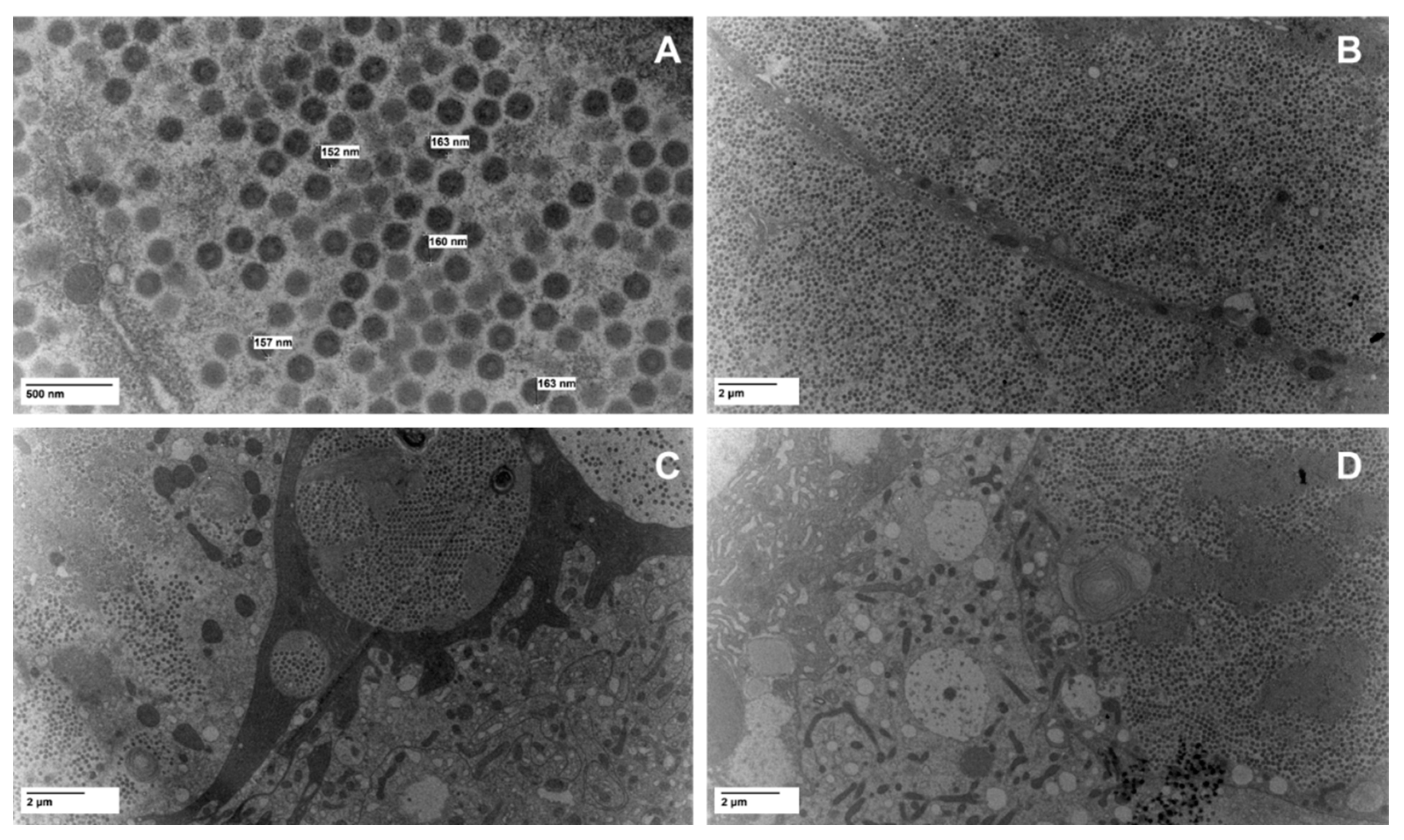
2.2. Viral Imaging Via Electron Microscopy

To capture images of virus particles and confirm active infection of CrIV within diseased crickets, we dissected the fat body from a cricket with an overt infection from the diseased colony. Samples were fixed in 2% paraformaldehyde/2.5% glutaraldehyde/0.05 M NaCacodylate/0.005 M CaCl2 (pH = 7). They were post fixed in 1% Osmium for two hours and then processed in graded alcohols, propylene oxide and LX112 (Ladd Research, Williston, VT, USA) resin with 48 h polymerization and shipped to the USDA-ARS Microscopy Services Laboratory at the National Animal Disease Center (Ames, Iowa, United States) for further processing. Thick sections (1 µm) were performed on select samples and a toluidine blue stain and basic fucsin stain were applied. Polaroid photos were taken of these images and the area of interest for thin sections was identified. A uranyl acetate and Reynold’s lead stain were performed on the thin section before being examined with a ThermoFisher FEI Tecnai G2 BioTWIN electron microscope (ThermoFisher FEI Co., Hillsboro, OR, USA) and images were taken with a Nanosprint12 camera (AMT Corp., Woburn, MA, USA) [59].

2.3. RNA Extraction and cDNA Synthesis

We extracted RNA from whole body homogenates of individual crickets from each population (20 crickets/population). Previously frozen (−80 °C) crickets were placed individually in tubes with 1 mL sterile 1x PBS (pH 7.2) and two 3.2 mm diameter sterile stainless-steel beads and macerated using a TissueLyser II (Qiagen, Hilden, Germany). The resulting liquid homogenate was removed (about 0.9 mL) and placed in a new sterile tube for RNA extraction. RNA was extracted from 100 µL of cricket homogenate using the RNeasy Mini prep kit (Qiagen) following the “Purification of Total RNA from Animal Tissues” protocol. The concentration of RNA within each sample was estimated via a NanoDrop OneC Microvolume UV-Vis Spectrophotometer (ThermoFisher, Waltham, MA, USA) and 260/280 and 260/230 values were above 1.8 for all samples. RNA from each sample was diluted to 1 µg, treated with DNA Wipeout, and then converted to cDNA using the QuantiTect Reverse Transcription Kit (Qiagen) prior to conducting reverse transcriptase quantitative PCR assays. All cDNA samples were stored at −20 °C until further use.

2.4. Gene Target-Specific Primer Design

We searched for the target genes in the head transcriptome of G. sigillatus [60] using the BLAST+ command-line application [61,62,63]. Specifically, protein sequences from the mosquito Aedes aegypti or nucleotide sequences of the cricket A. domesticus were used as a reference, using blastx and megablast, respectively with default settings to find G. sigillatus sequences. The resulting cricket sequences were blasted to the nr database to confirm gene identity [64,65], and sequences were manually trimmed to remove potential chimeric sequences. Subsequently, the coding regions of these sequences were translated to their respective proteins in Ugene [66], and were aligned with homologous proteins from a representative set of insects (the moth Bombyx mori, the bee Apis mellifera, the fruit fly Drosophila melanogaster, the mosquito Aedes aegypti, the beetle Tribolium castaneum, the termite Zootermopsis nevadensis, the grasshoppers Locusta migratoria and Schistocerca gregaria, and the cricket Gryllus bimaculatus), as available on the ncbi protein database [65]. Alignments were performed and visualized in Ugene using the MUSCLE algorithm [67] with default settings (Supplementary Figures S1–S13). For Dicer-2, Argonaute-2, Relish, Dorsal and Dif, phylogenetic trees were made to further confirm sequence identity (Supplementary Figures S14–S16). Protein sequences were aligned using MUSCLE with default settings on a linux machine, and Maximum Likelihood trees were subsequently made using RAxML v. 8.2.12 [68]. Trees were visualized using the Interactive Tree of Life (iTOL) [69]. All sequences were deposited in GenBank (see Table 1 for accession numbers).
All primers used in the present study were designed using Primer-BLAST (NCBI) (all primers from IDT, Inc., Coralville, IA, USA). For quantification purposes, we designed primers targeting the 18S and 16S ribosomal RNA (Table 1) as invariant housekeeping genes. These were selected based on their performance/ranking via RefFinder [70,71], which uses the algorithm from major computational programs such as geNorm, Normfinder, and Best-Keeper to compare and rank candidate reference genes. We then calculated the geometric mean for expression of these two genes for each individual and used this as our reference target for gene expression. To evaluate whether fungal or bacterial load could contribute to differential viral loads between populations, we quantified the 16s rRNA for bacteria (16SrRNA-Fw 5′-TCCTACGGGAGGCAGCAGT-3′ and 16SrRNA-Rv 5′ GGACTACCAGGGTATCTAATCCTGTT-3′) and the 18s rRNA (18SrRNA-Fw 5′-AGATACCGTCGTAGTCTTAACCATAAACT-3′ and 18SrRNA-Rv 5′-TTCAGCCTTGCGACCATACT-3′) for fungi from crickets across both populations.

2.5. Reverse Transcriptase Quantitative PCR (RT-qPCR) Detection and Quantification

RT-qPCR reactions were run on a Quant-Studio 6 Real-Time PCR instrument (Thermo Fisher Scientific, Waltham, MA, USA), and included a melt-curve stage to confirm product specificity. One microliter of cDNA product was used in a 10 µL RT-qPCR reaction using gene specific primers (Table 1) and PowerUp SYBR green Master mix kit (Qiagen). RT-qPCR cycling conditions consisted of holding at 50 °C for 2 min and 95 °C for 2 min and 40 cycles of 1 s at 95 °C and 30 min at 60 °C. The identities of targets were confirmed by mapping sequences to the reference target genes using default settings in Geneious Prime® following Sanger sequencing.

2.6. Statistical Analysis

Gene expression profiles were evaluated using the ΔΔCt method [72]. We used an unpaired t-test with Welch’s correction for each immune gene target to compare expression of healthy and diseased crickets between populations, in addition to comparing microbial loads. All expression data were log2-transformed to fit normality assumptions. All analyses and graphs were performed and created using GraphPad Prism 9 (version 9.0.0).

3. Results

3.1. TEM Imaging

TEM analysis revealed substantial quantities of virions and confirmed the presence of large (~160 nm) icosahedral viruses in cells from the dissected fat body of a cricket from the diseased population (Figure 1). Cricket cells were packed with virions that often formed small paracrystalline arrays (e.g., Figure 1C).

3.2. Immune Signaling Pathways

Within the Toll pathway, the relative expression for the transcription factors Dorsal (Welch’s corrected t(26.06) = 3.626, p = 0.0012) and Dif (Welch’s corrected t(27.50) = 5.779, p < 0.0001) were significantly higher in the diseased population than in the healthy population. Similarly, the expression of Cactus, a negative regulator, was higher in diseased crickets (Welch’s corrected t(22.26) = 4.058, p = 0.0005). The adapter molecule MyD88 did not differ between populations (Welch’s corrected t(35.32) = 1.285, p = 0.207; Figure 2A).
For targets within the Imd pathway, both the transcription factor Relish (Welch’s corrected t(19.95) = 6.361, p < 0.0001) and the adapter molecule Imd (Welch’s corrected t(27.25) = 3.507, p = 0.0016) were more highly expressed in the diseased population. However, expression of the pathogen recognition receptor PGRP-LC was similar across populations (Welch’s corrected t(28.84) = 0.6128, p = 0.5448).
Finally, the relative expression of all targets measured in the Jak/STAT pathway were significantly higher in the diseased population than in the healthy population, including the receptor Domeless (Welch’s corrected t(35.31) = 5.525, p < 0.0001), the transcription factor STAT5B (Welch’s corrected t(24.21) = 4.728, p < 0.0001), and the negative regulator PIAS (Welch’s corrected t(24.49) = 5.424, p < 0.0001) (Figure 2C).

3.3. RNAi Pathway

All targets measured within the RNAi pathway, Argonaute-2 (Welch’s corrected t(19.62) = 9.101, p < 0.0001), R2D2 (Welch’s corrected t(19.39) = 5.933, p < 0.0001), and Dicer-2 (Welch’s corrected t(27.77) = 11.19, p < 0.0001), were more highly expressed in the diseased population than in the healthy population (Figure 3A).

3.4. Lysozyme

The relative expression of lysozyme (Welch’s corrected t(16.03) = 7.504, p < 0.0001; Figure 3B) was significantly higher in the diseased population compared with the healthy population.

3.5. Microbial Load

Both fungal load (Welch’s corrected t(28.85) = 1.152, p = 0.2588) and bacterial load (Welch’s corrected t(68.00) = 1.348, p = 0.1821) were similar between healthy and diseased populations based on relative quantification of fungal 18s rRNA and bacterial 16s rRNA, respectively (Figure 4).

4. Discussion

Despite its importance in host response to pathogens, we still know little about the molecular basis of cricket immunity, and even less about immune responses within the context of covert infections. To improve our understanding of host–virus interactions in crickets, we evaluated canonical immune signaling pathways that have been shown in other arthropod systems to be involved in immunity to microbial organisms, including viruses. By quantifying gene expression across several facets of the invertebrate immune system, we have begun to characterize the immune response to overt cricket iridovirus (CrIV) infections in the popularly reared G. sigillatus cricket. Specifically, we found that crickets infected with high levels of cricket iridovirus had higher gene expression across the Toll, Imd, and Jak/STAT immune signaling pathways as well as within the RNAi pathway.
Activation of the Toll and Imd pathways are typically associated with defenses against pathogenic fungi, bacteria, and protozoa. Furthermore, these two pathways have been linked with the antiviral response in Diptera [73,74], but less is known about their role in orthopterans. Both Jak/STAT and RNAi pathways are known to play important roles in antiviral immunity and so it is not surprising that targets across these two pathways were upregulated in crickets with large CrIV viral loads in our study. Our study also evaluated an important antimicrobial effector, lysozyme, which has been found to have antiviral activity against dengue virus in mosquitoes [75] as well as against other viruses infecting eukaryotic hosts [51]. Our transcript level analyses show that the population of diseased crickets had significantly higher expression of lysozyme compared with the healthy population. This suggests that lysozyme might play a significant role in the cricket’s efforts to control the systemic replication of CrIV. Taken together, we can conclude that crickets with overt CrIV infection have an immune profile exhibiting strong induction of critical immune pathway components across Toll, Imd, Jak/STAT, and RNAi. At the same time, it begs the question of whether crickets tolerate viruses when they occur at a lower concentration and fully engage the immune system only when viral loads surpass a certain threshold. Unfortunately, our study is unable to fully answer this question given that our control (healthy population) also carried CrIV, albeit at significantly lower levels.
A few previous studies have identified immune related genes in Orthoptera, including crickets; however, none to our knowledge have investigated an antiviral response in this order. An enzyme similar to the AMP prolixicin was discovered in A. domesticus suspected of being infected with a gregarine parasite and found to occur at higher concentrations in juveniles compared with adults [76]. In the black field cricket (Teleogryllus emma), researchers identified 58 differentially expressed unigenes and several AMPs following inoculation with E. coli [77]. In Gryllus bimaculatus, 4 inducible lysozymes and 6 AMPs were identified with similarities to defensin and diptericin, as well as pyrrhocoricin, prolixicin, and hemiptericin [78]. A comparative transcriptomic analysis of the immune response of migratory locusts challenged with Metarhizium fungi identified immune related unigenes including those involved with Toll, Imd, and Jak/STAT pathways, with 58 and 3 differentially expressed in the fat body and hemocytes, respectively [79]. It also found higher expression of lysozyme transcripts post-infection. While it is difficult to draw direct comparisons across these few studies, some patterns emerge, including the roles that the canonical immune signaling pathways and their effectors play in the Orthopteran immune system.
Our findings add to the current understanding of the insect host immune response to iridovirus in crickets. Previous work has shown that lab reared Gryllus texensis crickets infected with an iridovirus have significantly lower phenoloxidase activity than uninfected crickets [80]. This contrasts with our findings of increased immune gene expression, but we did not assess any genes involved directly in the phenoloxidase cascade and phenoloxidase activity has been shown to be negatively associated with other components of immunity or reduced following immune activation in other insects [81]. Although, to our knowledge, there are no reports of studies that have investigated molecular markers of immune activation following an infection with CrIV, several studies have characterized host response to the closely related Invertebrate Iridescent virus 6 (IIV-6) in Drosophila [13,82]. From these, we know that the IIV-6 genome encodes for proteins that can inhibit insect host immune responses, including RNA silencing by the RNAi pathway (e.g., 340 L) [83], which is the primary defense against IIV-6 [84,85]. In our study, both Dicer-2 and Argonaute-2 were upregulated in crickets with overt CrIV infections, suggesting that the RNAi pathway is also important in antiviral defense for G. sigillatus. IIV-6 was also found to be able to inhibit both Imd and Toll pathways [86]. Interestingly, while there is no evidence that the Jak/STAT pathway confers immunity against IIV-6 infection in Drosophila [85], our study indicates significant induction of Jak/STAT pathway components in response to CrIV. Importantly, we found no evidence of viral inhibition of these responses at the transcriptional level in the present study.
Why individuals from one population suffer from overt CrIV infections while the other maintains covert, asymptomatic infections remains an open question. One possibility is that the diseased population was exposed to an undetected microbe (e.g., bacteria or fungi) that made it more susceptible to an overt viral infection. Indeed, previous studies have demonstrated that co-infection with IIV-6 and a Gram-negative bacterium result in more rapid mortality in Drosophila [86]. Although we did not find differences in total microbial loads (Figure 4), we did not characterize microbiomes and therefore cannot rule out the possibility that bacterial or fungal composition are different between populations. Future studies will characterize the microbiome between populations with overt and covert infections to determine if the microbiome may play a role in promoting (or inhibiting) overt infections. While we previously found evidence of low viral loads of AdDNV in both populations of crickets [13], AdDNV has not previously been associated with disease in G. sigillatus [15]. Still, we did find that crickets with overt infections of CrIV had significantly higher (albeit relatively low) AdDNV viral loads. Additional studies will evaluate the impact of viral dynamics on infection outcomes. Another possibility is that intrinsic (e.g., inbreeding) or extrinsic (e.g., rearing environment) factors may impair immune function leading to the opportunistic reactivation of covert infections [87]. However, crickets with high levels of CrIV were able to mount an immune response across multiple pathways and so it is unlikely that crickets from the diseased population have a dysfunctional immune response, at least at the transcription level. Further studies probing post-transcriptional and post-translational outcomes will be essential to understanding the role immunity plays in regulating viral loads.
In the present study, we assessed genes that are known to be important in immunity in other model insects (e.g., mosquitos, flies, moths). Future studies (e.g., RNAi knockdown experiments) will determine the importance of specific pathways on clearing or decreasing viral loads in these crickets. Furthermore, a comparison of complete transcriptomes between populations is required to obtain the global gene expression repertoire of infected crickets. These studies will be essential to fully characterize the defensive strategy of crickets at the transcript level and will improve our understanding of how crickets can tolerate low levels of CrIV and maintain covert infections. Hampering these efforts is the fact that few genetic resources for crickets currently exist [88], including the absence of a complete and annotated genome of G. sigillatus. Advancements in this field will greatly aid research efforts, including those seeking to improve production of reared beneficial insects.

5. Conclusions

By evaluating the induction of immune-related genes across populations of crickets with an overt or covert infection, we can begin to understand the immune responses of G. sigillatus, a popularly reared cricket and a model insect across multiple branches of research. Crickets with an overt infection of the highly pathogenic CrIV presented significantly higher induction of multiple genes across all canonical immune signaling pathways, in addition to the RNAi pathway, compared to crickets with a covert infection. Our data suggest that G. sigillatus can tolerate low levels of viral infection and are able to mount an immune response when faced with an overt viral infection.

Supplementary Materials

The following supporting information can be downloaded at: https://www.mdpi.com/article/10.3390/v14122712/s1, Figure S1: Partial protein alignment of Dicer-2; Figure S2: Protein alignment of Argonaute-2; Figure S3: Protein alignment of Dorsal; Figure S4: Protein alignment of Relish; Figure S5: Protein alignment of MyD88; Figure S6: Protein alignment of Cactus; Figure S7: Protein alignment of PGRP-LC; Figure S8: Protein alignment of Imd; Figure S9: Protein alignment of Domeless; Figure S10: Protein alignment of PIAS; Figure S11: Protein alignment of STAT5B; Figure S12: Protein alignment of R2D2; Figure S13: Protein alignment of Lysozyme; Figure S14: Maximum likelihood tree of Dicer proteins; Figure S15: Maximum likelihood tree of Argonaute and Piwi proteins; Figure S16: Maximum likelihood tree of Dorsal and Relish proteins.

Author Contributions

Conceptualization, K.R.D. and J.L.R.; methodology, K.R.D., J.L.R., B.F. and J.A.S.; validation, K.R.D. and J.L.R.; formal analysis, K.R.D.; resources, J.H., B.M.S. and S.K.S.; data curation, K.R.D.; writing—original draft preparation, K.R.D., B.F. and J.L.R.; writing—review and editing, all authors; visualization, K.R.D. and J.A.S.; supervision, J.L.R. All authors have read and agreed to the published version of the manuscript.

Funding

This research was supported in part by the U.S. Department of Agriculture, Agricultural Research Service. B.F., S.K.S. and B.M.S. were supported by a grant from the National Science Foundation (IOS 16–54028). J.H. was supported by a grant from the Australian Research Council (DP180101708).

Institutional Review Board Statement

Not applicable.

Informed Consent Statement

Not applicable.

Data Availability Statement

The datasets generated during the current study will be available in the GenBank repository upon acceptance.

Acknowledgments

We warmly thank Kylie Hampton and Haley Gore for their lab assistance. Mention of trade names or commercial products in this publication is solely for the purpose of providing specific information and does not imply recommendation or endorsement by the U.S. Department of Agriculture. USDA is an equal opportunity provider and employer.

Conflicts of Interest

The authors declare no conflict of interest.

References

  1. Defoliart, G.R. Edible Insects as Minilivestock. Biodivers. Conserv. 1995, 4, 306–321. [Google Scholar] [CrossRef]
  2. Sørensen, J.G.; Addison, M.F.; Terblanche, J.S. Mass-rearing of insects for pest management: Challenges, synergies and advances from evolutionary physiology. Crop. Prot. 2012, 38, 87–94. [Google Scholar] [CrossRef]
  3. Bosch, G.; Zhang, S.; Oonincx, D.G.A.B.; Hendriks, W.H. Protein quality of insects as potential ingredients for dog and cat foods. J. Nutr. Sci. 2014, 3, E29. [Google Scholar] [CrossRef] [PubMed] [Green Version]
  4. Makkar, H.P.S.; Tran, G.; Heuzé, V.; Ankers, P. State-of-the-art on use of insects as animal feed. Anim. Feed. Sci. Technol. 2014, 197, 1–33. [Google Scholar] [CrossRef]
  5. van Huis, A.; Van Itterbeeck, J.; Klunder, H.; Mertens, E.; Halloran, A.; Muir, G.; Vantomme, P. Edible Insects: Future Prospects for Food and Feed Security; FAO Forestry Paper 171; Food and Agriculture Organization of the United Nations (FAO): Rome, Italy, 2013; Volume 171. [Google Scholar]
  6. Hahn, T.; Tafi, E.; Paul, A.; Salvia, R.; Falabella, P.; Zibek, S. Current state of chitin purification and chitosan production from insects. J. Chem. Technol. Biotechnol. 2020, 95, 2775–2795. [Google Scholar] [CrossRef]
  7. Surendra, K.C.; Tomberlin, J.K.; van Huis, A.; Cammack, J.A.; Heckmann, L.H.L.; Khanal, S.K. Rethinking organic wastes bioconversion: Evaluating the potential of the black soldier fly (Hermetia illucens (L.))(Diptera: Stratiomyidae)(BSF). J. Waste Manag. 2020, 117, 58–80. [Google Scholar] [CrossRef]
  8. Ortiz, J.C.; Ruiz, A.T.; Morales-Ramos, J.A.; Thomas, M.; Rojas, M.G.; Tomberlin, J.K.; Yi, L.; Han, R.; Giroud, L.; Jullien, R.L. Chapter 6—Insect Mass Production Technologies, in Insects as Sustainable Food Ingredients. In Insects as Sustainable Food Ingredients; Dossey, A.T., Morales-Ramos, J.A., Rojas, M.G., Eds.; Academic Press: San Diego, CA, USA, 2016; pp. 153–201. ISBN 978-0-12-802856-8. [Google Scholar]
  9. Bertola, M.; Mutinelli, A.F. Systematic Review on Viruses in Mass-Reared Edible Insect Species. Viruses 2021, 13, 2280. [Google Scholar] [CrossRef]
  10. Maciel-Vergara, G.; Jensen, A.B.; Lecocq, A.; Eilenberg, J. Diseases in edible insect rearing systems. J. Insects Food Feed. 2021, 7, 621–638. [Google Scholar] [CrossRef]
  11. Maciel-Vergara, G.; Ros, V.I. Viruses of insects reared for food and feed. J. Invertebr. Pathol. 2017, 147, 60–75. [Google Scholar] [CrossRef]
  12. de Miranda, J.R.; Granberg, F.; Onorati, P.; Jansson, A.; Berggren, Å. Virus prospecting in crickets—Discovery and strain divergence of a novel iflavirus in wild and cultivated Acheta domesticus. Viruses 2021, 13, 364. [Google Scholar] [CrossRef]
  13. Duffield, K.R.; Hunt, J.; Sadd, B.M.; Sakaluk, S.K.; Oppert, B.; Rosario, K.; Behle, R.W.; Ramirez, J.L. Active and Covert Infections of Cricket Iridovirus and Acheta domesticus Densovirus in Reared Gryllodes sigillatus Crickets. Front. Microbiol. 2021, 12, 780796. [Google Scholar] [CrossRef] [PubMed]
  14. de Miranda, J.R.; Granberg, F.; Low, M.; Onorati, P.; Semberg, E.; Jansson, A.; Berggren, Å. Virus Diversity and Loads in Crickets Reared for Feed: Implications for Husbandry. Front. Vet. Sci. 2021, 8, 510. [Google Scholar] [CrossRef] [PubMed]
  15. Weissman, D.B.; Gray, D.A.; Pham, H.T.; Tijssen, P. Billions and billions sold: Pet-feeder crickets (Orthoptera: Gryllidae), commercial cricket farms, an epizootic densovirus, and government regulations make for a potential disaster. Zootaxa 2012, 3504, 67–88. [Google Scholar] [CrossRef]
  16. Sorrell, I.; White, A.; Pedersen, A.B.; Hails, R.S.; Boots, M. The evolution of covert, silent infection as a parasite strategy. Proc. R. Soc. B 2009, 276, 2217–2226. [Google Scholar] [CrossRef]
  17. Adamo, S.A.; Parsons, N.M. The emergency life-history stage and immunity in the cricket, Gryllus texensis. Anim. Behav. 2006, 72, 235–244. [Google Scholar] [CrossRef]
  18. Reginald, K.; Wong, Y.R.; Shah, S.M.R.; Teh, K.F.; Jalin, E.J.F.; Khan, N.A. Investigating immune responses of the house cricket, Acheta domesticus to pathogenic Escherichia coli K1. Microbes Infect 2021, 23, 104876. [Google Scholar] [CrossRef]
  19. Cho, Y.; Cho, S. Hemocyte-hemocyte adhesion by granulocytes is associated with cellular immunity in the cricket, Gryllus bimaculatus. Sci. Rep. 2019, 9, 18066. [Google Scholar] [CrossRef] [Green Version]
  20. Drayton, J.M.; Jennions, M.D. Inbreeding and measures of immune function in the cricket Teleogryllus commodus. Behav. Ecol. 2011, 22, 486–492. [Google Scholar] [CrossRef] [Green Version]
  21. Adamo, S.A.; Jensen, M.; Younger, M. Changes in lifetime immunocompetence in male and female Gryllus texensis (formerly G. integer): Trade-offs between immunity and reproduction. Anim. Behav. 2001, 62, 417–425. [Google Scholar] [CrossRef]
  22. Hampton, K.J.; Duffield, K.R.; Hunt, J.; Sakaluk, S.K.; Sadd, B.M. Male and female genotype and a genotype-by-genotype interaction mediate the effects of mating on cellular but not humoral immunity in female decorated crickets. Heredity 2021, 126, 477–490. [Google Scholar] [CrossRef]
  23. Piñera, A.V.; Charles, H.M.; Dinh, T.A.; Killian, K.A. Maturation of the immune system of the male house cricket, Acheta domesticus. J. Insect Physiol. 2013, 59, 752–760. [Google Scholar] [CrossRef]
  24. Ferguson, L.V.; Dhakal, P.; Lebenzon, J.E.; Heinrichs, D.E.; Bucking, C.; Sinclair, B.J. Seasonal shifts in the insect gut microbiome are concurrent with changes in cold tolerance and immunity. Funct. Ecol. 2018, 32, 2357–2368. [Google Scholar] [CrossRef] [Green Version]
  25. Duffield, K.R.; Hampton, K.J.; Houslay, T.M.; Hunt, J.; Sadd, B.M.; Sakaluk, S.K. Inbreeding alters context-dependent reproductive effort and immunity in male crickets. J. Evol. Biol. 2019, 32, 731–741. [Google Scholar] [CrossRef] [PubMed]
  26. Duffield, K.R.; Hampton, K.J.; Houslay, T.M.; Rapkin, J.; Hunt, J.; Sadd, B.M.; Sakaluk, S.K. Macronutrient intake and simulated infection threat independently affect life history traits of male decorated crickets. Ecol. Evol. 2020, 10, 11766–11778. [Google Scholar] [CrossRef] [PubMed]
  27. Kerr, A.M.; Gershman, S.N.; Sakaluk, S.K. Experimentally induced spermatophore production and immune responses reveal a trade-off in crickets. Behav. Ecol. 2010, 21, 647–654. [Google Scholar] [CrossRef]
  28. Letendre, C.; Duffield, K.R.; Sadd, B.M.; Sakaluk, S.K.; House, C.M.; Hunt, J. Genetic covariance in immune measures and pathogen resistance in decorated crickets is sex and pathogen specific. J. Anim. Ecol. 2022, 91, 1471–1488. [Google Scholar] [CrossRef]
  29. Gershman, S.N.; Barnett, C.A.; Pettinger, A.M.; Weddle, C.B.; Hunt, J.; Sakaluk, S.K. Inbred decorated crickets exhibit higher measures of macroparasitic immunity than outbred individuals. Heredity 2010, 105, 282–289. [Google Scholar] [CrossRef] [Green Version]
  30. Simmons, L.W. Resource allocation trade-off between sperm quality and immunity in the field cricket, Teleogryllus oceanicus. Behav. Ecol. 2011, 23, 168–173. [Google Scholar] [CrossRef] [Green Version]
  31. Adamo, S.A. Estimating disease resistance in insects: Phenoloxidase and lysozyme-like activity and disease resistance in the cricket Gryllus texensis. J. Insect Physiol. 2004, 50, 209–216. [Google Scholar] [CrossRef]
  32. Bascuñán-García, A.P.; Lara, C.; Córdoba-Aguilar, A. Immune investment impairs growth, female reproduction and survival in the house cricket, Acheta domesticus. J. Insect Physiol. 2010, 56, 204–211. [Google Scholar] [CrossRef]
  33. Park, Y.; Stanley, D. Physiological trade-off between cellular immunity and flight capability in the wing-dimorphic sand cricket, Gryllus firmus. J. Asia Pac. Entomol. 2015, 18, 553–559. [Google Scholar] [CrossRef]
  34. Duffield, K.R.; Hampton, K.J.; Houslay, T.M.; Hunt, J.; Rapkin, J.; Sakaluk, S.K.; Sadd, B.M. Age-dependent variation in the terminal investment threshold in male crickets. Evolution 2018, 72, 578–589. [Google Scholar] [CrossRef] [PubMed] [Green Version]
  35. Miyashita, A.; Lee, T.Y.M.; McMillan, L.E.; Easy, R.; Adamo, S.A. Immunity for nothing and the eggs for free: Apparent lack of both physiological trade-offs and terminal reproductive investment in female crickets (Gryllus texensis). PLoS ONE 2019, 14, e0209957. [Google Scholar] [CrossRef] [PubMed]
  36. Sorrell, M.R.; Killian, K.A. Innate immune system function following systemic RNA-interference of the Fragile X Mental Retardation 1 gene in the cricket Acheta domesticus. J. Insect Physiol. 2020, 126, 104097. [Google Scholar] [CrossRef]
  37. Gillespie, J.P.; Kanost, M.R.; Trenczek, T. Biological mediators of insect immunity. Annu. Rev. Entomol. 1997, 42, 611–643. [Google Scholar] [CrossRef]
  38. Hoffmann, J.A. The immune response of Drosophila. Nature 2003, 426, 33–38. [Google Scholar] [CrossRef]
  39. Kingsolver, M.B.; Huang, Z.; Hardy, R.W. Insect antiviral innate immunity: Pathways, effectors, and connections. J. Mol. Biol. 2013, 425, 4921–4936. [Google Scholar] [CrossRef] [Green Version]
  40. Casanova-Torres, Á.M.; Goodrich-Blair, H. Immune signaling and antimicrobial peptide expression in Lepidoptera. Insects 2013, 4, 320–338. [Google Scholar] [CrossRef] [Green Version]
  41. Sim, S.; Jupatanakul, N.; Dimopoulos, G. Mosquito immunity against arboviruses. Viruses 2014, 6, 4479–4504. [Google Scholar] [CrossRef] [Green Version]
  42. Gottar, M.; Gobert, V.; Michel, T.; Belvin, M.; Duyk, G.; Hoffmann, J.A.; Ferrandon, D.; Royet, J. The Drosophila immune response against Gram-negative bacteria is mediated by a peptidoglycan recognition protein. Nature 2002, 416, 640–644. [Google Scholar] [CrossRef]
  43. Horng, T.; Medzhitov, R. Drosophila MyD88 is an adapter in the Toll signaling pathway. Proc. Natl. Acad. Sci. USA 2001, 98, 12654–12658. [Google Scholar] [CrossRef] [PubMed] [Green Version]
  44. Meng, X.; Khanuja, B.S.; Ip, Y.T. Toll receptor-mediated Drosophila immune response requires Dif, an NF-κB factor. Genes Dev. 1999, 13, 792–797. [Google Scholar] [CrossRef] [PubMed] [Green Version]
  45. Choe, K.M.; Werner, T.; Stoven, S.; Hultmark, D.; Anderson, K.V. Requirement for a peptidoglycan recognition protein (PGRP) in Relish activation and antibacterial immune responses in Drosophila. Science 2002, 296, 359–362. [Google Scholar] [CrossRef] [PubMed] [Green Version]
  46. Silverman, N.; Zhou, R.; Stöven, S.; Pandey, N.; Hultmark, D.; Maniatis, T. A Drosophila IκB kinase complex required for Relish cleavage and antibacterial immunity. Genes Dev. 2000, 14, 2461–2471. [Google Scholar] [CrossRef] [PubMed] [Green Version]
  47. Yan, R.; Small, S.; Desplan, C.; Dearolf, C.R.; Darnell Jr, J.E. Identification of a Stat gene that functions in Drosophila development. Cell 1996, 84, 421–430. [Google Scholar] [CrossRef] [PubMed] [Green Version]
  48. Betz, A.; Lampen, N.; Martinek, S.; Young, M.W.; Darnell Jr, J.E. A Drosophila PIAS homologue negatively regulates stat92E. Proc. Natl. Acad. Sci. USA 2001, 98, 9563–9568. [Google Scholar] [CrossRef] [Green Version]
  49. Feng, M.; Fei, S.; Xia, J.; Labropoulou, V.; Swevers, L.; Sun, J. Antimicrobial peptides as potential antiviral factors in insect antiviral immune response. Front. Immunol. 2020, 11, 2030. [Google Scholar] [CrossRef]
  50. Schneider, P.M. Purification and properties of three lysozymes from hemolymph of the cricket, Gryllus bimaculatus (De Geer). Insect Biochem. 1985, 15, 463–470. [Google Scholar] [CrossRef]
  51. Chen, T.T.; Tan, L.R.; Hu, N.; Dong, Z.Q.; Hu, Z.G.; Jiang, Y.M.; Chen, P.; Pan, M.H.; Lu, C. C-lysozyme contributes to antiviral immunity in Bombyx mori against nucleopolyhedrovirus infection. J. Insect Physiol. 2018, 108, 54–60. [Google Scholar] [CrossRef]
  52. Wang, X.H.; Aliyari, R.; Li, W.X.; Li, H.W.; Kim, K.; Carthew, R.; Atkinson, P.; Ding, S.W. RNA interference directs innate immunity against viruses in adult Drosophila. Science 2006, 312, 452–454. [Google Scholar] [CrossRef]
  53. Paradkar, P.N.; Trinidad, L.; Voysey, R.; Duchemin, J.B.; Walker, P.J. Secreted Vago restricts West Nile virus infection in Culex mosquito cells by activating the Jak-STAT pathway. Proc. Natl. Acad. Sci. USA 2012, 109, 18915–18920. [Google Scholar] [CrossRef] [Green Version]
  54. Rand, T.A.; Ginalski, K.; Grishin, N.V.; Wang, X. Biochemical identification of Argonaute 2 as the sole protein required for RNA-induced silencing complex activity. Proc. Natl. Acad. Sci. USA 2004, 101, 14385–14389. [Google Scholar] [CrossRef] [PubMed] [Green Version]
  55. Williams, T. Covert iridovirus infection of blackfly larvae. Proc. R. Soc. Lond. B Biol. Sci. 1993, 251, 225–230. [Google Scholar]
  56. Williams, T. Natural invertebrate hosts of iridoviruses (Iridoviridae). Neotrop. Entomol. 2008, 37, 615–632. [Google Scholar] [CrossRef] [Green Version]
  57. Tonka, T.; Weiser, J. Iridovirus infection in mayfly larvae. J. Invertebr. Pathol. 2000, 76, 229–231. [Google Scholar] [CrossRef] [PubMed]
  58. Jupatanakul, N.; Sim, S.; Dimopoulos, G. The insect microbiome modulates vector competence for arboviruses. Viruses 2014, 6, 4294–4313. [Google Scholar] [CrossRef] [PubMed]
  59. Bozzola, J.; Russell, L. Electron Microscopy; Jones and Bartlett Publishers: Boston, MA, USA, 1992; ISBN 9780867201260. [Google Scholar]
  60. Foquet, B.; Rapkin, J.; Sharma, M.D.; Sadd, B.M.; Sakaluk, S.K.; Hunt, J. Transcriptomic responses of females to consumption of nuptial food gifts as a potential mediator of sexual conflict in decorated crickets. J. Evol. Biol. 2022. [Google Scholar] [CrossRef] [PubMed]
  61. Altschul, S.F.; Gish, W.; Miller, W.; Myers, E.W.; Lipman, D.J. Basic local alignment search tool. J. Mol. Biol. 1990, 215, 403–410. [Google Scholar] [CrossRef] [PubMed]
  62. Altschul, S.F.; Madden, T.L.; Schäffer, A.A.; Zhang, J.; Zhang, Z.; Miller, W.; Lipman, D.J. Gapped BLAST and PSI-BLAST: A new generation of protein database search programs. Nucleic Acids Res. 1997, 25, 3389–3402. [Google Scholar] [CrossRef] [PubMed] [Green Version]
  63. Camacho, C.; Coulouris, G.; Avagyan, V.; Ma, N.; Papadopoulos, J.; Bealer, K.; Madden, T.L. BLAST+: Architecture and applications. BMC Bioinform 2009, 10, 1–9. [Google Scholar] [CrossRef] [PubMed] [Green Version]
  64. Johnson, M.; Zaretskaya, I.; Raytselis, Y.; Merezhuk, Y.; McGinnis, S.; Madden, T.L. NCBI BLAST: A better web interface. Nucleic Acids Res. 2008, 36, W5–W9. [Google Scholar] [CrossRef] [PubMed]
  65. Wheeler, D.L.; Barrett, T.; Benson, D.A.; Bryant, S.H.; Canese, K.; Chetvernin, V.; Church, D.M.; DiCuccio, M.; Edgar, R.; Federhen, S.; et al. Database resources of the national center for biotechnology information. Nucleic Acids Res. 2007, 36, D13–D21. [Google Scholar] [CrossRef] [PubMed] [Green Version]
  66. Okonechnikov, K.; Golosova, O.; Fursov, M.; Ugene Team. Unipro UGENE: A unified bioinformatics toolkit. Bioinformatics 2012, 28, 1166–1167. [Google Scholar] [CrossRef] [PubMed] [Green Version]
  67. Edgar, R.C. MUSCLE: Multiple sequence alignment with high accuracy and high throughput. Nucleic Acids Res. 2004, 32, 1792–1797. [Google Scholar] [CrossRef] [PubMed] [Green Version]
  68. Stamatakis, A. RAxML version 8: A tool for phylogenetic analysis and post-analysis of large phylogenies. Bioinformatics 2014, 30, 1312–1313. [Google Scholar] [CrossRef] [Green Version]
  69. Letunic, I.; Bork, P. Interactive Tree Of Life (iTOL) v5: An online tool for phylogenetic tree display and annotation. Nucleic Acids Res. 2021, 49, W293–W296. [Google Scholar] [CrossRef]
  70. Silver, N.; Best, S.; Jiang, J.; Thein, S.L. Selection of housekeeping genes for gene expression studies in human reticulocytes using real-time PCR. BMC Mol. Biol. 2006, 7, 33. [Google Scholar] [CrossRef] [Green Version]
  71. Xie, F.; Xiao, P.; Chen, D.; Xu, L.; Zhang, B. miRDeepFinder: A miRNA analysis tool for deep sequencing of plant small RNAs. Plant Mol. Biol. 2012, 80, 75–84. [Google Scholar] [CrossRef]
  72. Livak, K.J.; Schmittgen, T.D. Analysis of Relative Gene Expression Data Using Real-Time Quantitative PCR and the 2−ΔΔCT Method. Methods 2001, 25, 402–408. [Google Scholar] [CrossRef]
  73. Xi, Z.; Ramirez, J.L.; Dimopoulos, G. The Aedes aegypti toll pathway controls dengue virus infection. PLoS Pathog. 2008, 4, e1000098. [Google Scholar] [CrossRef]
  74. Costa, A.; Jan, E.; Sarnow, P.; Schneider, D. The Imd pathway is involved in antiviral immune responses in Drosophila. PLoS ONE 2009, 4, e7436. [Google Scholar] [CrossRef] [PubMed]
  75. Ramirez, J.L.; Souza-Neto, J.; Torres Cosme, R.; Rovira, J.; Ortiz, A.; Pascale, J.M.; Dimopoulos, G. Reciprocal tripartite interactions between the Aedes aegypti midgut microbiota, innate immune system and dengue virus influences vector competence. PLoS Negl. Trop. Dis. 2012, 6, e1561. [Google Scholar] [CrossRef] [PubMed]
  76. Oppert, B.; Perkin, L.C.; Lorenzen, M.; Dossey, A.T. Transcriptome analysis of life stages of the house cricket, Acheta domesticus, to improve insect crop production. Sci. Rep. 2020, 10, 3471. [Google Scholar] [CrossRef] [PubMed] [Green Version]
  77. Lee, J.H.; Markkandan, K.; Kim, I.W.; Kim, S.Y.; Seo, M.; Kim, M.A.; Kim, S.H.; Hwang, J.S. De novo assembly and functional annotation of the emma field cricket (Teleogryllus emma) transcriptome. J. Asia Pac. Entomol. 2019, 22, 1–5. [Google Scholar] [CrossRef]
  78. Johnston, P.R.; Paris, V.; Rolff, J. Immune gene regulation in the gut during metamorphosis in a holo- versus a hemimetabolous insect. Philos. Trans. R Soc. B 2019, 374, 20190073. [Google Scholar] [CrossRef] [PubMed] [Green Version]
  79. Zhang, W.; Chen, J.; Keyhani, N.O.; Zhang, Z.; Li, S.; Xia, Y. Comparative transcriptomic analysis of immune responses of the migratory locust, Locusta migratoria, to challenge by the fungal insect pathogen, Metarhizium acridum. BMC Genom. 2015, 16, 867. [Google Scholar] [CrossRef] [PubMed] [Green Version]
  80. Adamo, S.A.; Kovalko, I.; Easy, R.H.; Stoltz, D. A viral aphrodisiac in the cricket Gryllus texensis. J. Exp. Biol. 2014, 217, 1970–1976. [Google Scholar] [CrossRef] [PubMed] [Green Version]
  81. Rao, X.-J.; Ling, E.; Yu, X.-Q. The role of lysozyme in the prophenoloxidase activation system of Manduca sexta: An in vitro approach. Dev. Comp. Immunol. 2010, 34, 264–271. [Google Scholar] [CrossRef] [Green Version]
  82. Papp, T.; Marschang, R.E. Detection and characterization of invertebrate iridoviruses found in reptiles and prey insects in Europe over the past two decades. Viruses 2019, 11, 600. [Google Scholar] [CrossRef] [Green Version]
  83. Bronkhorst, A.W.; van Cleef, K.W.; Venselaar, H.; van Rij, R.P. A dsRNA-binding protein of a complex invertebrate DNA virus suppresses the Drosophila RNAi response. Nucleic Acids Res. 2014, 42, 12237–12248. [Google Scholar] [CrossRef] [Green Version]
  84. Bronkhorst, A.W.; van Cleef, K.W.; Vodovar, N.; İnce, İ.A.; Blanc, H.; Vlak, J.M.; Saleh, M.C.; van Rij, R.P. The DNA virus Invertebrate iridescent virus 6 is a target of the Drosophila RNAi machinery. Proc. Natl. Acad. Sci. USA 2012, 109, E3604–E3613. [Google Scholar] [CrossRef] [PubMed]
  85. Kemp, C.; Mueller, S.; Goto, A.; Barbier, V.; Paro, S.; Bonnay, F.; Dostert, C.; Troxler, L.; Hetru, C.; Meignin, C.; et al. Broad RNA interference–mediated antiviral immunity and virus-specific inducible responses in Drosophila. J. Immunol. 2013, 190, 650–658. [Google Scholar] [CrossRef] [PubMed] [Green Version]
  86. West, C.; Rus, F.; Chen, Y.; Kleino, A.; Gangloff, M.; Gammon, D.B.; Silverman, N. IIV-6 Inhibits NF-κB Responses in Drosophila. Viruses 2019, 11, 409. [Google Scholar] [CrossRef] [PubMed] [Green Version]
  87. Williams, T.; Virto, C.; Murillo, R.; Caballero, P. Covert infection of insects by baculoviruses. Front. Microbiol. 2017, 8, 1337. [Google Scholar] [CrossRef] [PubMed] [Green Version]
  88. Nakamura, T.; Ylla, G.; Extavour, C.G. Genomics and genome editing techniques of crickets, an emerging model insect for biology and food science. Curr. Opin. Insect Sci. 2022, 50, 100881. [Google Scholar] [CrossRef]
Figure 1. TEM images of fat body tissues dissected from a diseased cricket infected with cricket iridovirus (CrIV) at (A) 30,000× magnification and (BD) 4800× magnification.
Viruses 14 02712 g001
Figure 2. Gene expression profiles of immune signaling pathways (A) Toll, (B) Imd, and (C) Jak/STAT in the whole bodies of Gryllodes sigillatus adults from two populations (sample size for MyD88, Domeless, and PGRP-LC: diseased n = 20 and healthy n = 19; sample size for Cactus, Dorsal, Dif, Imd, Relish, PIAS, and STAT5B: diseased n = 20, healthy n = 20). Each dot represents a single cricket with horizontal lines representing mean expression with 95% confidence intervals. The statistical significance of fold change values was determined on log2-transformed values via unpaired t-test with Welch’s correction. ** p < 0.01, *** p < 0.001, **** p < 0.0001, ns = not significant.
Viruses 14 02712 g002
Figure 3. Gene expression profiles of (A) the RNAi pathway and (B) lysozyme in the whole bodies of Gryllodes sigillatus adults from two populations (sample size: diseased n = 20, healthy n = 20). Each dot represents a single cricket with horizontal lines representing mean expression with 95% confidence intervals. The statistical significance of fold change values was determined on log2-transformed values via unpaired t-test with Welch’s correction. **** p < 0.0001.
Viruses 14 02712 g003
Figure 4. Fungal and bacterial loads via relative quantification of fungal 18s rRNA and bacterial 16s rRNA, respectively, in the whole bodies of Gryllodes sigillatus adults from two populations (sample size: diseased n = 20, healthy n = 20). Each dot represents the microbiocidal load value from a single cricket and the horizontal black bar indicates the mean microbial load with 95% confidence intervals. The statistical significance of fold change values was determined on log2-transformed values via unpaired t-test with Welch’s correction; ns = not significant.
Viruses 14 02712 g004
Table 1. Primers used to quantify gene expression in Gryllodes sigillatus adults in this study.
TargetGene IDPrimer SequenceAmplicon
Size (bp)
EfficiencyR2
DorsalON081012GGTAGGGGCTCTCTTTGGTC10798%0.9978
CGTTCTGCTGGCTCTATTCC
DifON081011TATGAATGCGAAGGGAGGTC13098%0.9963
ACAGCACGACCCTGATAACC
CactusON081013GTGTGACCAGCGTAAGTGGA7592%0.9979
CCTCAGCAGTGTGTTGCATT
MyD88ON081014AACGGCTCCAGCATCTAAAA11590%0.9949
TGGTGGATCTGTCAAGCAAG
PGRP-LCON081023AATAGCCAGAGGAGCAGCAA99100%0.9982
GGCCAAACTGGAGATACCAA
ImdON081024ATTCCTCGCATCAACACTCC14396%0.9839
TCAGGTGATGGTGATTTGGA
RelishON081022GGCAGTTTCACCTTCCACAT11896%0.9999
GCTGCAGATGGCTCTAAAGG
STAT5BON081015GCCCCATACCATGTCCTAGA10991%0.9971
TATGTGCACAATCCCCTCAA
PIASON081016GGTCACAAAGCCTTCAGGAG82100%0.9973
AGTTCTCTGGACGTGCCAAT
DomelessON081017CCATTCAGGCACCAGAAGAT12499%0.9995
TGCCAAAAGAACCAGTTTCC
Argonaute-2ON081018TGCATGTTCATCCCTTGAAA13595%0.9976
GTTCCCGGCAAGACATTAAA
Dicer-2ON081020CCCTTTCTCCATGACTTCCA78100%0.9992
CCTCCAATTTTCAGCACCAC
R2D2ON081019ATGTCTGCCTGTTGGGAAAC9999%0.9986
GCGCTCACGTGTACTGTTGT
LysozymeON081021TTACGACTACGGCCTGTTCC8498%0.9994
TCGCACTTCATCTTGCAATC
18S rRNAKR904053GCCGTTCTTAGTTCGTGGAG13097%0.9979
CGCCTGTCCCTCTAAGAAGA
16S rRNAAF514593TCGTCACCCCAACCAAATAC10696%0.9984
TAATGGGGGACGAGAAGACC
Publisher’s Note: MDPI stays neutral with regard to jurisdictional claims in published maps and institutional affiliations.

