Mark Powell Profile picture
Jan 30 20 tweets 8 min read
West and East facing sides of the limestone gravestone to Mercy Ann Smith, died 1911. The latest stone to be scrutinised in detail. 24 lichen spp. present, over a third of which would have been unrecognised or likely misinterpreted at the start of this century.
Thread ImageImage
7/24 are members of the Verrucariaceae, a generally poorly understood and misinterpreted family. Caloplaca dichroa was described as new to science in 2006. V. elaeina and V. ochrostoma were lost to the consciousness of most lichenologists until into the present century. Image
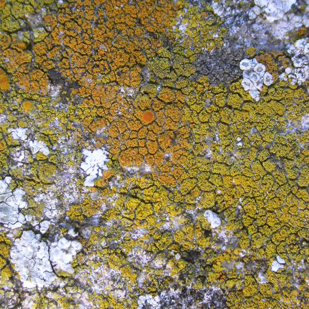
First for a couple of well known and uncontroversial lichens, both Caloplaca (despite appearances). C. flavescens and C. teicholyta. ImageImage
Caloplaca flavescens again, beside the lemon yellow Candelariella medians. Image
Caloplaca dichroa, living up to its name with orange and yellow individuals. Described as new to science in 2006 (despite being the common blastidiate Caloplaca on tops of limestone memorials across the country, and having distinctive spores). Image
What about this dark scurfy crust with minute black apothecia. I had a hunch in the field but had to examine microscopically to confirm Catillaria chalybeia (more common than generally realised on nutrient-enriched limestone). Image
A minute black perithecium (just above centre) sits at the edge of an area of bare stone, cleared of almost all lichens, algae and cyanobacteria. The clearing is caused by this strange fungus/lichen? Sarcopyrenia gibba (which has bizarre propeller shaped spores). Image
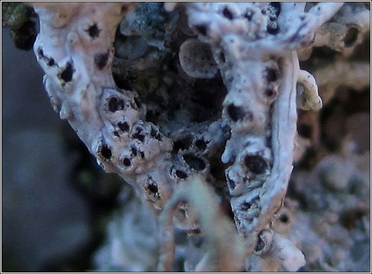
Verrucaria ochrostoma, a lichen I revived from obscurity (left half) with grey superficial thallus, and V. hochstetteri (right half) with its thallus immersed in the stone. Image
Verrucaria hochstetteri (lower half, immersed thallus + perithecia). Upper half has a similar lichen (looks like the stone has woodworm) but with a raised 'lithocortex'. This could be one of several things in the field, microscopy proved it to be Polyblastia albida. Image
Lecanora horiza (upper half) with L. campestris (heavily chewed) below. These two lichens were not distinguished by churchyard recorders until after my work with Jiri Malicek in 2013. Image
Here is one of the large, richly muriform Polyblastia albida spores. Difficult to see the septation in fresh material but a few moments heating over a tea light candle flame quickly 'cleared' the spore. ImageImage
Catillaria chalybeia in section. Some individuals lack the dark green pigment and then they may be confused with the var. chloropoliza which is a different sort of thing occurring in more specialised habitat. ImageImage
During my early microscope investigations, I noticed two features of C. chalybeia not mentioned in the books. The rounded hyphae of the hypothecium are somewhat distinctive. At the time I first saw the pale patches on the photobiont cells, I had no idea they were haustoria. ImageImage
The Lecanora albescens has familiar but mysterious 'Ribena' spots. For the first time ever I found a possible culprit as I will illustrate in the next tweet. Image
First the pigmentation in section, colourful, slightly dulls but mainly unchanged in K. ImageImage
After staining I found strange clusters of minute cells (little over 1 micron) probably arising from hyphae ascending from the lichen tissue. Have I seen the first glimpse of whatever it is that causes purple spots on Lecanora albescens? ImageImage
A distinctive basal zone is much more a feature of sandstone gravestones but the base of this limestone one has Caloplaca citrina, Opegrapha ochrocheila and Verrucaria elaeina, not found anywhere else (this zone often neglected during churchyard surveys). Image
There is one taxon on the stone that I think is still much misunderstood (Verrucaria nigrescens f. tectorum). We seem to lump at least two separate entities under this name.
I broke the thread so click on this to continue.
Well actually there is another taxon on the gravestone that seems to contain unrecognised complexity:

• • •

Missing some Tweet in this thread? You can try to force a refresh
 

Keep Current with Mark Powell

Mark Powell Profile picture

Stay in touch and get notified when new unrolls are available from this author!

Read all threads

This Thread may be Removed Anytime!

PDF

Twitter may remove this content at anytime! Save it as PDF for later use!

Try unrolling a thread yourself!

how to unroll video
  1. Follow @ThreadReaderApp to mention us!

  2. From a Twitter thread mention us with a keyword "unroll"
@threadreaderapp unroll

Practice here first or read more on our help page!

More from @obfuscans3

Feb 1
I have been sent a very interesting query from a correspondent who found this lichen on a fragment of ironstone lying on the ground. This thread will illustrate the value of recording what you observe, even if you can't name the lichen. Image
My correspondent assigned a provisional genus - Lecania. I think it belongs in a different genus but it is useful to make an initial hypothesis and then to keep an open mind. Image
Perhaps my correspondent was led astray by a misinterpretation of the spores as being septate. Spores in fresh material are difficult to interpret without 'clearing' and while iodine is a useful stain (used here), it is not harsh enough on its own to clear spores. Image
Read 14 tweets
Jan 31
Today's gravestones, a headstone and a footstone, both limestone, Eliz. Packwood, 1917. A couple of dozen species, one of which a lichenicolous fungus.
Thread: Image
Plus Verrucaria polysticta on the footstone. 10/24 are members of the Verrucariaceae. C. dichroa described as new in 2006. V. obfuscans added to British list in 2015. Image
Three lichens, all common on the headstone, none of which would have been named correctly (or at all) twenty years ago. Caloplaca dichroa and Verrucaria ochrostoma on left side. But what about the ashy grey expanse on the right? Image
Read 15 tweets
Jan 31
We tend to record Diploscistes muscorum when parasitic on Cladonia (as here, from dorsetnature.co.uk/pages-lichen/l…) and D. scruposus when growing directly on rock. There are supposed to be microscopic differences but my observations seem difficult to resolve with the literature.
Thread: Image
Today I came across a colony growing on moss, no Cladonia in sight, on a gravestone. The number of spores per ascus fits muscorum while the septation of the spores fits scruposus! This not the first time I have noticed this problem. Image
The asci are consistently 4-spored which is supposed to be a D. muscorum trait. ImageImage
Read 5 tweets
Jan 31
Catillaria nigroclavata is familiar to me these days on twigs and branches in my region. Will I learn any more about it if I attempt to make some drawings of it? Image
At first sighting, C. nigroclavata (in lower part of image) is very similar to Amandinea punctata (above). With careful study differences in appearance of both thallus and apothecia become apparent. Image
The thallus of A. punctata consists of minute grey warts, its apothecia become somewhat convex with maturity. C. nigroclavata has a thin, scurfy, dark, dull green thallus; the apothecia have narrow margins and discs remain flatter with age. Image
Read 7 tweets
Jan 31
When sectioning fruiting bodies which are immersed in stone, the fine edge of the blade is soon blunted. How do I cut numerous sections of saxicolous material while minimising waste of razor blades?
Thread
There are two basic types, single edged above which are safer and more robust but without the ultra-fine edge that I prefer in the double edged type below.
It is best to snap (or cut) the double edged type in half lengthwise (careful) before use. The disadvantages of this type are rounded ends of the blade (makes picking up sections more difficult) and the fine edge is very easily ruined by slightest touch against hard substratum.
Read 7 tweets
Jan 30
A thread with a few grey 'parmelioids' from my walk today.
Punctelia subrudecta (left), Parmelia sulcata (right) Image
Hypotrachyna revoluta (left) with erect lobes, the apices covered in fine soredia.
Parmelia sulcata (right) with angular lobes and white tipped ridges. Image
Parmotrema perlatum, may be confused with H. revoluta (last tweet) at first glance (both have fine soredia on apices of erect lobes) but with a little experience they are quite easily distinguished. Image
Read 6 tweets

Did Thread Reader help you today?

Support us! We are indie developers!


This site is made by just two indie developers on a laptop doing marketing, support and development! Read more about the story.

Become a Premium Member ($3/month or $30/year) and get exclusive features!

Become Premium

Don't want to be a Premium member but still want to support us?

Make a small donation by buying us coffee ($5) or help with server cost ($10)

Donate via Paypal

Or Donate anonymously using crypto!

Ethereum

0xfe58350B80634f60Fa6Dc149a72b4DFbc17D341E copy

Bitcoin

3ATGMxNzCUFzxpMCHL5sWSt4DVtS8UqXpi copy

Thank you for your support!

:(