Trichophyton rubrum - Classification, History, Habitat, Morphology, Culture, Identification
Introduction to Trichophyton rubrum
Trichophyton rubrum is an anthropophilic saprotroph, an exclusively clonal, dimorphic fungus that colonizes the upper layers of dead skin. Worldwide, it is the most common cause of athlete's foot, fungal infection of nails, jock itch, and ringworm. It is characterized by the presence of both smooth-walled macro- and microconidia.
Trichophyton rubrum is penicillin produces, both in vitro and in vivo but does not produce urease.
Classification of Trichophyton rubrum
Trichophyton rubrum is classified as follows:
Kingdom: Fungi
Division: Ascomycota
Class: Eurotiomycetes
Order: Onygenales
Family: Arthrodermataceae
Genus: Trichophyton
Species: T. rubrum
History of Trichophyton rubrum
In 1845, Trichophyton rubrum was first described by Malmsten. Sabour in 1911 named the fungi Trichophyton rubrum.
Habitat of Trichophyton rubrum
Trichophyton rubrum has two distinct strains on the basis of biogeographical subpopulations. The Trichophyton rubrum which clinically manifests as tinea corporis and tinea capitis is restricted to the parts of Africa and southern Asia. In contrast, Trichophyton rubrum that clinically manifests as tinea pedis and tinea unguium are globally distributed.
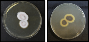
Trichophyton rubrum frontside (left) and reverse side (right) on SDA (Source: ResearchGate)
Morphology of Trichophyton rubrum
The typical colony of Trichophyton rubrum is white and cottony on the surface while the underside of the colony is red, yellowish, or brownish. In culture, it grows slowly with sparse production of teardrop or peg-shaped microconidia laterally on fertile hyphae.
Macroconidia, if produced by some isolates, are narrowly club-shaped, thin, and smooth-walled with 3 to 8 septa.
In the presence of compounds such as nitrogen, sulfur, and phosphorous, Trichophyton rubrum growth is inhibited.
Culture of Trichophyton rubrum
Like most fungi, Trichophyton rubrum can be cultured in a laboratory.
It grows on Sabouraud’s Dextrose Agar (SDA) incorporated with chloramphenicol and cycloheximide and incubated at 25 to 30°C aerobically for 1 to 3 weeks.
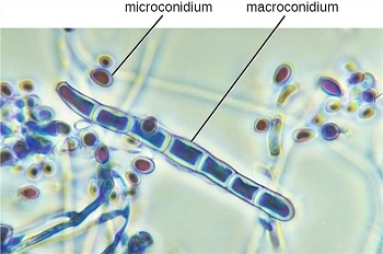
Trichophyton rubrum microconidia, macroconidia (Source: Creative biolabs)
Identification of Trichophyton rubrum
The fungi Trichophyton rubrum is identified on the basis of colony morphology, color, pigment production, and presence/absence of microconidia or macroconidia.
Fresh culture extracted from the agar plate using cellophane tape and placed on a slide containing a drop of Lacto Phenol Cotton Blue (LPCB). The preparation is viewed under a microscope to demonstrate the presence/absence of microconidia or macroconidia.
Trichophyton rubrum colonies are cottony which later turns velvety with red pigment on the reverse side. Numerous microconidia are numerous, teardrop-shaped, and present along the side of the hyphae. Microconidia are usually absent but if present are few in number, smooth, thin-walled, and pencil-shaped.
Bromocresol purple (BCP) milk solid glucose agar test can also be used to identify Trichophyton rubrum. In this test, until 7 to 10 days after inoculation, the medium containing Trichophyton rubrum remains sky blue. This is due to Trichophyton rubrum producing less amount of ammonium ion which maintains neutral pH.
 milk solid glucose agar test.jpg)
Bromocresol purple (BCP) milk solid glucose agar test of Trichophyton rubrum (Source: Wikimedia Commons)