
Manuscript accepted on :12-10-2023
Published online on: 23-11-2023
Plagiarism Check: Yes
Reviewed by: Dr. Audrey Jacob and Dr. Preeti Yaduvanshi Parmar
Second Review by: Dr. Randa Salah Gomez
Final Approval by: Dr. Patorn Piromchai
Putu Indah Budiapsari1* , Ida Bagus Bramantya Dwinanta Kartha2, Putu Ayu Jaya Switi2, Komang Trinia Amanda Riadi2, Evellyne Natania2 and Anak Agung Raka Dyah Nindyaswari2
1Department of Parasitology Faculty of Medicine and Health Sciences, Warmadewa University, Denpasar, Indonesia.
2Faculty of Medicine and Health Sciences, Warmadewa University, Denpasar, Indonesia.
Corresponding Author E-mail:putuindah51@yahoo.com
DOI : https://dx.doi.org/10.13005/bpj/2799
Abstract
Background: Various types of nutritious plants owned by Indonesia can be used as traditional medicine by the community because they are considered more economical and cause fewer side effects than chemical drugs. For example, the dadap plant (Erythrina subumbrans) and the cumin plant (Plectranthus amboinicus) which have potential as anti-inflammatory drugs. Dadap leaves contain active ingredients, such as alkaloids, flavonoids, and tannins. Meanwhile, cumin leaves contain saponins, flavonoids, polyvenols, and essential oils. Flavonoids work by inhibiting the action of COX-2 which causes inflammation. Objective: This study aimed to determine the effectiveness of the extract gel of dadap leaves and cumin leaves as anti-inflammatory in male Balb/c mice. Methods: The research was carried out through the stages of making extracts, making gel formulas, and induction of inflammation in male mice by use caragens. The data analysis method used was the ANOVA test which was obtained by observing the tumor, calor, rubor, and dolor of male mice. Conclusion: Dadap leaves and cumin leaves can be used as anti-inflammatory gels on the extremities. The three gel formulations of dadap leaves and cumin leaves can relieve inflammation. Results: From the test on mice, the most effective gel formulation was the formulation of 7.5% dadap leaf extract and 17.5% cumin leaf extract.
Keywords
Cumin; Dadap; Flavonoids; Gel; Inflammation
Download this article as:| Copy the following to cite this article: Budiapsari P. I, Kartha I. B. B. D, Switi P. A. J, Riadi K. T. A, Natania E, Nindyaswari A. A. R. D. Effect of Gel from Erythrina Subumbrans And Plectranthus Amboinicus Leaf Extracts as Anti-Inflammatory Agent in Male Balb/c Mice. Biomed Pharmacol J 2023;16(4). |
| Copy the following to cite this URL: Budiapsari P. I, Kartha I. B. B. D, Switi P. A. J, Riadi K. T. A, Natania E, Nindyaswari A. A. R. D. Effect of Gel from Erythrina Subumbrans And Plectranthus Amboinicus Leaf Extracts as Anti-Inflammatory Agent in Male Balb/c Mice. Biomed Pharmacol J 2023;16(4). Available from: https://bit.ly/3SUp79v |
Introduction
Indonesia has various types of plants that are efficacious to be used as traditional medicines. Indonesian people are known for their traditional medicine from the past until now.1 Traditional medicine is more in demand by the people of Indonesia because it is more economical, easy to obtain, and causes fewer side effects than chemical drugs. For example, the dadap plant (Erythrina subumbrans) and the cumin plant (Plectranthus amboinicus). Plants as traditional medicines are usually used by pounding and then affixing them to places that are sick.2
Inflammation is the body’s response to infection and tissue damage caused by trauma or infection.3 This response aims to eliminate the cause of tissue damage.4 Inflammation could be divided by acute and chronic inflammation.5 Acute inflammation occurs within minutes to days and is characterized by increased blood flow and edema.6 While chronic inflammation occurs in the long term that triggers an increase in cell type turnover.7 Inflammation begins with an increase in capillary permeability, then continues with the recruitment of leukocytes in the injured area for phagocytosis that causes tissue damage, and ends with termination.8 Inflammation is characterized by calor (heat), dolor (pain), tumor (swelling), and rubor (redness).9 Rubor and calor occur simultaneously, where rubor is caused by dilatation of the arteries that supply blood to the injured area, resulting in increased blood flow.10 Then, the presence of a tumor is caused by an increase in capillary permeability so that plasma leaves the blood vessels.11 The release of chemicals or inflammatory mediators, such as histamine, prostaglandins, and serotonin can cause pain.12 Dadap leaves are one of the plants that contain active ingredients, such as alkaloids, flavonoids, and tannins.13 While cumin leaves contain saponins, flavonoids, polyphenols, and essential oils.14 Both plants have the potential as anti-inflammatory drugs. Several countries in Southeast Asia use the stems and leaves of dadap as a medicine for infections and joint pain.15 From this description, dadap leaves and cumin leaves have the potential as a traditional alternative in anti-inflammatory in the extremities which are formulated in the form of a gel.16 This research is expected to be able to provide scientific information related to the effectiveness of dadap leaves and cumin leaves as anti-inflammatories in further research activities and provide benefits to the community to overcome inflammation in the extremities.17 In addition, increasing the use value of natural resources in Indonesia.18 The purpose of this study was to determine the effectiveness of the extracted gel of dadap leaves and cumin leaves as anti-inflammatory.
Material And Methods
The research activity was carried out at the Biomedical Laboratory and the Animal Care Laboratory, Faculty of Medicine, Warmadewa University for 2 months starting in July 2021. Research was conducted by using several tools and materials. The tools used are a chopper, filter, oven, analytical balance, beaker, glass jar, evaporator, water bath, hot plate, glass stirrer, stone pulverizer, plethismometer, measuring cup, and thermometer.19 Meanwhile, the ingredients used were dadap leaves, cumin leaves, 90% ethanol, aluminum foil, filter paper, dadap leaf extract, cumin leaf extract, Na-CMC, glycerin, propylene glycol, aqua dest, 20 male mice, dadap leaf extract gel. and cumin, syringes, alcohol swabs, 0.9% NaCl, and carrageenan.20
Research Procedure
This experimental study used mice as animal model that modified to imitate the inflammation sign. The method used was collected by literature study, such as journals, books, and scientific articles. Primary data was obtained from the results of trials and observations of male mice species Mus musculus.21 The first step of study was making extracts and making gel formulas from extracts of dadap leaves and cumin leaves. Furthermore, the experiment was conducted on 20 male mice. The test animals were divided into 4 groups and each group consisted of 5 male mice. The groups were given different treatments, namely the negative control group, treatment group 1, treatment group 2, and treatment group 3.22 The data analysis method used was the ANOVA test which was obtained by observing the tumor and heat of male mice.22 Furthermore, data analysis was carried out using the SPSS 27 program to determine the effectiveness of gel from dadap leaves and cumin leaves as an anti-inflammatory in the extremities.
Identification/Authentication of plants
Erythrina Subumbrans characterized by medium-sized tree, reaching 15–20 m high and 50–60 cm tall. The young, smooth bark has green, gray, light brown or whitish vertical stripes; Stems usually with small (1–2 mm) black sticky spines. The canopy is like an umbrella or loosely rounded, dropping leaves in the dry season. The compound leaves have three leaves, green to light green, leaf axils with 10–40 cm long stalks. Child leaves round egg upside down, triangular, to a rhombus shape with a blunt tip; the largest leaflets in size, 9–25 × 10–30 cm. The flowers are arranged in conical bunches, beside or at the ends of bare branches, usually appearing when the leaves fall, attracting many birds to pollinate them. The crown is orange-red to dark red; flag 5.5–8 × 8 cm, with short nails, no white stripe.[3] Thick, dark pods, narrow between the seeds, 15–20 cm × 1.5–2 cm, containing 5–10 ovoid, brown, red, or purple glossy seeds.1,2
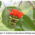 |
Figure 1: Erythrina Subumbrans (Dadap leaves). |
Plectranthus Amboinicus (Cumin leaf) is an annual herb with an often woody, ascending base and reaching 1 m in height. The trunk is jointed, and what touches the ground will come out of it. The leaves are single, fleshy, oval in shape, the tip and base are pointed with jagged/ringgitated edges, except at the base. The leaf bones are pinnate, and the branches form a net-like shape. The surface is thick hairy, like white velvet with a length of 5-7 cm, and a width of 4-6 cm and the color is light green, smells good when squeezed. The inflorescence is compound in the form of bunches with a length of 20 cm, coming out of the ends of the branches, and the axils of the leaves with a purplish-blue color. The seeds are hard, flattened, and light brown in color.15
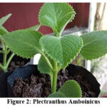 |
Figure 2: Plectranthus Amboinicus |
Preparation of Erythrina Subumbrans and Cumin Extract
Extracts were made by the maceration method. The first step is to clean the dadap and cumin leaves using clean water and dry them in the oven for 3 days. Then, each leaf is crushed using a chopper.17 The crushed leaves will be sieved first. After being sifted, the dadap leaves and cumin leaves were put into different beakers and then ethanol was added up to 1 knuckle above the leaves. The leaves were placed in a glass jar covered with aluminium foil and allowed to stand for 3 days. After 3 days, it was filtered using filter paper to obtain the filtrate. The filtrate was evaporated and thickened using an evaporator and a water bath at a temperature of 70°C.22
Preparation of Dadap and Cumin Leaf Extract Gel
Starting with weighing the ingredients for making the gel according to the formula based on the standard gel based on Sodium Carboxymethyl Cellulose (Na-CMC), composed with Na-CMC 5%, glycerine 10%, propylene glycol 5%, 100% aqua dest. Gel formulation 1 was consist of Dadap leaf extracts 12.5% and cumin leaf 12.5%, Dadap leaf extract 0.3125 g, Cumin leaf extract 0.3125 g. Gel formulation 2 was consist of Dadap leaf extracts 17.5% and cumin leaf 7.5%, Dadap leaf extract 0.4375 g, Cumin leaf extract 0.1875 g. Gel formulation 3 was consist of Dadap leaf extract 7.5% and cumin leaf 17.5%, Dadap leaf extract 0.1875 g, Cumin leaf extract 0.4375 g. Each formula was dissolved with distilled water, then heated at a temperature of 50°C. After that, add 0.125 grams of Na-CMC, 0.25 grams of glycerine, and 0.125 grams of propylene glycol, then stir regularly until a gel is formed.1
Gel Test Method on Mice
It was carried out using 20 healthy male (Mus musculus) mice that had been adapted to their environment for 1-2 weeks. After that, one leg of each mouse was injected intraplantar using 1% carrageenan which had been homogenized with 0.9% NaCl. Then the 20 male mice were divided into 4 groups and each group consisted of 5 male mice. Once every 30 minutes for 2 hours, each group of mice was given a different treatment, namely: Negative control group: the feet of mice were not treated.5 Treatment group 1: mice’s feet were treated with gel formula 1. Treatment group 2: mice’s feet were treated with gel formula 2. Treatment group 3: mice’s feet were treated with gel formula 3. Measurements of leg volume, and feet temperature observations in each group of mice to test the effectiveness of dadap and cumin leaf gel extracts as anti-inflammatories were carried out before and after the treatment.11 Tumor measurements were examined by the volume of swelling feet of mice by dipping the feet to the ankles into the water on a plethysmometer. The temperature of feet also examined by using a thermometer. After induction of inflammation, then each group was given treatment for second time according to the formula. 13
Results and Discussion
The results show combination of dadap and cumin in the formula I produced 2.25 grams gel, formula II produced 2.12 grams gel, and formula III produced 2.09 grams gel. The physical stability of each gel was evaluated by organoleptic analysis. Organoleptic analysis was carried out through observations in terms of clarity, colour, odour, and amount of gel extracts. The result of observation was described in table 1. We found the formula 1 was less odour compared with formula 2 and 3. The strong smell of Erythrina Subumbrans was found in formula 2, and the strong smell of cumin found in formula 3. The consistency of each formula appear to be same solid and minimal liquid.
Table 1: Result of Gel Analyses
|
Formula |
Observation |
||
|
Color |
Smell |
Form |
|
|
Formula1 |
Dark green |
Sharp, like extract |
Solid, minimal liquid |
|
Formula 2 |
Green |
Sharp, like extract, leaves, strong smell of Erithrina |
Solid, minimal liquid |
|
Formula 3 |
Black green |
Sharp, like extract, leaves, strong smell of Cumin |
Solid, minimal liquid |
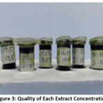 |
Figure 3: Quality of Each Extract Concentration. |
The quality of each extract was described as smell, color and concistency. There were no difference concistency of all formula, furthermore formula 2 which more dominantly composed by eritryna colored more green, than formula 3 composed dominantly by Cumin more dark. Twenty male mice aged 7-8 weeks weight 25-30 gram was used as experimented animal. Each mouse was adapted for 7 days to environmental conditions. The condition of male mice before intervention was healthy, there were no abnormalities in behavior and clinical appearance. The cage was maintained to be clean so the mice were not contaminated. At the first day inflammation induction, we measure the leg volume and temperature of feet before treated by gel formula. The result was described in table 2.
Table 2: Mean temperature and Volume of mouse leg before treatment
|
Variable |
Temperature (°C) mean |
Leg Volume (ml) mean |
|
Negative control |
36.58 |
0.14 |
|
Treatment 1 |
36.6 |
0.13 |
|
Treatment 2 |
36.61 |
0.12 |
|
Treatment 3 |
36.7 |
0.12 |
Table 3: Mean temperature and Volume of mouse leg after induction of inflammation
|
Variable |
First 30 minute temperature (°C) |
First 30 minute Volume (ml) |
After 60 minute temperature (°C) |
After 60 minute Volume (ml) |
|
Negative control |
37.3 |
0.21 |
37.5 |
0.22 |
|
Treatment 1 |
37.4 |
0.19 |
37.5 |
0.24 |
|
Treatment 2 |
37.3 |
0.23 |
37.2 |
0.23 |
|
Treatment 3 |
37.2 |
0.15 |
37.6 |
0.24 |
The mean of leg temperature before and after treatment shown to be same range, this result may be caused by two possible reason, first the dose of carangeenans must be low so could not induce massive increasing of temperature, second time observation too short, so increasing of temperature not reached optimum level.
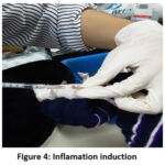 |
Figure 4: Inflamation induction |
 |
Figure 5: Gel application in mice. |
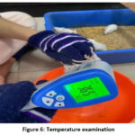 |
Figure 6: Temperature examination |
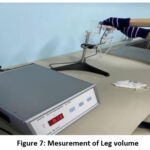 |
Figure 7: Mesurement of Leg volume |
Table 4: Mean temperature and Volume of mouse leg after gel application.
|
Variable |
After 30 minute temperature (°C) |
After 30 minute Volume (ml) |
After 60 minute temperature (°C) |
After 60 minute Volume (ml) |
After 90 minute temperature (°C) |
After 90 minute Volume (ml) |
|
Negative control |
37.2 |
0.18 |
37.1 |
0.19 |
37.3 |
0.19 |
|
Treatment 1 |
36.9 |
0.13 |
36.9 |
0.12 |
36.5 |
0.12 |
|
Treatment 2 |
37.3 |
0.13 |
37 |
0.13 |
36.8 |
0.13 |
|
Treatment 3 |
37 |
0.12 |
36.6 |
0.13 |
36.5 |
0.13 |
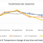 |
Figure 8: Temperature change of any time and treatment |
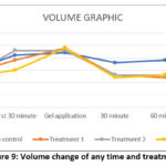 |
Figure 9: Volume change of any time and treatment. |
Table 5: The Difference of leg temperature and volume before intervention, after inflammation and after gel application.
|
Variable |
Time Observation |
Mean |
SD |
P |
95% Confidence Interval |
|
|
|
Lower |
Upper |
||||
|
Temperature |
Before Intervention |
36.6 |
0.10 |
0.01 |
36.66 |
36.58 |
|
After Inflammation |
37.3 |
0.15 |
|
37.42 |
37.34 |
|
|
After Gel application |
36.9 |
0.33 |
|
36.86 |
37.012 |
|
|
Leg Volume |
Before Intervention |
0.12 |
0.01 |
0.01 |
0.13 |
0.12 |
|
After Inflammation |
0.21 |
0.03 |
|
0.22 |
0.20 |
|
|
After Gel application |
0.13 |
0.02 |
|
0.14 |
0.13 |
|
*Significance at p<0.05, Temperature: p 0.01, Volume: p 0.01
One-way Anova test was using to analyze the mean difference of mice temperature before intervention, after inflammation induction and after gel application. The result show there was significant statistically (p<0.05). The leg volume before intervention, after inflammation induction and after gel application also significant statistically (p<0.05). Although the mean difference are not represented the fever.
Table 6: Result of Anova Effect of Temperature and Leg Volume change after Gel Application.
|
Observation After Gel Application |
Group |
N |
Mean |
SD |
95% Confidence Interval |
Min |
Max |
P |
|
|
Lower |
Upper |
|
|||||||
|
30’ Temperature |
Neg |
6 |
37,20 |
0,12 |
37,0 |
37,3 |
37,0 |
37,4 |
0.01 |
|
1 |
7 |
36,90 |
0,14 |
36,7 |
37,0 |
36,7 |
37,1 |
|
|
|
2 |
8 |
37,30 |
0,17 |
37,1 |
37,4 |
37,0 |
37,6 |
|
|
|
3 |
8 |
37,00 |
0,16 |
36,8 |
37,1 |
36,8 |
37,2 |
|
|
|
30’ Leg Volume |
Neg |
6 |
0,18 |
0,013 |
0,17 |
0,19 |
0,17 |
0,20 |
0.01 |
|
1 |
7 |
0,13 |
0,013 |
0,12 |
0,14 |
0,12 |
0,15 |
|
|
|
2 |
8 |
0,12 |
0,007 |
0,11 |
0,13 |
0,11 |
0,13 |
|
|
|
3 |
8 |
0,12 |
0,010 |
0,11 |
0,13 |
0,11 |
0,14 |
|
|
|
60’ Temperature |
Neg |
6 |
37,10 |
0,08 |
37,0 |
37,1 |
37,0 |
37,2 |
0.01 |
|
1 |
7 |
36,98 |
0,30 |
36,7 |
37,2 |
36,7 |
37,4 |
|
|
|
2 |
8 |
37,00 |
0,16 |
36,8 |
37,1 |
36,7 |
37,3 |
|
|
|
3 |
8 |
36,60 |
0,13 |
36,4 |
36,7 |
36,4 |
36,8 |
|
|
|
60’ Leg Volume |
Neg |
6 |
0,19 |
0,010 |
0,18 |
0,20 |
0,18 |
0,21 |
0.01 |
|
1 |
7 |
0,12 |
0,01 |
0,11 |
0,13 |
0,11 |
0,14 |
|
|
|
2 |
8 |
0,12 |
0,00 |
0,12 |
0,13 |
0,12 |
0,14 |
|
|
|
3 |
8 |
0,13 |
0,00 |
0,12 |
0,13 |
0,12 |
0,14 |
|
|
|
90’ Temperature |
Neg |
6 |
37,30 |
0,15 |
37,1 |
37,4 |
37,1 |
37,5 |
0.01 |
|
1 |
7 |
36,50 |
0,08 |
36,4 |
36,5 |
36,4 |
36,6 |
|
|
|
2 |
8 |
36,80 |
0,22 |
36,6 |
36,9 |
36,5 |
37,1 |
|
|
|
3 |
8 |
36,75 |
0,59 |
36,2 |
37,2 |
36,3 |
37,7 |
|
|
|
90’ Leg Volume |
Neg |
6 |
0,19 |
0,01 |
0,18 |
0,20 |
0,18 |
0,21 |
0.01 |
|
1 |
7 |
0,12 |
0,01 |
0,11 |
0,13 |
0,11 |
0,14 |
|
|
|
2 |
8 |
0,12 |
0,00 |
0,12 |
0,13 |
0,12 |
0,14 |
|
|
|
3 |
8 |
0,13 |
0,00 |
0,12 |
0,13 |
0,12 |
0,14 |
|
|
*Significance level at p< 0.05
Discussion
The result of one way Anova test show the were significance difference of mice temperature before intervention, after inflammation and after gel application. Formula 1 was slightly most effective in decreasing temperature compared with formula 2 and 3. This result accordance with previous research, Erytrina gel application had cooling down effect that work as same as ice compression after inflammation or injury. Dadap leaves and cumin leaves contain active compounds that can act as anti-inflammatories. Dadap leaves contain alkaloids, flavonoids, and tannins.4 Meanwhile, cumin leaves contain saponins, flavonoids, polyphenols, and essential oils.5 Flavonoids are a typical compound that is usually found in green plants. Flavonoid bio-actives are considered the most important phytochemicals that have broad biological benefits for humans such as anti-inflammatory, antioxidant, and antimicrobial. Flavonoids are phenolic compounds that can inhibit the work of COX-2, causing a decrease in the production of prostaglandins and inhibiting of the release of arachidonic acid. Arachidonic acid and prostaglandins are mediators of inflammation. Saponins work through interactions with membrane lipids, such as phospholipids which are precursors of prostaglandins and other inflammatory mediators.12 Flavonoids are well-known phytochemicals for their anti-inflammatory activity by inhibitingcyclooxygenase and lipoxygenase.
Short observation 30’ 60’ 90’ was conducted to evaluate fast action of gel extract that could be apllicated in real case of acute injury. The anti-inflammatory assay of Dadap leaves extract was carried out using the paw edema method by observing the test substance’s ability to prevent swelling due to carrageenan induction. Inflammation volume changes was analysed by Anova test and shown to be significantly difference with negative control group, each formula were proven to be effective as antiinflmatory agent but the most effective seems in formula 2.21
Previous research showed that cumin leaf extract contains phenols, flavonoids, alkaloids, and saponins which have been shown to be anti-convulsants in experimental animals and contain carene, terpinene, camphor, and carvacrol compounds which function as anti-rheumatic arthritis. Phytochemical analysis of cumin leaf extract contained isopropyl phenol, squalene, caryophelen, and phytol. Phytopharmacological analysis of the cumin plant has active ingredients in the form of caryophyllene, cavacrol, and forskolin which have antinephropathy and antioxidant activity.19 Another study stated that cumin leaf extract containing rosmarinic acid (CHM9102) has been shown to be anti-inflammatory and inhibits activator protein-1 (AP-1) binding which is responsible for cellular processes of inflammation, stress response, cell differentiation, and tumor formation. Several studies have found that the content of the active compounds in cumin leaf extract is quite varied and consistent with other studies, this shows the level of uniformity of the active compounds present in cumin leaves and different geographical conditions determine the composition of the compounds contained in the cumin plant. Wei et al also mentioned anti-Inflammatory Effects of Cumin Essential Oil by Blocking JNK, ERK, and NF-κB Signaling Pathways in LPS-Stimulated RAW 264.7 Cells. According to this study, other research also found at doses of 250, 500, 1000 mg / Kgbw it has an anti-inflammatory effect by significantly reducing the edema volume of 90-330 minutes (p <0.05). Phytochemical compounds of black cumin seed extract are flavonoids, tannins, saponins, triterpenes, and steroids.23
Conclusion
From the results of this study, we can conclude that dadap leaves and cumin leaves can be used as anti-inflammatory gels on the extremities. The three formulations of dadap leaf and cumin leaf gel formulations can relieve inflammation. The results of the test on mice showed that the most effective gel formulation was the formulation of 7.5% dadap leaf extract and 17.5% cumin leaf extract. Scientific conjectures for future research, namely: Finding the right level of flavonoid and alkaloid test so that it is effectively used as an anti-inflammatory in the extremities. Applying the formulation of dadap leaves and cumin leaves in other forms that have a higher probability of effectiveness than the gel form. Testing the effectiveness of the gel from extracts of dadap leaves and cumin leaves on other areas of the body other than the extremities.
Ethical Approval
This research has been approved by Ethical Comitte of Medical faculty of Udayana University by number: 2177/UN14.2.2.VII.14/LT/2021
Acknowledgement
This research activity cannot be separated from the support and contributions of various parties. The researcher expresses his deepest gratitude to Kementerian Riset and Technology, Pendidikan Tinggi, Republic of Indonesia for the support and opportunities provided in funding the 2021 PKM research activities. In addition, the researchers would like to thank all lecturers and staff of Warmadewa University as well as all parties who contributed to the project. research and preparation of this scientific article.
Conflict of Interest
There is no conflict of interest.
Funding Sources
This research is funded by Kementerian Riset and Technology, Pendidikan Tinggi, Republic of Indonesia Grant number is 1949/E2/KM.05.01/2021.
References
- Dewi, Ni Luh Kade Arman Anita Et Al. In Vivo Anti-Inflammatory Activity Of Dadap Leaves (Erythrina Subumbrans (Hassk.) Merr). International Journal Of Biosciences And Biotechnology, [S.L.], V. 9, N. 1, P. 24 – 31, Sep. 2021. Issn 2655-9994. Available At: <Https://Ojs.Unud.Ac.Id/Index.Php/Jbb/Article/View/78198>. Date Accessed: 09 May 2023. Doi: Https://Doi.Org/10.24843/Ijbb.2021.V09 .I01.P03.
CrossRef - Wardani, I. G. A. A. K., Udayani, N. N. W. ., Cahyaningsih, E., Hokor, M. D. T., & Suena, N. M. D. S. (2023). Effectiveness of Cream from Dadap Serep (Erythrina subumbrans (Hassk.) Merr.) Leaf Extract as Anti- inflammatory. Jurnal Ilmiah Medicamento, 9(1), 36–41. https://doi.org/10.36733/medicamento.v9i1.5257.
CrossRef - Ganesh S, & Vijey Aanandhi M. (2020). Macroscopic And Microscopic Diagnostic Features Of Erythrina Subumbrans (Hassk.) Merr. International Journal of Research in Pharmaceutical Sciences, 11((SPL 4), 2531–2536. Retrieved from https://ijrps.com/index.php/home/ article/view/2612.
CrossRef - Kumar, A., et al. “Erythrina variegata Linn: A review on morphology, phytochemistry, and pharmacological aspects.” Pharmacognosy Reviews, vol. 4, no. 8, July-Dec. 2010, p. 147. Gale OneFile: Health and Medicine. Accessed 9 May 2023.
CrossRef - V. R. Krishna Raju Mantena, G. Tejaswini. Anti Inflammatory Activity Of Erythrina Variegata. Int J Pharm Pharm Sci, Vol 7, Issue 4, 386-388.
- Vanny Eka Septiana1, Ratna Wijayatri 2, Imron Wahyu Hidayat. Formulation of Dadap Serep Leaf Extract Balm(Erythrina Subumbrans (Hassk.) Merr). The 14th University Research Colloqium 2021 Sekolah Tinggi Ilmu Ekonomi Muhammadiyah Cilacap
- Alemu A, Tamiru W, Nedi T, Shibeshi W. Analgesic and Anti-Inflammatory Effects of 80% Methanol Extract of Leonotis ocymifolia (Burm.f.) Iwarsson Leaves in Rodent Models. Evidence-Based Complement Altern Med. Published online 2018:1-8.
CrossRef - Rukachaisirikul T., Saekee A., Tharibun C., Watkuolham S., Suksamran A. Biological Activities of the chemical constituents of Erythrina stricta and Erythrina subumbrans. Arch. Pharm. Res. 2007;30:1398–1403.
CrossRef - Rukachaisirikul T., Innok P., Aroonrerk N., Boonamnuaylap W., Limrangsun W., Boonyon C., Woonjina U., Suksamrarn A. Antibacterial pterocarpans from Erythrina subumbrans. J. Ethnopharmacol. 2007;110:171–175.
CrossRef - Rukachaisirikul T., Innok P., Suksamrarn A. Erythrina alkaloids and a pterocarpan from the bark of Erythrina subumbrans. J. Nat. Prod. 2008;71:156–158.
CrossRef - Rukachaisirikul T., Chokchaisiri S., Suksamran A. Chemical constituents of the roots of Erythrina subumbrans. Chem. Nat. Compd. 2014;49:1127–1128.
CrossRef - Hanáková, Z., Hošek, J., Kutil, Z., Temml, V., Landa, P., Vaněk, T., Schuster, D., Dall’Acqua, S., Cvačka, J., Polanský, O., & Šmejkal, K. (2017). Anti-inflammatory Activity of Natural Geranylated Flavonoids: Cyclooxygenase and Lipoxygenase Inhibitory Properties and Proteomic Analysis. Journal of Natural Products, 80(4), 999–1006. https://doi.org/10.1021/acs.jnatprod.6b01011
CrossRef - Wang, B., Wu, L., Chen, J., Dong, L., Chen, C., Wen, Z., Hu, J., Fleming, I., & Wang, D. W. (2021). Metabolism pathways of arachidonic acids: mechanisms and potential therapeutic targets. Signal Transduction and Targeted Therapy, 6(1). https://doi.org/10.1038/s41392-020-00443-w.
CrossRef - Chiu YJ, Huang TH, Chiu CS, Lu TC, Chen YW, Peng WH, Chen CY. Analgesic and Antiinflammatory Activities of the Aqueous Extract from Plectranthus amboinicus (Lour.) Spreng. Both In Vitro and In Vivo. Evid Based Complement Alternat Med. 2012;2012:508137. doi: 10.1155/2012/508137. Epub 2011 Sep 11. PMID: 21915187; PMCID: PMC3170901.
CrossRef - Duraisamy, P., Manikandan, B., Koodalingam, A. et al. Anti-inflammatory, anti-nociceptive and anti-oxidant activities of carvacrol containing leaf extracts of edible Indian borage plant Plectranthus amboinicus: an in vivo and in vitro approach. Comp Clin Pathol 30, 397–413 (2021). https://doi.org/10.1007/s00580-021-03230-3
CrossRef - Wohn-Jenn Leu, Jui-Ching Chen and Jih-Hwa Guh. Extract from Plectranthus amboinicus Inhibit Maturation and Release of Interleukin 1β Through Inhibition of NF-κB Nuclear Translocation and NLRP3 Inflammasome Activation. Front. Pharmacol., Sec. Inflammation Pharmacology. Volume 10–2019 https://doi.org/10.3389/fphar. 2019. 00573.
CrossRef - Yung-Jia Chiu, Tai-Hung Huang, Chuan-Sung Chiu, Tsung-Chun Lu, Ya-Wen Chen, Wen -Huang Peng, and Chiu-Yuan Chen. Analgesic andAntiinflammatory Activities of the Aqueous Extract from Plectranthus amboinicus (Lour.) Spreng. Both In Vitroand In Vivo. Hindawi Publishing Corporation. Evidence-Based Complementary and Alternative Medicine. Volume 2012, Article ID 508137, 11 pages. doi:10.1155/2012/508137.
CrossRef - Ananthi T, & Ramya V. (2021). HPTLC analysis and anti-inflammatory activity of Plectranthus amboinicus (Lour.) leaves. International Journal of Research in Pharmaceutical Sciences, 12(3), 2077–2082. Retrieved from https://ijrps.com/index.php/home/article/view/196.
CrossRef - Solfaine R, Muniroh L, Sadarman, Apriza and Irawan A (2021). Anti-inflammatory effect of Coleus amboinicus leaves extract on uric acid-induced nephrotoxicity rats. Adv. Anim. Vet. Sci. 9(10): 1553-1558.
CrossRef - Yuan-Siao Chen, Hui-Ming Yu, Jiun-Jie Shie, Ting-Jen Rachel Cheng, Chung-Yi Wu, Jim-Min Fang, Chi-Huey Wong. Chemical constituents of Plectranthus amboinicus and the synthetic analogs possessing anti-inflammatory activity, Bioorganic & Medicinal Chemistry,Volume 22, Issue 5,2014,Pages 1766-1772.
CrossRef - Dewi, Ni Luh Kade Arman Anita Et Al. In Vivo Anti-Inflammatory Activity Of Dadap Leaves (Erythrina subumbrans (Hassk.) Merr). International Journal of Biosciences and Biotechnology, [S.l.], v. 9, n. 1,
p. 24 – 31, sep. 2021. ISSN 2655-9994. Available at: <https://ojs.unud. ac.id /index.php/ jbb/article/view/78198>. Date accessed: 07 aug. 2023. doi: https://doi.org/10.24843/IJBB.2021.v09.i01.p03.
CrossRef - Wardani IGAAK, Udayani NNW, Cahyaningsih E, Hokor MDT, Suena NMDS. Effectiveness of Cream from Dadap Serep (Erythrina subumbrans (Hassk.) Merr.) Leaf Extract as Anti- inflammatory. JINTO [Internet]. 2023 Mar. 30 [cited 2023 Aug. 7];9(1):36-41. Available from: https://e-journal.unmas.ac.id/index.php/ Medicamento/ article/view/ 5257
CrossRef - Wei J, Zhang X, Bi Y, Miao R, Zhang Z, Su H. Anti-Inflammatory Effects of Cumin Essential Oil by Blocking JNK, ERK, and NF-κB Signaling Pathways in LPS-Stimulated RAW 264.7 Cells. Evid Based Complement Alternat Med. 2015;2015:474509. doi: 10.1155/2015/474509.
CrossRef

