
Loimia davidi, Martin & Capa & Martínez & Costa, 2022
|
publication ID |
https://doi.org/ 10.5852/ejt.2022.833.1887 |
|
publication LSID |
lsid:zoobank.org:pub:F2C142A4-94C0-4D5E-86AD-925F23103A95 |
|
DOI |
https://doi.org/10.5281/zenodo.6958891 |
|
persistent identifier |
https://treatment.plazi.org/id/D10C6790-B176-4686-B7D3-E7235953AD99 |
|
taxon LSID |
lsid:zoobank.org:act:D10C6790-B176-4686-B7D3-E7235953AD99 |
|
treatment provided by |
Felipe |
|
scientific name |
Loimia davidi |
| status |
sp. nov. |
Loimia davidi View in CoL sp. nov.
urn:lsid:zoobank.org:act:D10C6790-B176-4686-B7D3-E7235953AD99
Figs 1–9 View Fig View Fig View Fig View Fig View Fig View Fig View Fig View Fig View Fig , Tables 1–4 View Table 1 View Table 2 View Table 3 View Table 4 , Supp. file 1
Diagnosis
Species of Loimia with two pairs of lappets on segments 1 and 3; first pair ventrolateral, with ventral margins in contact midventrally; second pair smaller, lateral. 14–15 ventral shields from segment 2, fused on segments 2 and 3; reddish-brown, with same width in first nine segments, deeply dark brown in following six segments, then progressively narrowing, giving an overall triangular appearance. Ventral shields smooth on segments 2–3 to 10 and with transverse grooves on segments 11 to 16. Uncini pectinate, arranged in a single row on segments 5–10 and in double rows on segments 11–20 (back-toback), all with a single tooth row over main fang. Thoracic uncini with three and abdominal with four teeth over the main fang (smaller specimens) or all with five teeth over main fang (larger specimen). Thoracic capillary notochaetae alimbate and unilimbate (smaller specimens) or alimbate, unilimbate and bilimbate (larger specimen). Pygidium with either sixteen small, cirriform (smaller specimens) or seven (five dorsolateral, two ventral) long conical (larger specimen) marginal papillae surrounding anus.
Etymology
The specific epithet is a homage to David Martin, the first author’s second brother, who recently cheated death and recovered from serious psychological illness, but also for his professional and personal achievements and, mainly, for being the person he is.
Material examined
Holotype PORTUGAL • 1 ♂ specimen (complete, in three fragments); Açores Archipelago , São Miguel Island , Ilhéu de São Roque – Rostro de Cão ; 37°44′37″ N, 25°38′17″ W; 8 m depth; 11 Jul. 2017; D. Martin and M. Capa leg.; anterior and posterior fragments fixed in 4% formalin/seawater solution, preserved in 70% ethanol; mid- abdominal fragment fixed and preserved in 96% ethanol; CEAB A.P. 935A. GoogleMaps
Paratypes PORTUGAL • 1 specimen (complete, in two fragments); same collection data as for holotype; fixed in 4% formalin/seawater solution, preserved in 70% ethanol; CEAB A.P. 935B GoogleMaps • 1 specimen (incomplete); same collection data as for holotype; fixed in 4% formalin/seawater solution, preserved in 70% ethanol; MNCN 16.01/19140 GoogleMaps • 9 specimens; same collection data as for holotype; fixed and preserved in 96% ethanol; CEAB A.P. 935C GoogleMaps • 4 specimens; same collection data as for holotype; fixed in 4% formalin/ seawater solution, preserved in 70% ethanol; CEAB A.P. 935D GoogleMaps • 4 specimens; same collection data as for holotype; fixed in 4% formalin/seawater solution, preserved in 70% ethanol; MNCN 16.01/19141 GoogleMaps .
Comparative material of L. gigantea (as L. ramzega )
FRANCE • 2 specs; English Channel, Brittany, Landéda Beach ; 48°36′37.7″ N 04°36′24.5″ W; intertidal; 25 Jan. 2012; preserved in 70% ethanol; ARC- Loimia -IND2 and -IND5 GoogleMaps .
Description
Holotype
Complete specimen divided in three fragments, measuring 310 mm long in vivo, with 147 segments; thorax 57 mm long, 12 mm wide when preserved. Body pale brownish in vivo, uniformly beige when preserved ( Figs 1A–C View Fig , 2A View Fig ; Supp. file 1: video S1); thorax with ill-defined segmentation dorsally; first three abdominal segments dorsally similar to thoracic ones; remaining abdominal segments with well-marked segmentation and a posterior whitish swelling linking neuropodia dorsally, more visible in posterior-most segments ( Figs 1A View Fig , 2E View Fig ). Tentacles long, pale beige in vivo, almost whitish when preserved, with a deep ciliated groove. Tentacular membrane well-defined, increasing in length dorsally, laterally hidden by first pair of lateral lappets ( Fig. 2B–C View Fig ). Eyespots absent. Upper lip conical, with rounded tip, wider than longer; pale brownish, well projecting forward in vivo ( Fig. 1C View Fig ; Supp. file 1: video S1), pale beige, not projecting over first pair of lateral lappets when preserved ( Fig. 2B, D View Fig ). Lower lip not covered by membrane joining first pair of lappets in vivo ( Fig. 1C View Fig ; Supp. file 1: video S1); small, square, covered by membrane joining first pair of lappets when preserved ( Fig. 2D View Fig ). Lateral lappets large, pale brownish to whitish in vivo ( Fig. 1A–C View Fig ; Supp. file 1: video S1), pale beige when preserved ( Fig. 2A–D View Fig ), two pairs, on segment 1 (ventrolateral) and segment 3 (lateral, oblique, with wavy edges, smaller), elephant ear-shaped; first pair laterally reaching notopodia level, ventrally joined by a poorly-developed membrane; second pair separated from base of first pair, laterally hiding segment 2, covering base of first and second branchiae, ending ventrally between first and second ventral shields ( Fig. 2B–D View Fig ). Branchiae on segments 2–4, arborescent, very long, first pair ca 1/6 longer and third pair ⅛ shorter than body width in vivo, with thick stalks and numerous dendritic branches in eight levels, dark red, showing rhythmic contractions in vivo ( Fig. 1A–C View Fig ; Supp. file 1: video S1); whitish when preserved ( Fig. 2A–D View Fig ). Nephridial papillae not seen. Ventral shields on segment 2–9 reddish brown, smooth, with same width; on segments 10–16 deeply dark brown, with transverse grooves, progressively narrowing posteriorly, giving an overall triangular appearance ( Fig. 1C View Fig ; Supp. file 1: video S1). Ventral shields fused on segments 2–3, smooth on segments 2–10 and with transverse grooves on segments 11–16, two on 11–12, 3 on 13, 4 on 14–16) ( Fig. 2A View Fig ). Notopodia from segments 4–20 (17 segments) as swollen, conspicuous lobes, all except first one pale beige to whitish in vivo ( Fig. 1A–C View Fig ; Supp. file 1: video S1), first eleven surrounded by whitish glandular patches ( Fig. 1A–B View Fig ), pale beige when preserved ( Fig. 2A View Fig ). Capillary notochaetae numerous, as long as chaetal lobes, smooth, of three types: alimbate, and uni- and bilimbate ( Fig. 3A–C View Fig ), in J-shaped arrangement. Thoracic neuropodia from segment 5 well developed, pale brownish to whitish in vivo ( Fig. 1A–B View Fig ; Supp. file 1: video S1), uniformly pale beige when preserved ( Fig. 2A–C View Fig ), with numerous uncini arranged in single rows in segments 5–10 and in double rows (back-to-back) in segments 11–20 ( Fig. 3D–E View Fig ), uncini rows ranging from 4 to 6 mm
long. Abdominal neuropodia narrow (first abdominal ca 3.6 times as narrow as last thoracic), as long as wide, projecting posteriorly, pale brownish to whitish in vivo ( Fig. 1A–B View Fig ; Supp. file 1: video S1), uniformly pale beige when preserved ( Fig. 2A View Fig ), with uncini in single rows until body end ( Fig. 3H View Fig ). Thoracic uncini measuring ca 120 µm long and 60 µm wide, pectinate, with a crest of five teeth in a single row over main fang, with a curved back three times as long as prow, and reduced heel and dorsal button, with anterior filament long, projected downwards ( Figs 3E–F View Fig , 4A View Fig ). Abdominal uncini pectinate, measuring ca 105 µm long and 55 µm wide, with a crest of five teeth in a single row over main fang ( Fig. 3I View Fig ), connected to basis of parapodia by long, hyaline ligaments ( Fig. 3H View Fig ), similar in shape to thoracic ones, with a less curved back, 2.5 times as long as prow, heel inconspicuous, and strongly reduced dorsal button ( Fig. 3I View Fig ). Regenerating posterior end, abruptly differing from previous segments, with shorter and narrower segments, dark reddish with pale beige posterior swellings linking bases of neuropodia ( Fig. 2E–F View Fig ). Pygidium with terminal anus, surrounded by eighteen small, almost cirriform terminal papillae, dorso-laterally broadly grouped in pairs (12), ventrolaterally individual (6) ( Fig. 2F View Fig ). Tube at least four times as long as body length, formed by aggregated sand grains, shell fragments and
other calcareous debris covering a thick, smooth, inner mucus layer ( Fig. 1D View Fig ), partly hidden under big boulders. Coelom filled with oocytes measuring ca 60 µm in diameter.
Paratypes
Based on paratype CEAB A.P. 936B (with variation in the other small paratypes between brackets). Body divided in two fragments, 41 mm long with 77 segments in total (other paratypes were all anterior fragments, including thorax and several abdominal segments). Thorax 19 mm (8–20 mm) long and 3.6 mm (2–5 mm) wide; with ill-defined segmentation dorsally; first six abdominal segments dorsally similar to thoracic ones, but segmentation better defined; remaining abdominal segments well marked, long, pale reddish, with a posterior whitish swelling dorsally linking neuropodia ( Fig. 5A View Fig ). Tentacles few in number, with U-shaped cross-section. Tentacular membrane well defined, poorly developed on ventral side; laterally hidden by first pair of lateral lappets ( Fig. 5D View Fig ); eyespots present in some specimens, progressively decreasing in diameter when more dorsal ( Fig. 5F View Fig ). Upper lip well projecting forward, wider than long; thicker at base, almost completely hidden ventrally by first pair of lateral lappets
( Fig. 5B, D–E View Fig ). Lower lip ¼ times as long as upper lip, swollen, with conical tip, hidden ventrally by membrane connecting first pair of lateral lappets ( Fig. 5E View Fig ). Lateral lappets large, discontinuous, two pairs, on segments 1 and 3 ( Fig. 5A–E View Fig ); first pair quadrangular, laterally reaching notopodia level, with a well-developed joining membrane; second pair separated from base of first pair, ⅔ times as large as first, laterally hiding segment 2, covering base of first branchiae, ending ventrally between first and second ventral shields. Three pairs of branched branchiae ( Fig. 5C View Fig ) whitish (preserved material), starting from segment 2; first pair ca ⅓ as long as body width, third pair ca 0.8 times as long as body width; branchiae with thick stalks, with many dendritic branches arranged in four levels. Nephridial papillae not seen. First twelve notopodia surrounded by whitish glandular patches ( Fig. 5A View Fig ); fourteen ventral shields, starting from segment 2, fused on segments 2–3, wider than long on segments 2–11; on segments 2–10 smooth, all about the same size; on segments 11–13 (11–12 in some specimens) with one transverse groove, then two transverse grooves on segment 14 and more than two on segment 15 (non-distinguishable in smallest specimen); abdomen smooth ventrally until pygidium ( Fig. 5A View Fig ). Notopodia from segment 4, extending through segment 20 as swollen, conspicuous lobes ( Fig. 5A View Fig ). Notochaetae of two types within same fascicle, alimbate and narrowly unilimbate capillaries, similar in length ( Fig. 6A–B View Fig ), in J-shaped arrangement. Thoracic neuropodia starting from segment 5, first seven ⅔ times as large as posterior ones, with uncini arranged in single rows in segments 5–10, and in double rows (back-to-back position)
in segments 11–20 ( Fig. 6C View Fig ). Abdominal neuropodia narrow (first abdominal ca 4.1 times as broad as last thoracic), half as long as wide, projecting posteriorly, with uncini in single rows until pygidium. Thoracic uncini measuring ca 60 µm long and 35 µm wide, pectinate, with a crest of three teeth in a single row over main fang, with a curved back twice as long as prow, well-marked heel and reduced dorsal button, with anterior filament long, projected downwards ( Figs 4C View Fig , 6D View Fig ). Abdominal uncini ca 46 µm long and 30 µm wide, similar in shape to thoracic ones, with a crest of four teeth in a single row over main fang ( Fig. 6E View Fig ). Pygidium with terminal, rounded anus, surrounded by seven long, conical terminal papillae with a well-defined base, forming two clearly separated groups of five dorsolateral and two ventral papillae ( Fig. 5G–H View Fig ). Tube not seen.
Remarks
Larger vs smaller specimens of L. davidi sp. nov.
The specimens of L. davidi sp. nov. show obvious morphological differences, which in other circumstances could have been considered as representing different species ( Table 2 View Table 2 ). This is the reason why we present the comparisons of both morphotypes with other species of the genus separated in the next two sections. However, most of these differences appear to be size-related to some extent, since the largest specimen always shows larger or more numerous structures, such as branchiae, capillary chaetae, and uncini. The only differences apparently non size-related are the presence of eyes and the length of terminal pygidial papillae. However, eyes are subdermal and may become hidden by the thicker tegument of the larger specimen. As for the pygidial papillae, they clearly have distinct shapes, but also are more numerous in the largest specimen and proportionally longer in the smaller ones. Nevertheless, the giant specimen was regenerating its posterior end, having the last ca 29 segments thinner and shorter than the previous ones ( Figs 1A View Fig , 2E View Fig ). Although we suggest that the shape and smaller size of its terminal pygidial papillae ( Fig. 2F View Fig ) may be related to the regenerating process, this cannot be confirmed because there is only one large specimen avilable.
Once dissected, the parapodia of the largest specimen show lateral zones with growing uncini both in the thorax ( Fig. 3D–E View Fig ) and in the abdomen ( Fig. 3H View Fig ). In thoracic parapodia, the uncini are arranged in single rows in the growing zones, even in those parapodia with double rows of normal uncini ( Fig. 3E View Fig ). Similar growing zones are not seen in the small specimens, likely because they are too small to be distinguished. The living largest specimen rhythmically contracted its branchial tips (Supp. file 1: video S1), likely increasing water renewal. Branchial contractions cannot be confirmed for our small specimens (none of them were observed in vivo), and neither have any been previously reported for other giant specimens of Loimia ( Montagu 1819; Lavesque et al. 2017). Conversely, Wilson (1928) reported contractile branchiae and clearly visible red blood pulsations through the blood vessels in the early benthic stages of Loimia from the English Channel.
It would be interesting to examine more material – especially large animals – to further assess the variation/homogeneity of all these particular morphological characters, which in most cases have been considered as key for species diagnosis.
Larger specimen of L. davidi sp. nov. vs other species of Loimia Malmgren, 1866
The larger specimen of L. davidi sp. nov. is distinguished from other congeners by a unique combination of features: (1) two pairs of lappets on segments 1 and 3, first pair almost reaching each other midventrally and second pair laterally, not joining midventrally; (2) long arborescent branchiae with up to eight levels of branches; (3) three kinds of notochaetae in thoracic segments including alimbate, unilimbate and bilimbate capillaries; and (4) thoracic and abdominal uncini with five teeth in a single longitudinal row over main fang ( Table 3 View Table 3 ; Supp. file 1: Table S3 View Table 3 ). The holotype resembles the described specimens of L. gigantea from Brittany ( France) both in size and overall morphology, but also in bearing two additional types of capillary notochaetae together with the typical smooth ones, instead of none or one in all remaining species of Loimia ( Table 3 View Table 3 ; Supp. file 1: Table S3 View Table 3 ). However, the presence of three types of capillary chaetae is only known in these very large specimens of Loimia , whereas the smaller specimens of L. davidi sp. nov. show only two types, thus casting serious doubts on the taxonomic value of this character.
The larger specimen of L. davidi sp. nov. differs from L. gigantea in having the first pair of lateral lappets more developed (second pair more developed in L. gigantea ), lappets uniformly pale brownish to whitish in vivo (first pair with a red margin and second pair entirely red in L. gigantea ). Lateral lappets may show some variability among Terebellidae , although they have traditionally been used to distinguish species (e.g., Jirkov 2020). Thus, we consider the observed differences as relevant enough to be mentioned here. These two species also differ in the absence of abdominal dark spots (present in L. gigantea ), branchiae arranged in eight levels (five in L. gigantea ), fifteen ventral shields (sixteen in L. gigantea ), and uncini ca 120 µm long with slightly marked, round heel and upper-most tooth very small, often difficult to distinguish (100 µm, well-marked, angular heel and well-defined upper tooth in L. gigantea ) ( Fig. 4A–B View Fig ). Moreover, although being of doubtful value due to its regenerating posterior end, the larger specimen of L. davidi sp. nov. has 18 terminal pygidial papillae (14 in L. gigantea ). In addition, this specimen was found subtidally, partly hidden under big boulders, and its tube is composed of sand grains and shell remains, while L. gigantea occurred intertidally and, in addition to sand and shell fragments, their tubes characteristically show macroalgal filaments attached to the emerging portion (absent in the tube of the larger specimen of L. davidi sp. nov.). Considering that we only found one large specimen of L. davidi sp. nov., we cannot confirm whether the observed differences in tube structure can be considered species-specific.
We have found two additional morphological differences between the larger specimen of L. davidi sp. nov. and L. gigantea after the re-examination of the paratypes of the latter, not mentioned in its original description ( Lavesque et al. 2017). First, the segmentation in L. gigantea is clearly defined dorsally all along the body, with all segments transversally divided by several grooves and at least the
median one entirely splitting each segment. This is particularly evident in the abdominal segments, where the main transversal groove divides each segment into two equal parts. Also, there are no traces of a swelling dorsally linking the abdominal parapodia. In contrast, the body segmentation in the larger specimen of L. davidi sp. nov. is characteristically ill-defined dorsally in the thoracic and first three abdominal segments, and well-marked with a posterior whitish swelling dorsally linking the neuropodia
in all remaining abdominal segments (including those in the regenerating region). Second, in L. gigantea , the uncini are arranged in typical back-to-back double rows ( Fig. 7A View Fig ) and irregularly distributed in the abdominal parapodia ( Fig. 7B–C View Fig ), whereas in the larger specimen of L. davidi sp. nov. the abdominal uncini are arranged in a single row ( Fig. 3H View Fig ). The lateral zones with growing uncini observed in the larger specimen of L. davidi sp. nov. ( Fig. 3D–E, H View Fig ) are also present in L. gigantea ( Fig. 7A–B View Fig ) and typically also occur in the species of Axionice ( Jirkov & Leontovich 2017) .
Smaller specimens of L. davidi sp. nov. vs other species of Loimia Malmgren, 1866
The smaller specimens of L. davidi sp. nov. are distinguished from other congeners by a unique combination of features: (1) presence of eyespots in the tentacular membrane; (2) two pairs of similar-
sized lappets on segments 1 and 3, first pair ventral and almost reaching each other midventrally and second pair lateral; (3) two kinds of notochaetae in thoracic segments including alimbate and unilimbate capillaries; and (4) thoracic and abdominal uncini with three and four teeth, respectively, in a single longitudinal row over main fang ( Table 3 View Table 3 ; Supp. file 1: Table S3 View Table 3 ).
These small specimens resemble L. medusa , as redescribed by Hutchings & Glasby (1995), in the shape and size of uncini, although the latter has a dorsal button three times longer, relative to uncinus length ( Fig. 4C, G View Fig ). However, L. davidi sp. nov. lacks visible pigmented red spots on the tentacles (present in L. medusa ), possesses alimbate and unilimbate capillary notochaetae (narrow bilimbate in L. medusa ), has fourteen ventral shields (twelve in L. medusa ) and bears seven terminal papillae, forming two groups on the pygidium (absent in L. medusa ). The specimens from the Mexican Caribbean identified and described as L. medusa by Londoño-Mesa & Carrera-Parra (2005) were considered as morphologically indistinguishable from the upper Persian Gulf neotype designated by Hutchings & Glasby (1995). However, the number of ventral shields in these Caribbean specimens varied between eleven and sixteen (twelve in Persian L. medusa ), with those from segments 2, 3 and part of 4 being almost fused and all of them being entire, not divided by transversal grooves in the Caribbean specimens ( Londoño-Mesa & Carrera-Parra 2005) and entirely fused from 2 to 4 in the Persian specimens ( Hutchings & Glasby 1995). Moreover, the uncini of the Caribbean specimens were slightly longer than those of the Persian L. medusa and of L. davidi sp. nov. ( Fig. 4C, G–H View Fig ). Considering these differences and the distance between the Caribbean Sea and the Persian Gulf, we suggest that the Caribbean specimens probably represent a different species that merits further analysis.
The smaller specimens of L. davidi sp. nov. resemble Loimia salazari Londoño-Mesa & Carrera-Parra, 2005 and Loimia minuta Treadwell, 1929 in having two types of capillary notochaetae which, in addition to the arrangement of the lateral lappets and number of uncinal teeth, clearly distinguishes these species from other congeners ( Table 3 View Table 3 ). However, L. davidi sp. nov. has equally long unilimbate and alimbate capillary notochaetae (longer narrowly bilimbate and shorter alimbate in L. salazari ), fourteen ventral shields (eighteen in L. salazari ) transversally grooved from the ninth shield (thirteenth in L. salazari ), and seven terminal pygidial papillae in two groups (fourteen small papillae in L. salazari ). Loimia salazari was described as the only Loimia bearing uncini with posterior processes in the anterior thoracic neuropodia, a feature that is instead typical of genera such as Pista Malmgren, 1866 , Lanicides Hessle, 1917 , Eupistella Chamberlin, 1919 , or Opisthopista Caullery, 1944 . Also, the shape of the uncini was originally described as pectinate ( Londoño-Mesa & Carrera-Parra 2005) and later as avicular ( Lavesque et al. 2017). Accordingly, the generic assignment of L. salazari is here considered doubtful. Loimia minuta has ten ventral shields, transversally grooved from the seventh one, and a pygidium with six long digitate terminal papillae. Moreover, the species descriptions across geographical areas markedly differed in the presence of different types of notochaetae. The type material (from Florida) had asymmetrically bilimbate capillaries with two different lengths ( Londoño-Mesa 2009), whereas the Mexican Caribbean specimens had long, thick bilimbate and short, thin, pointed alimbate capillaries ( Londoño-Mesa & Carrera-Parra 2005). Whether this is connected with the size of the respective specimens is not discussed by Londoño-Mesa & Carrera-Parra (2005) and, thus, neither herein. Finally, the uncini of L. davidi sp. nov. are overall similar in shape and size to those L. minuta , from both Florida and Mexican Caribbean populations ( Fig. 4C–F View Fig ).
Distribution
Sandy patches among boulders and seaweeds, in shallow subtidal waters of São Miguel Island, Açores, Portugal (Atlantic Ocean). It represents the first report of the genus Loimia in the Açores Archipelago.
Molecular analyses
The alignment of 31 sequences of cox1 of Loimia and the outgroup is 402 bp long and includes 138 parsimony informative sites ( Fig. 8 View Fig ). The three sequences from the Açores nest in a well-supported clade, internally separated by short branches ( Fig. 8 View Fig , Table 4 View Table 4 ). Indeed, the genetic divergences within this clade are 1.1% (between the small specimens) and 2.1‒2.2% (between large and small specimens). In contrast, they diverge by 16.4–36.1% from the other species of Loimia included in the analyses ( Table 4 View Table 4 ).
Sister group relationships between the Azorean clade and the rest of the sequences from other congeners are poorly supported, but there are some indications (although bootstrap values are around 60) that the sequences from a specimen identified as L. medusa from an unknown locality ( Siddall et al. 2001), and the Chinese specimens of L. arborea Moore, 1903 and L. bandera Hutchings, 1990 seem to be closely related ( Fig. 8 View Fig ).
PTP analyses group the 31 sequences of Loimia in eleven clusters, with those from the small and large morphotypes of our new species lumping in a single entity. The other congeners are delimited as outlined into the main clades after ML analyses. The mPTM model lumps all sequences in the large clade branching off at the base of the tree (including the largest and small specimens of our new species together with specimens identified as L. medusa 2 from an unknown locality, L. arborea 1 from the Yellow Sea and L. bandera from Taiwan Strait) in a single entity, a result that we interpret as an underestimation of the actual diversity of the group, given the sequence divergence (> 17%) and their disjoint geographic origins.
The identity of some of the cox1 sequences of Loimia available at GenBank needs a taxonomic revision. For instance, the sequence assigned to Loimia ingens ( Grube, 1878) from Phuket, Thai Andaman Sea ( Colgan et al. 2001) always nests with the unidentified sequences of Loimia sp. 1 from the eastern (Goa, Arabian Sea) and western (Pondycherry, Bay of Bengal) coasts of India. They show an intraspecific genetic variability of 0.3% ( Table 4 View Table 4 ), suggesting they belong to the same species. Conversely, the South China Sea sequences also assigned to L. ingens are in an independent clade to those from Thailand. Members of this clade are geographically closer to Bohol, Philippines (the type locality of the species),
but given the levels of variability we are here reporting, their identity would be reasonably confirmed only after being able to include sequences of Bohol specimens in the analysis. Moreover, L. ingens , also recorded along the Australian coasts, exhibits considerable morphological variation, being thus regarded as a species complex ( Hutchings & Glasby 1988). Similarly, L. arborea , originally described from Suruga Bay (Pacific coast of Japan) ( Moore 1903), shows the British Columbia ( Carr et al. 2011) and the Yellow Sea ( Wang et al. 2020) sequences attributed to this species as being unrelated in our tree ( Fig. 8 View Fig ). The same occurs with two non-related specimens identified as L. medusa ( Siddall et al. 2001) ( Fig. 8 View Fig ).
Morphological analyses
We have successfully reconstructed the morphospace for 28species of Loimia based on twelve traditionally used taxonomical characters ( Fig. 9 View Fig ). We observe a polarization of the morphospace space according to the body size, although this polarization is considerably reduced if body size is not included in the hypervolume calculation (Supp. file 1: Fig. S1 View Fig ). The descriptive metrics of richness and dispersion are smaller in the hypervolume of the larger morphotypes, somehow indicating less morphological disparity amongst those than amongst the smaller ones, while the evenness of the large species morphospace was instead larger than that of the small ones, indicating a more homogenous species distribution within the morphospace in the former. Yet, our analysis relies only on the available published data, which does not account for all intraspecific variability. Both hypervolumes show a considerable overlapping, with a value of total beta diversity of 0.92.
The Euclidian distance in the morphospace between the larger and smaller specimens of our new species is 0.285, well within the range of variation observed across other pairs of species of Loimia (i.e., 0.02–0.63). These differences are not only affected by body size but probably also by the presence of comparatively rare characters in the small individuals of our new species, such as the presence of eyespots.
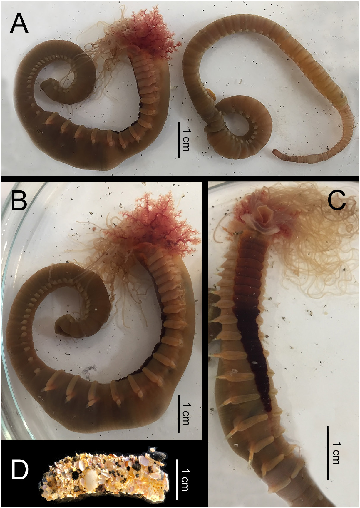
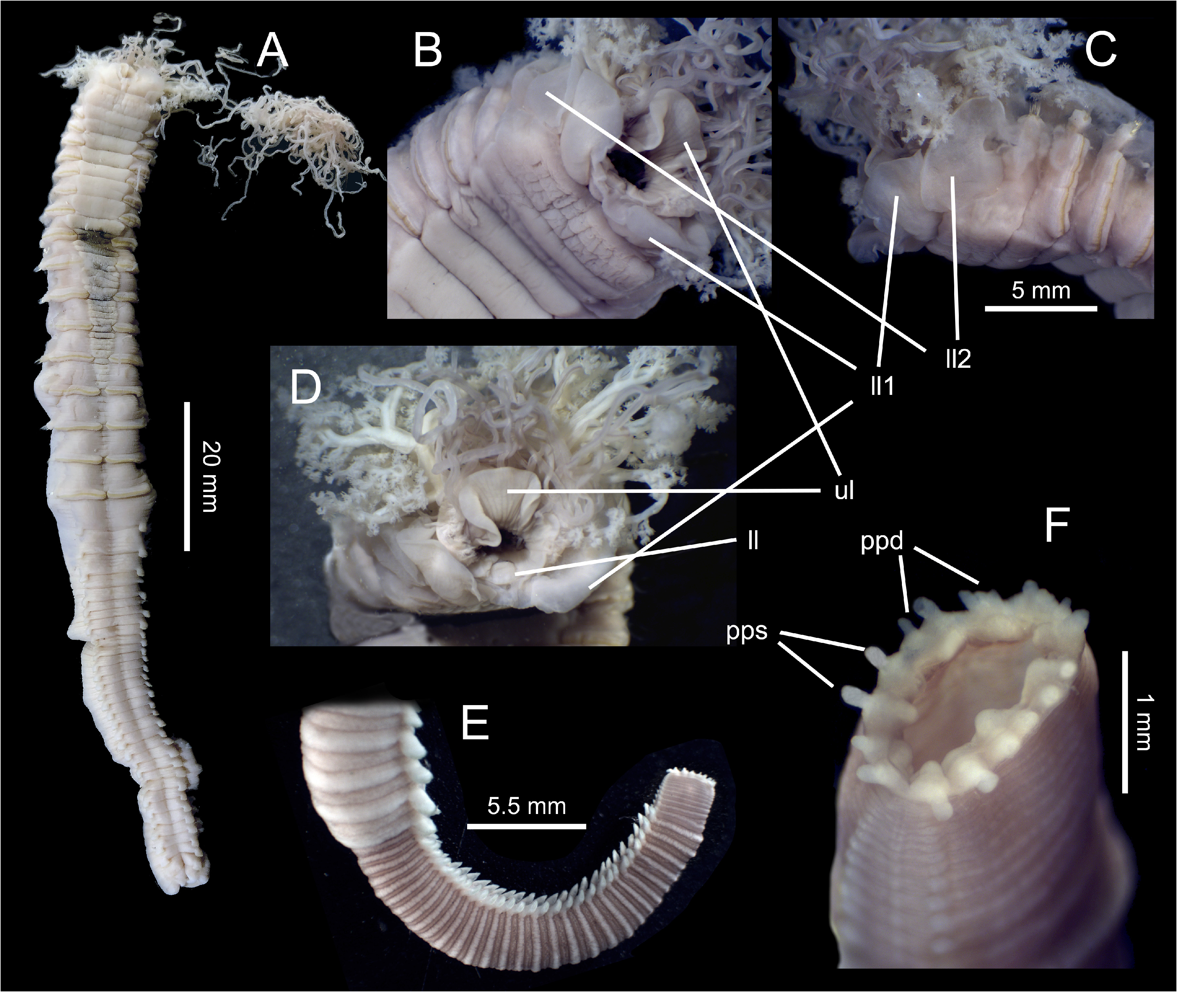
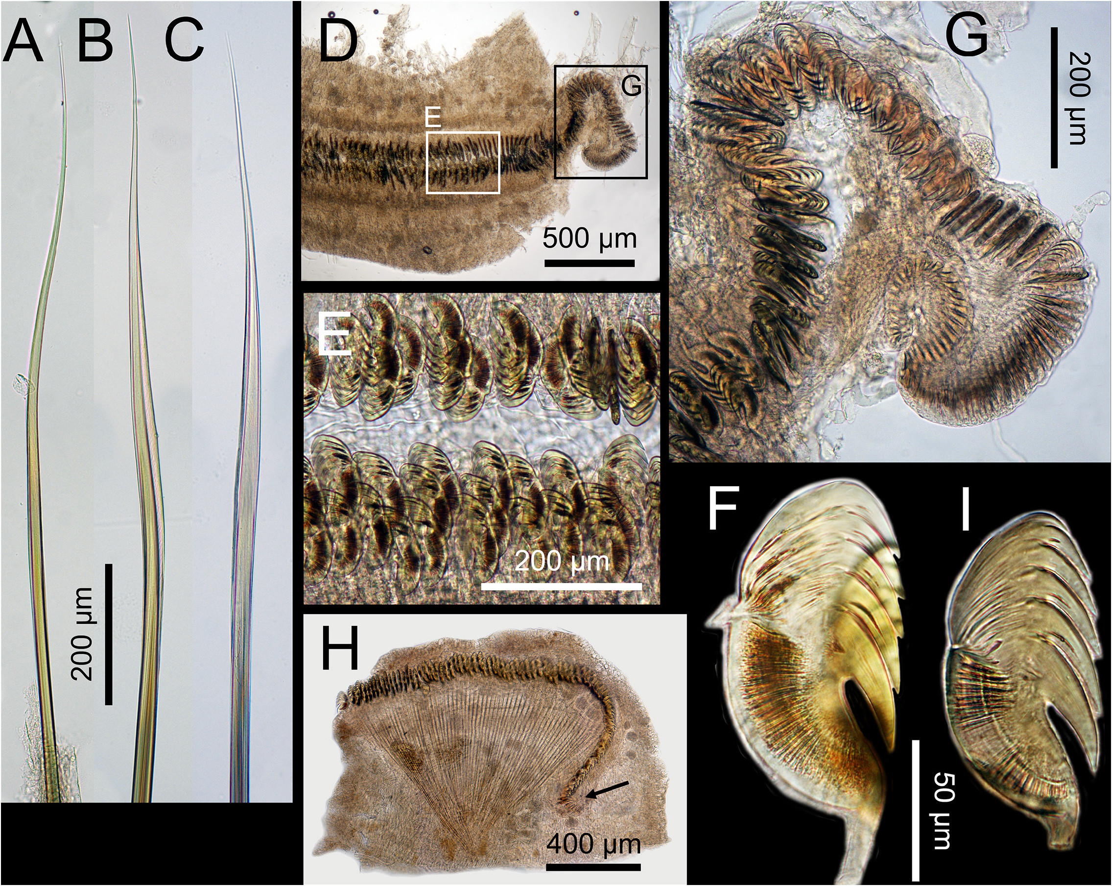
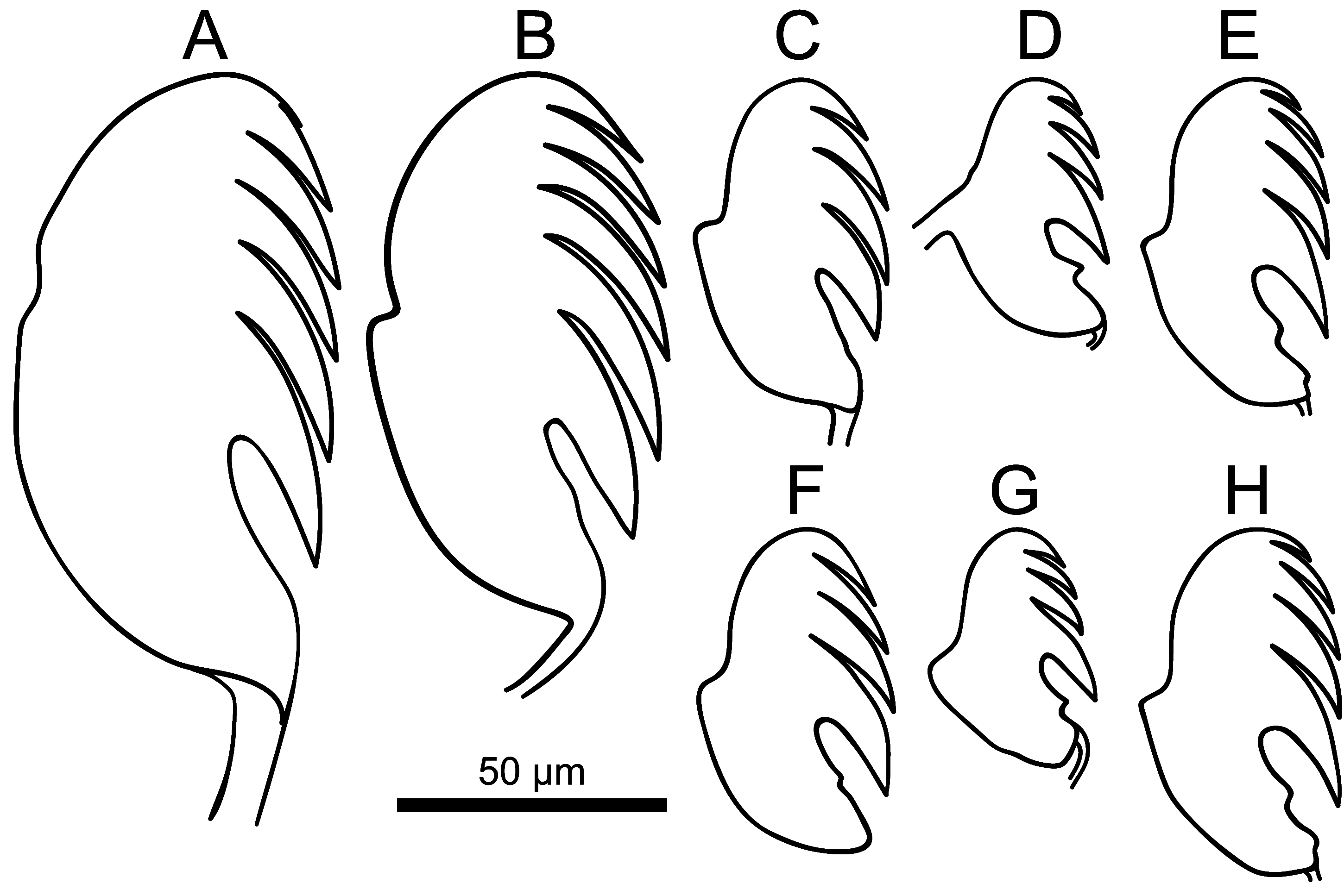
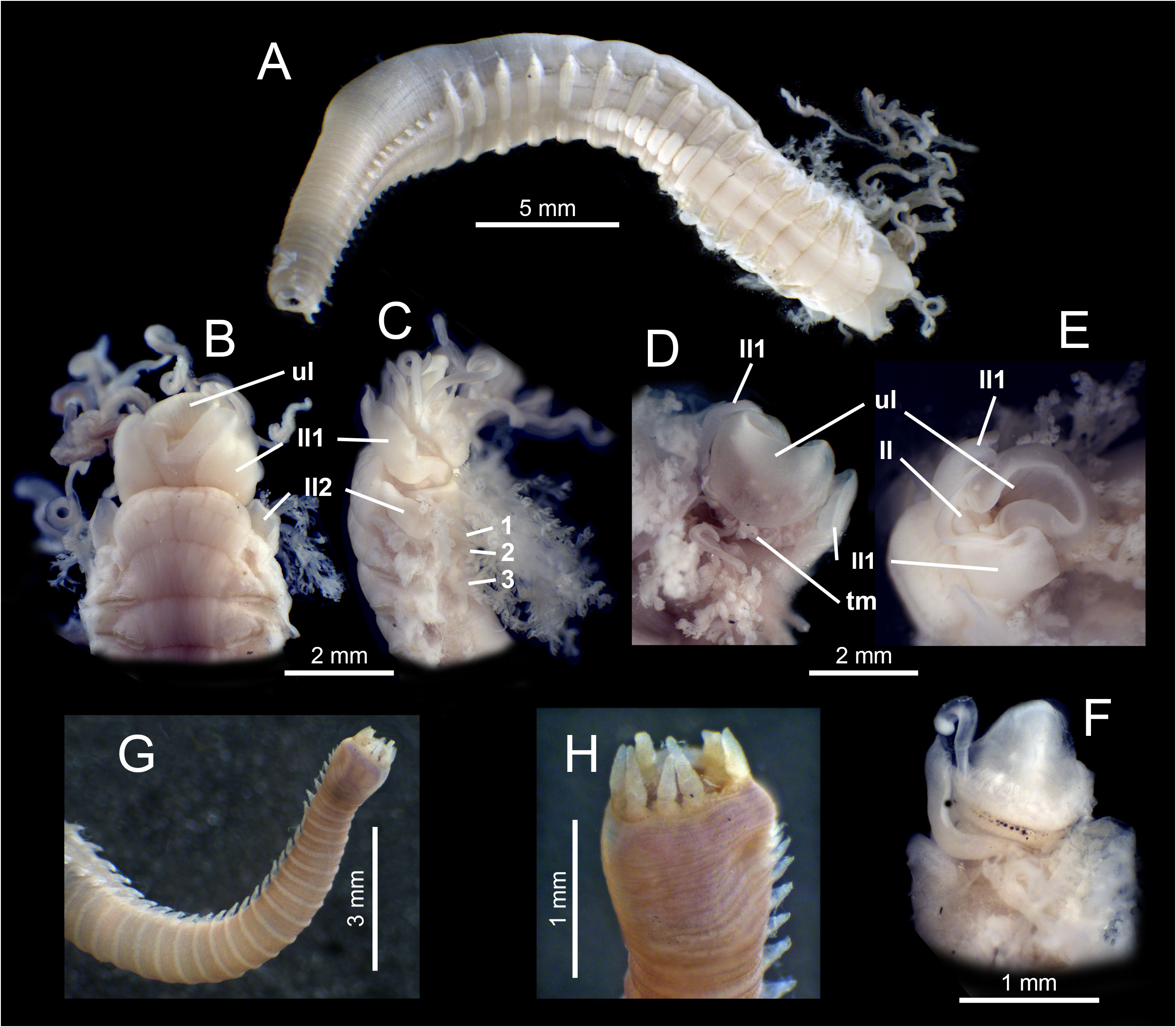
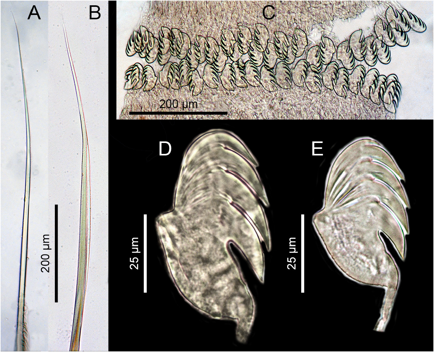
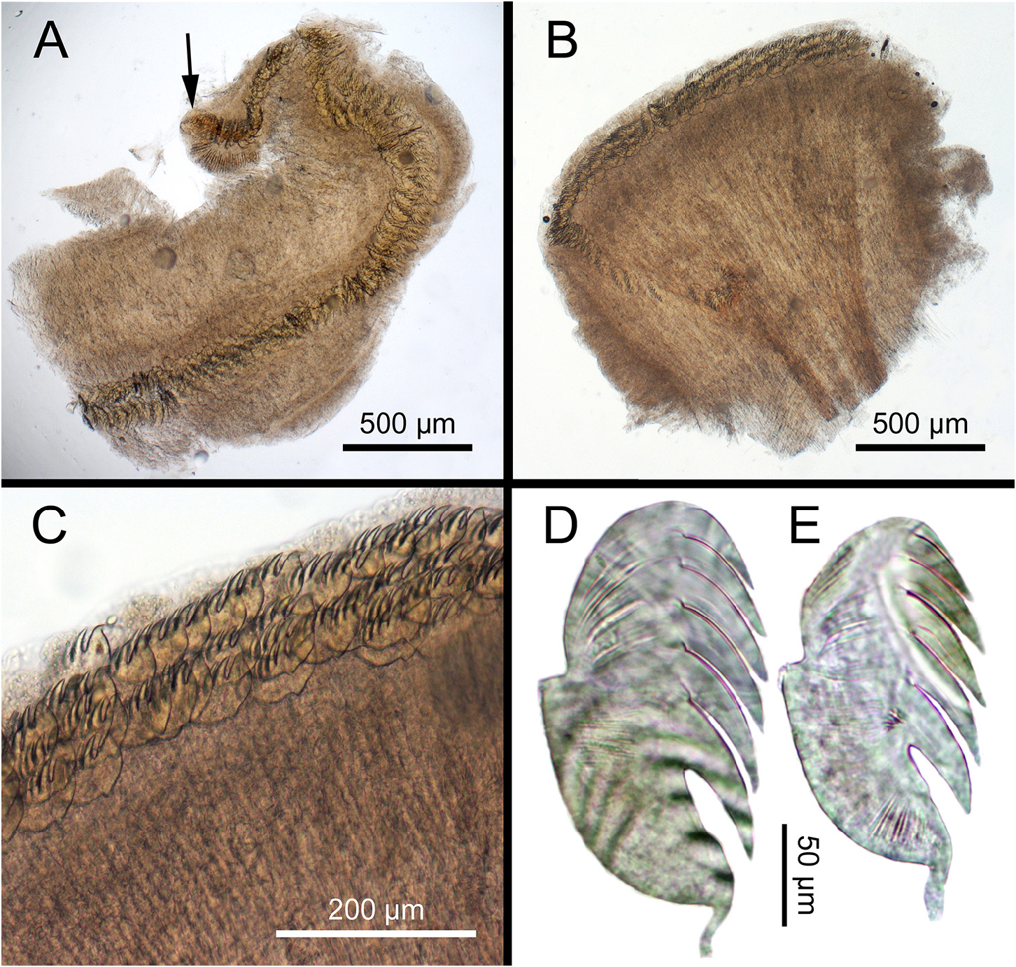
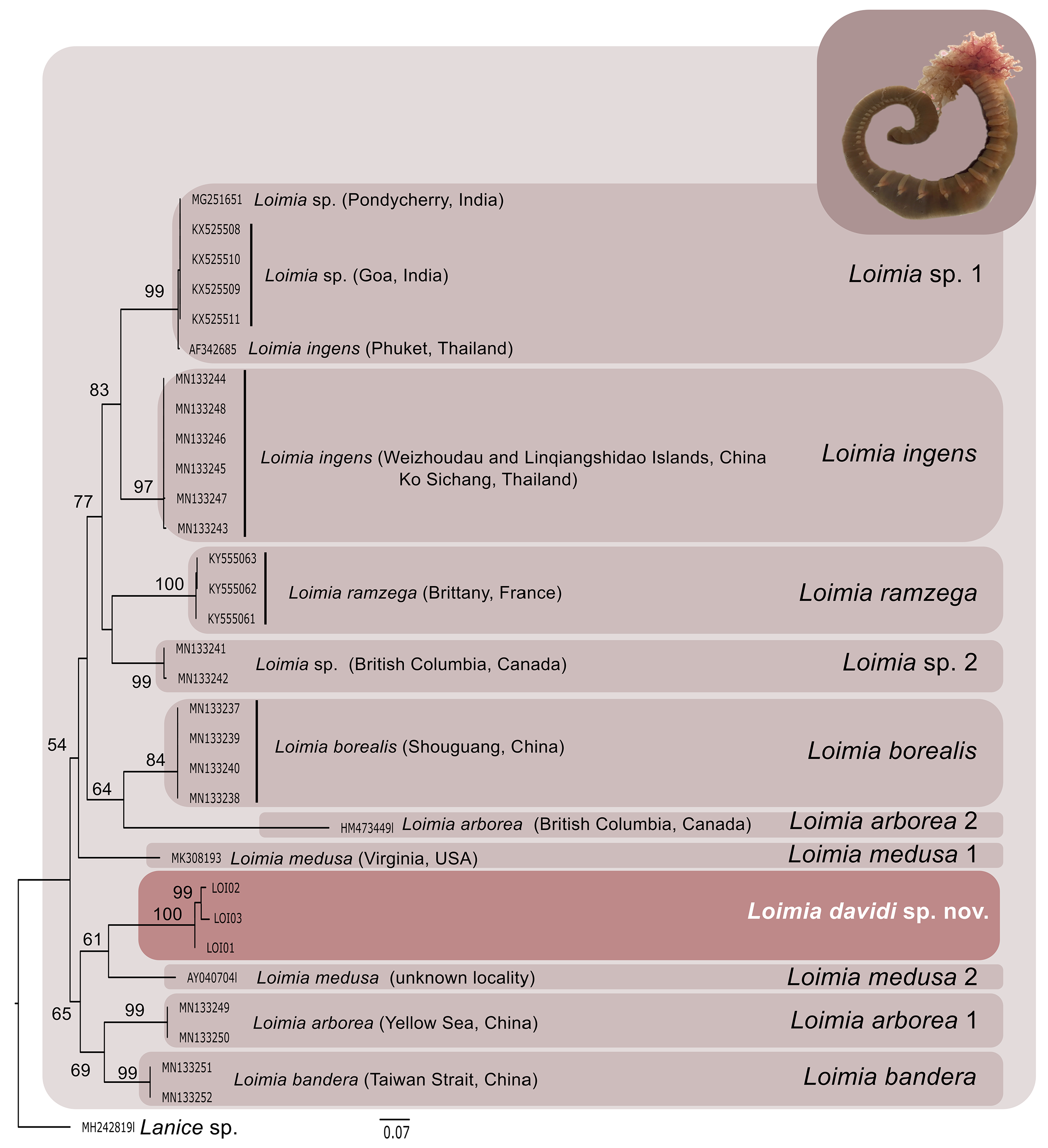
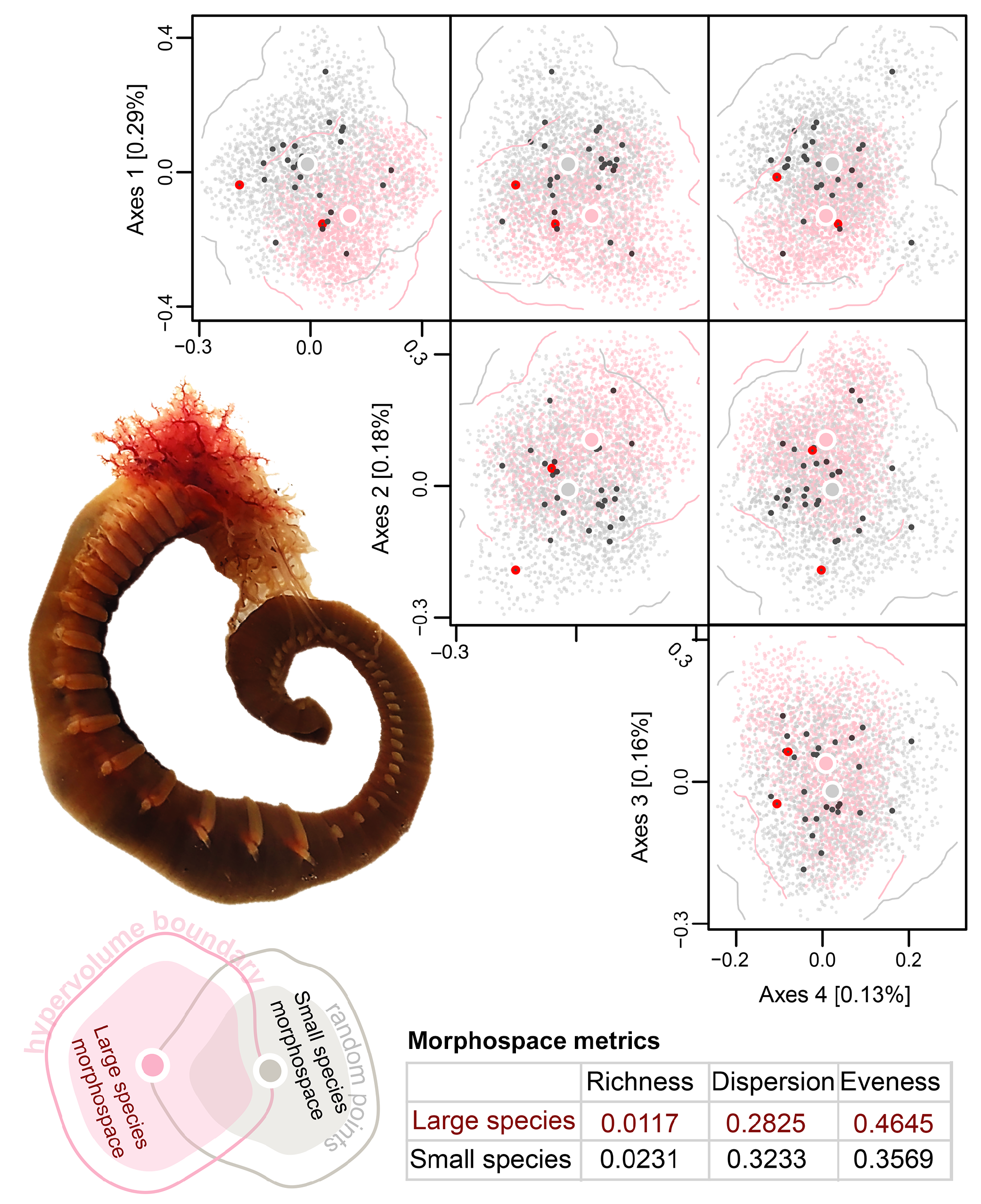
Table 2. Summary of the morphological characters diferring between larger and smaller specimens of Loimia davidi sp. nov.
| Characters | Larger | Smaller |
|---|---|---|
| Thorax length | 57 mm | 19 mm |
| Thorax width | 12 mm | 3.6 mm |
| Tentacles | very abundant | scarce |
| Eyespots | absent | present in some specimens, numerous, diameter progressively decreasing dorsalwards |
| Upper lip | tongue-shaped, wider than longer, not projecting over first pair of lateral lappets | spoon-like, well projecting forward, wider than longer; thicker at base, almost completely hidden ventrally by first pair of lateral lappets |
| Lower lip | square | swollen, with conical tip |
| Lateral lappets | elephant ear-shaped, similar in size, first pair with a poorly-developed joining membrane | quadrangular, with different size, first pair with a well-developed joining membrane |
| Branchiae | dendritic branches arranged in 8 levels | dendritic branches arranged in 4 levels |
| Presence of ventral shields | from segment 2, on 15 segments | from segment 2, on 14 segments |
| Anterior ventral shields | 9, similar width, wider than longer | 10, similar width, wider than longer, but 5–7 clearly narrower |
| Posterior ventral shields | 6, darker, progressively narrowing, giving an overall triangular appearance | 4, whitish, progressively narrowing, giving an overall triangular appearance |
| Fused ventral shields | segments 2–3 | segments 2–3 |
| Transversally non-grooved ventral shields | segments 2–10 | segments 2–10 |
| Ventral shields with 2 transversal grooves | segments 11–12 | segments 11–13 |
| Ventral shields with 3 transversal grooves | segment 13 | segment 14 |
| Ventral shields with> 3 transversal grooves | segments 14–16 | segment 15 |
| Notopodial whitish glandular patches | on first 11 segments | on first 12 segments |
| Types of capillary notochaetae | 3, alimbate, and uni- and bilimbate | 2, alimbate and unilimbate |
| Neuropodia | from segment 5, well-developed | from segment 5, 7 anterior pairs smaller |
| Size of thoracic uncini | 120 µm long, 60 µm wide | 60 µm long, 35 µm wide |
| Crest of thoracic uncini | 5 teeth over main fang, upper one difficult to see | 3 teeth over main fang |
| Heel of thoracic uncini | round, reduced | well-defined, triangular |
| Dorsal button of thoracic uncini | present, very reduced | well-marked |
| Abdomen | single specimen regenerating posterior end | all specimens except one incomplete posteriorly |
| Anterior abdominal segments | 3, similar to thoracic segments | 6, similar to thoracic segment, with better defined segmentation |
| Size of abdominal uncini | 105 µm long, 55 µm wide | 46 µm long, 30 µm wide |
| Crest of abdominal uncini | 5 teeth over main fang, upper one much smaller | 4 teeth over main fang |
| Back of abdominal uncini | slightly curved, 2.5 times as long as prow | curved, 3 times longer as long as prow |
| Heel of abdominal uncini | inconspicuous | well-defined, triangular |
| Dorsal button of abdominal uncini | strongly reduced | well-marked |
| Anus | terminal, surrounded by 18 terminal papillae | terminal, round, surrounded by 7 terminal papillae |
| Anal terminal papillae | small, cirriform; 12 dorso-laterally, broadly grouped in pairs; 6 ventro-laterally, individual | long, conical, forming 2 clearly separated groups of 5 dorsolateral and 2 ventral papillae |
No known copyright restrictions apply. See Agosti, D., Egloff, W., 2009. Taxonomic information exchange and copyright: the Plazi approach. BMC Research Notes 2009, 2:53 for further explanation.
|
Kingdom |
|
|
Phylum |
|
|
Class |
|
|
Order |
|
|
Family |
|
|
Genus |