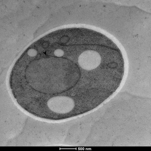
Wild type Saccharomyces cerevisiae cells (S228C background) were treated with 0.5 mM of sodium arsenite for 60 minutes. Then, samples were high pressure frozen and visualized through transmission electron microscopy (TEM). The images show the nucleus of the cells and the black arrow indicates nuclear envelope budding events.
Treated cells were cultured in YPD medium and high pressure frozen in a Wohlwend Compact 3 machine, followed by a short freeze substitution protocol in 2% Uranyl acetate and embedded in HM20 resin. Sections of 70 nm thickness were then contrast stained with 2% Uranyl acetate and Reynold's lead citrate. Pictures were aquired in a Tecnai T12 TEM with a Ceta CMOS 16M camera.